Review
. 2015 Nov 17;7(11):9538-57.
doi: 10.3390/nu7115479.

New Insights into the Pros and Cons of the Clinical Use of Vitamin K Antagonists (VKAs) Versus Direct Oral Anticoagulants (DOACs)

Affiliations
Review

New Insights into the Pros and Cons of the Clinical Use of Vitamin K Antagonists (VKAs) Versus Direct Oral Anticoagulants (DOACs)

Rick H van Gorp et al. Nutrients. .

Abstract

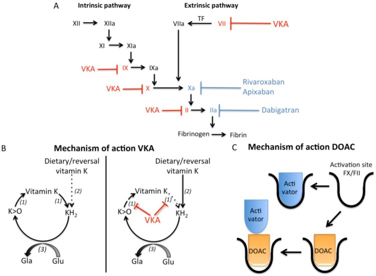
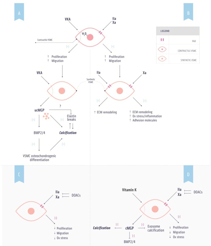
Vitamin K-antagonists (VKA) are the most widely used anticoagulant drugs to treat patients at risk of arterial and venous thrombosis for the past 50 years. Due to unfavorable pharmacokinetics VKA have a small therapeutic window, require frequent monitoring, and are susceptible to drug and nutritional interactions. Additionally, the effect of VKA is not limited to coagulation, but affects all vitamin K-dependent proteins. As a consequence, VKA have detrimental side effects by enhancing medial and intimal calcification. These limitations stimulated the development of alternative anticoagulant drugs, resulting in direct oral anticoagulant (DOAC) drugs, which specifically target coagulation factor Xa and thrombin. DOACs also display non-hemostatic vascular effects via protease-activated receptors (PARs). As atherosclerosis is characterized by a hypercoagulable state indicating the involvement of activated coagulation factors in the genesis of atherosclerosis, anticoagulation could have beneficial effects on atherosclerosis. Additionally, accumulating evidence demonstrates vascular benefit from high vitamin K intake. This review gives an update on oral anticoagulant treatment on the vasculature with a special focus on calcification and vitamin K interaction.

Keywords: DOACs; coumarin; oral anticoagulants; vascular calcification; vitamin K.


Figures

Figure 1
Figure 1
Effects of vitamin K antagonists and direct oral anticoagulants on coagulation. (A) The coagulation cascade can be activated by both the intrinsic and extrinsic pathway, which finally results in activation of thrombin and subsequently fibrin formation. Vitamin K antagonists (VKA) induce anticoagulation via inhibiting activation of the coagulation factors depicted in red (factors X, IX, VII, and II). Direct oral anticoagulants (DOACs) induce anticoagulation via blocking the activity of coagulation factors Xa (rivaroxaban and apixaban) and IIa (dabigatran) depicted in blue; (B) Vitamin K cycle is required to carboxylate, and thus activate, vitamin K dependent proteins. Vitamin K is converted to vitamin hydroquinone (KH2), which is oxidized by γ-glutamylcarboxylase (3) to convert glutamate (Glu) residues in γ-carboxyglutamate (Gla) residues. This reaction results in vitamin K epoxide (K > O), which is recycled to vitamin K through vitamin K epoxide reductase (1). VKA disrupts the vitamin K cycle by inhibiting vitamin K epoxide reductase (VKOR) leading to depletion of vitamin K and uncarboxylated vitamin K dependent proteins. In the liver, the inhibition of warfarin can be circumvented via NAD(P)H quinone reductase (2), which can convert vitamin K into KH2 even in the presence of VKA. In extra-hepatic tissues NAD(P)H quinone reductase activity is ca. 100 fold less, resulting in inactive vitamin K dependent proteins in the presence of VKA; (C) DOACs induce anticoagulation via inhibiting the activity of FXa and FIIa via binding to the activation site.
Figure 2
Figure 2
Effects of anticoagulants and vitamin K on vascular calcification. (A) Mechanism by which vitamin K antagonist (VKA) induces calcification. VKA induces phenotypic switching of contractile to synthetic vascular smooth muscle cells (VSMCs) by increasing oxidative stress resulting in increased proliferation and migration. Synthetic VSMCs secrete uncarboxylated matrix Gla protein (ucMGP) as a result of vitamin K depletion induced by VKA. ucMGP is unable to inhibit bone morphogenetic protein (BMP) 2 and 4, a marker for osteochondrogenic differentiation. Osteochondrogenic VSMCs are prone to calcification. Additionally, ucMGP is directly associated with increased calcification; (B) Thrombin and factor Xa induce non-hemostasis signaling via protease-activated receptors (PARs). Activation of PARs on contractile VSMCs can induce phenotypic switching resulting in increased proliferation and migration. PAR signaling in these synthetic VSMCs increases oxidative stress and adhesion molecules and induces extracellular (ECM) remodeling thereby facilitating calcification; (C) DOAC treatment in combination with (D) supplemental vitamin K administration has the potential to prevent both hypercoagulability and inhibit vascular calcification.
Figure 3
Figure 3
Similarities in bone metabolism and vascular calcification. The calcification process can be divided into three stages: initiation, nucleation and crystal growth. In order to initiate mineralization resting chondrocytes and contractile vascular smooth muscle cells (VSMCs) lose calcification inhibitors. Moreover, vesicles derived from chondrocytes and VSMCs form a nidus for calcification. In both bone metabolism and vascular calcification the matrix plays an important role in the nucleation stage. In bone metabolism, an osteoblast matrix results in proliferation of chondrocytes. Likewise, a calcifying matrix consisting of elastin, collagen and Ca2+ and P accompany vascular calcification. Additionally, contractile VSMCs undergo phenotypic switching resulting in synthetic VSMCs, which have increased proliferation and migration in comparison to contractile VSMCs. Finally, osteoblasts and osteochondrogenic VSMCs induce crystal growth in bone metabolism and vascular calcification, respectively.

References

    1. Mann K.G. Biochemistry and physiology of blood coagulation. Thromb. Haemost. 1999;82:165–174. - PubMed
    1. Macfarlane R.G. An enzyme cascade in the blood clotting mechanism, and its function as a biochemical amplifier. Nature. 1964;202:498–499. doi: 10.1038/202498a0. - DOI - PubMed
    1. Colman R.W. Are hemostasis and thrombosis two sides of the same coin? J. Exp. Med. 2006;203:493–495. doi: 10.1084/jem.20060217. - DOI - PMC - PubMed
    1. Badimon L. Atherosclerosis and thrombosis: Lessons from animal models. Thromb. Haemost. 2001;86:356–365. - PubMed
    1. Fuster V., Stein B., Ambrose J.A., Badimon L., Badimon J.J., Chesebro J.H. Atherosclerotic plaque rupture and thrombosis. Evolving concepts. Circulation. 1990;82:II47–II59. - PubMed

Publication types

MeSH terms