. 2016 Jan 11;55(2):550-4.
doi: 10.1002/anie.201508289. Epub 2015 Nov 23.

Probing the Lipid Annular Belt by Gas-Phase Dissociation of Membrane Proteins in Nanodiscs

Affiliations

Probing the Lipid Annular Belt by Gas-Phase Dissociation of Membrane Proteins in Nanodiscs

Michael T Marty et al. Angew Chem Int Ed Engl. .

Abstract

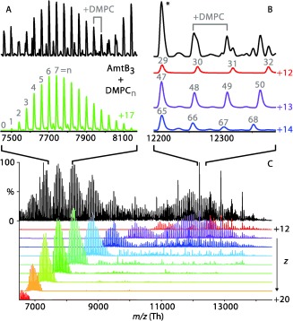
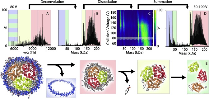
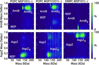
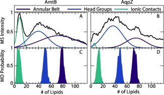
Interactions between membrane proteins and lipids are often crucial for structure and function yet difficult to define because of their dynamic and heterogeneous nature. Here, we use mass spectrometry to demonstrate that membrane protein oligomers ejected from nanodiscs in the gas phase retain large numbers of lipid interactions. The complex mass spectra that result from gas-phase dissociation were assigned using a Bayesian deconvolution algorithm together with mass defect analysis, allowing us to count individual lipid molecules bound to membrane proteins. Comparison of the lipid distributions measured by mass spectrometry with molecular dynamics simulations reveals that the distributions correspond to distinct lipid shells that vary according to the type of protein-lipid interactions. Our results demonstrate that nanodiscs offer the potential for native mass spectrometry to probe interactions between membrane proteins and the wider lipid environment.

Keywords: lipid annulus; mass spectrometry; membrane proteins; nanodiscs; protein-lipid interactions.


Figures

Figure 1
Figure 1
Representative spectrum (black) of AmtB nanodiscs with DMPC and MSP1D1(−) at 130 V CID. Deconvolved charge states from +12 to +20 are shown in various colors (C). Zoomed regions with their predominant charge states show a series of distinct peaks separated by a single DMPC (A and B). The number of lipids bound to the AmtB trimer is annotated in gray. The resonant peak in B is annotated with an asterisk.
Figure 2
Figure 2
Representative mass spectrum and deconvolution of AmtB nanodiscs with POPC and MSP1D1(−). A) Mass spectrum at 80 V CID deconvolves to yield B) the mass distribution. C) The mass distribution shifts to lower‐mass species with increasing collision energy. D) The average mass distribution over all CID states shows four distinct species, which are illustrated in (E): AmtB trimer with a broad mass distribution including tens of bound lipids (red), AmtB trimer with around nine bound lipids (yellow), AmtB monomer (green), and MSP (blue). These four states are similarly shaded in A, B, and D.
Figure 3
Figure 3
Mass defect analysis of membrane protein–nanodisc complexes. Representative plots are shown for a single sample averaged across all CID states for AmtB (top) and AqpZ (bottom) with POPC‐MSP1D1(−) (left), POPC‐MSP1E3D1(−) (middle), and DMPC‐MSP1D1(−) (right). The mass was rounded down to the nearest integer multiple of the lipid mass. Regions for MSP, membrane protein monomers, and membrane protein oligomers are annotated.
Figure 4
Figure 4
Lipid distribution from MS averaged across all three types of nanodiscs for A) AmtB and B) AqpZ compared with lipid distributions determined from MD (C and D, for AmtB and AqpZ, respectively) for the annular belt (purple), the head group shell (blue), and the ionic contacts (cyan). MS distributions were fit to three overlapping Gaussian distributions. MD distributions are a histogram of individual measurements for each frame, which are shown in Figure S6.

References

    1. None
    1. Contreras F. X., Ernst A. M., Wieland F., Brugger B., Cold Spring Harbor Perspect. Biol. 2011, 3, a004705; - PMC - PubMed
    1. Barrera N. P., Zhou M., Robinson C. V., Trends Cell Biol. 2013, 23, 1–8. - PubMed
    1. Bechara C., Robinson C. V., J. Am. Chem. Soc. 2015, 137, 5240–5247. - PubMed
    1. None

Publication types