. 2015 Oct 1;4(1):407-16.
doi: 10.1089/biores.2015.0032. eCollection 2015.

Melatonin Counteracts at a Transcriptional Level the Inflammatory and Apoptotic Response Secondary to Ischemic Brain Injury Induced by Middle Cerebral Artery Blockade in Aging Rats

Affiliations

Melatonin Counteracts at a Transcriptional Level the Inflammatory and Apoptotic Response Secondary to Ischemic Brain Injury Induced by Middle Cerebral Artery Blockade in Aging Rats

Sergio D Paredes et al. Biores Open Access. .

Abstract

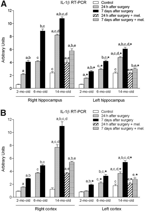
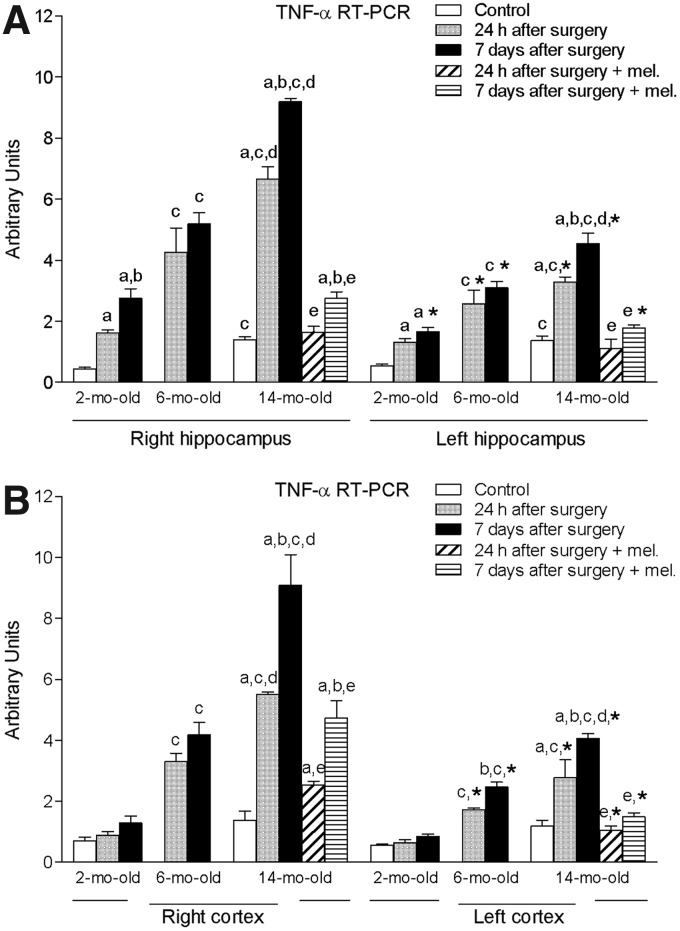
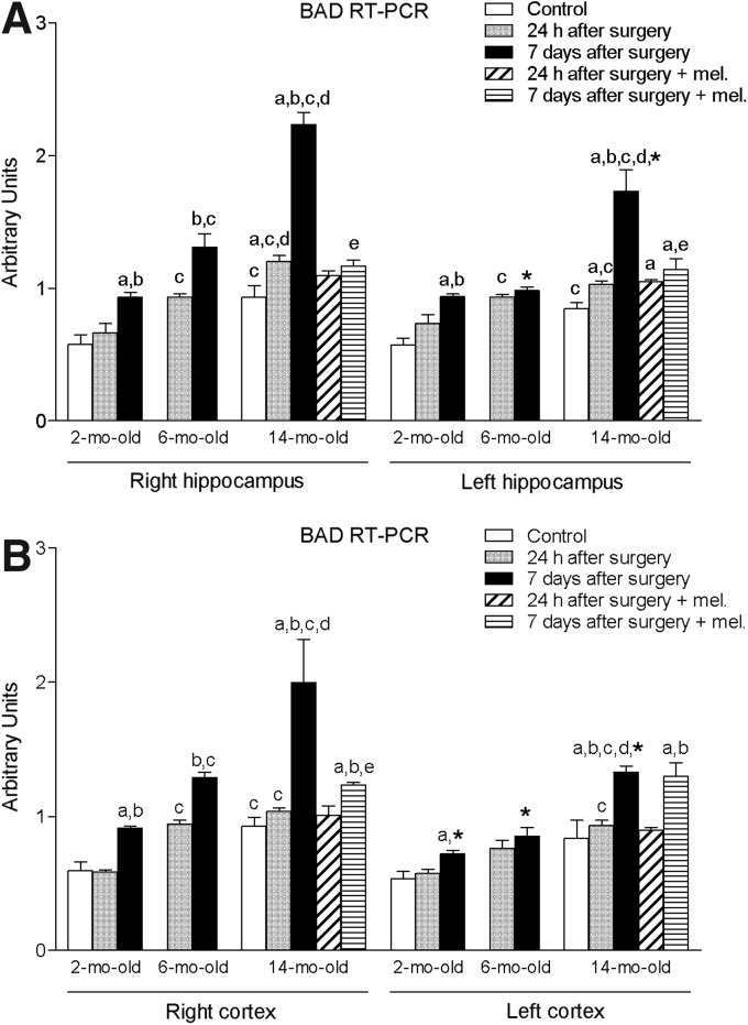
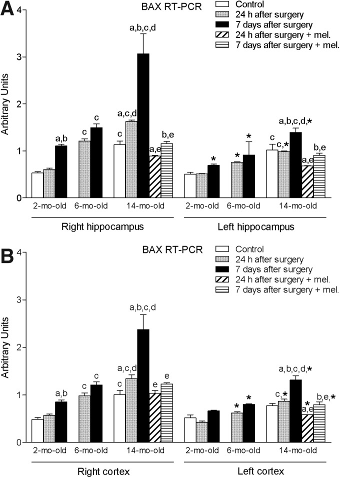
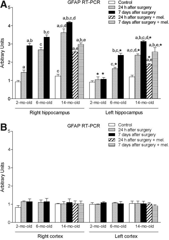
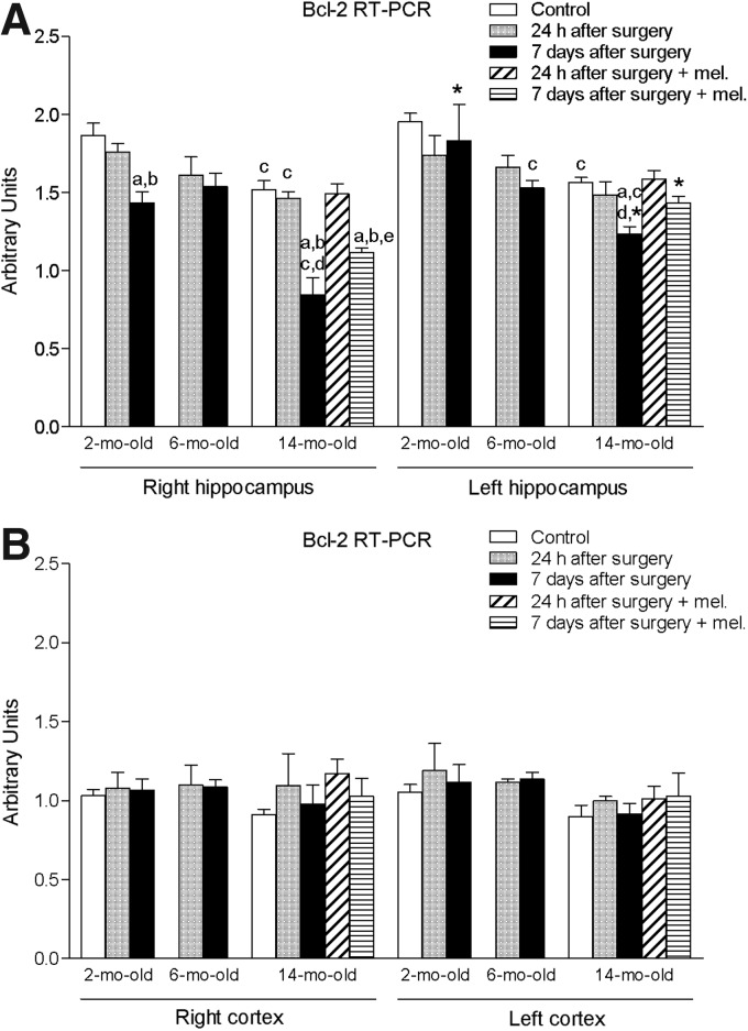
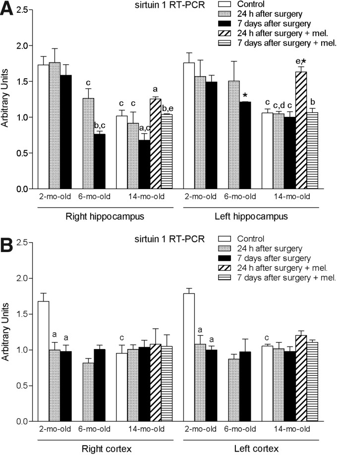
Aging increases oxidative stress and inflammation. Melatonin counteracts inflammation and apoptosis. This study investigated the possible protective effect of melatonin on the inflammatory and apoptotic response secondary to ischemia induced by blockade of the right middle cerebral artery (MCA) in aging male Wistar rats. Animals were subjected to MCA obstruction. After 24 h or 7 days of procedure, 14-month-old nontreated and treated rats with a daily dose of 10 mg/kg melatonin were sacrificed and right and left hippocampus and cortex were collected. Rats aged 2 and 6 months, respectively, were subjected to the same brain injury protocol, but they were not treated with melatonin. mRNA expression of interleukin-1 beta (IL-1β), tumor necrosis factor alpha (TNF-α), Bcl-2-associated death promoter (BAD), Bcl-2-associated X protein (BAX), glial fibrillary acidic protein (GFAP), B-cell lymphoma 2 (Bcl-2), and sirtuin 1 was measured by reverse transcription-polymerase chain reaction. In nontreated animals, a significant time-dependent increase in IL-1β, TNF-α, BAD, and BAX was observed in the ischemic area of both hippocampus and cortex, and to a lesser extent in the contralateral hemisphere. Hippocampal GFAP was also significantly elevated, while Bcl-2 and sirtuin 1 decreased significantly in response to ischemia. Aging aggravated these changes. Melatonin administration was able to reverse significantly these alterations. In conclusion, melatonin may ameliorate the age-dependent inflammatory and apoptotic response secondary to ischemic cerebral injury.

Keywords: aging; brain; ischemia; melatonin; middle cerebral artery blockade.


Figures

<b>FIG. 1.</b>
FIG. 1.
mRNA expression of interleukin-1 beta (IL-1β) in right and left hippocampus (A) and cortex (B) in nontreated rats aged 2 and 6 months and nontreated and melatonin-treated rats aged 14 months in control conditions and after 24 h or 7 days of being subjected to middle cerebral artery blockade. Each value represents the mean ± standard error of the mean of five determinations performed in duplicate. ap < 0.05 values obtained in the control group; bp < 0.05 values obtained at 24 h; cp < 0.05 values obtained in the 2-month-old group; dp < 0.05 values obtained in the 6-month-old group; ep < 0.05 values obtained in their respective nontreated group; *p < 0.05 values obtained in the corresponding contralateral tissue.
<b>FIG. 2.</b>
FIG. 2.
mRNA expression of tumor necrosis factor alpha (TNF-α) in right and left hippocampus (A) and cortex (B) in nontreated rats aged 2 and 6 months and nontreated and melatonin-treated rats aged 14 months in control conditions and after 24 h or 7 days of being subjected to middle cerebral artery blockade. Each value represents the mean ± standard error of the mean of five determinations performed in duplicate. ap < 0.05 values obtained in the control group; bp < 0.05 values obtained at 24 h; cp < 0.05 values obtained in the 2-month-old group; dp < 0.05 values obtained in the 6-month-old group; ep < 0.05 values obtained in their respective nontreated group; *p < 0.05 values obtained in the corresponding contralateral tissue.
<b>FIG. 3.</b>
FIG. 3.
mRNA expression of Bcl-2-associated death promoter (BAD) in right and left hippocampus (A) and cortex (B) in nontreated rats aged 2 and 6 months and nontreated and melatonin-treated rats aged 14 months in control conditions and after 24 h or 7 days of being subjected to middle cerebral artery blockade. Each value represents the mean ± standard error of the mean of five determinations performed in duplicate. ap < 0.05 values obtained in the control group; bp < 0.05 values obtained at 24 h; cp < 0.05 values obtained in the 2-month-old group; dp < 0.05 values obtained in the 6-month-old group; ep < 0.05 values obtained in their respective nontreated group; *p < 0.05 values obtained in the corresponding contralateral tissue.
<b>FIG. 4.</b>
FIG. 4.
mRNA expression of Bcl-2-associated X protein (BAX) in right and left hippocampus (A) and cortex (B) in nontreated rats aged 2 and 6 months and nontreated and melatonin-treated rats aged 14 months in control conditions and after 24 h or 7 days of being subjected to middle cerebral artery blockade. Each value represents the mean ± standard error of the mean of five determinations performed in duplicate. ap < 0.05 values obtained in the control group; bp < 0.05 values obtained at 24 h; cp < 0.05 values obtained in the 2-month-old group; dp < 0.05 values obtained in the 6-month-old group; ep < 0.05 values obtained in their respective nontreated group; *p < 0.05 values obtained in the corresponding contralateral tissue.
<b>FIG. 5.</b>
FIG. 5.
mRNA expression of glial fibrillary acidic protein (GFAP) in right and left hippocampus (A) and cortex (B) in nontreated rats aged 2 and 6 months and nontreated and melatonin-treated rats aged 14 months in control conditions and after 24 h or 7 days of being subjected to middle cerebral artery blockade. Each value represents the mean ± standard error of the mean of five determinations performed in duplicate. ap < 0.05 values obtained in the control group; bp < 0.05 values obtained at 24 h; cp < 0.05 values obtained in the 2-month-old group; dp < 0.05 values obtained in the 6-month-old group; ep < 0.05 values obtained in their respective nontreated group; *p < 0.05 values obtained in the corresponding contralateral tissue.
<b>FIG. 6.</b>
FIG. 6.
mRNA expression of B-cell lymphoma 2 (Bcl-2) in right and left hippocampus (A) and cortex (B) in nontreated rats aged 2 and 6 months and nontreated and melatonin-treated rats aged 14 months in control conditions and after 24 h or 7 days of being subjected to middle cerebral artery blockade. Each value represents the mean ± standard error of the mean of five determinations performed in duplicate. ap < 0.05 values obtained in the control group; bp < 0.05 values obtained at 24 h; cp < 0.05 values obtained in the 2-month-old group; dp < 0.05 values obtained in the 6-month-old group; ep < 0.05 values obtained in their respective nontreated group; *p < 0.05 values obtained in the corresponding contralateral tissue.
<b>FIG. 7.</b>
FIG. 7.
mRNA expression of sirtuin 1 in right and left hippocampus (A) and cortex (B) in nontreated rats aged 2 and 6 months and nontreated and melatonin-treated rats aged 14 months in control conditions and after 24 h or 7 days of being subjected to middle cerebral artery blockade. Each value represents the mean ± standard error of the mean of five determinations performed in duplicate. ap < 0.05 values obtained in the control group; bp < 0.05 values obtained at 24 h; cp < 0.05 values obtained in the 2-month-old group; dp < 0.05 values obtained in the 6-month-old group; ep < 0.05 values obtained in their respective nontreated group; *p < 0.05 values obtained in the corresponding contralateral tissue.

References

    1. Wong R, Bath PM, Kendall D, et al. . Progesterone and cerebral ischaemia: the relevance of ageing. J Neuroendocrinol. 2013;25:1088–1094 - PubMed
    1. Allen CL, Bayraktutan U. Risk factors for ischaemic stroke. Int J Stroke. 2008;3:105–116 - PubMed
    1. Rojas JI, Zurru MC, Romano M, et al. . Acute ischemic stroke and transient ischemic attack in the very old—risk factor profile and stroke subtype between patients older than 80 years and patients aged less than 80 years. Eur J Neurol. 2007;14:895–899 - PubMed
    1. Menken M, Munsat TL, Toole JF. The global burden of disease study: implications for neurology. Arch Neurol. 2000;57:418–420 - PubMed
    1. Alhusban A, Fagan SC. Secondary prevention of stroke in the elderly: a review of the evidence. Am J Geriatr Pharmacother. 2011;9:143–152 - PubMed

LinkOut - more resources