Review
. 2016 Jul;66(4):326-36.
doi: 10.3322/caac.21333. Epub 2015 Nov 23.

Multiparametric prostate magnetic resonance imaging in the evaluation of prostate cancer

Affiliations
Review

Multiparametric prostate magnetic resonance imaging in the evaluation of prostate cancer

Baris Turkbey et al. CA Cancer J Clin. 2016 Jul.

Abstract

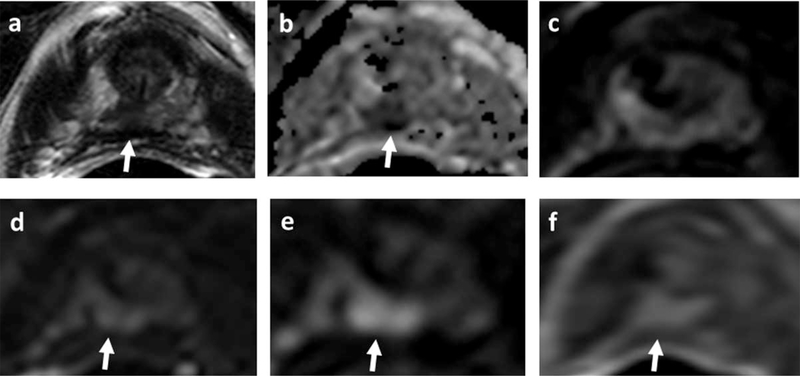
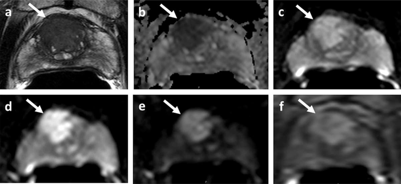
Imaging has traditionally played a minor role in the diagnosis and staging of prostate cancer. However, recent controversies generated by the use of prostate-specific antigen (PSA) screening followed by random biopsy have encouraged the development of new imaging methods for prostate cancer. Multiparametric magnetic resonance imaging (mpMRI) has emerged as the imaging method best able to detect clinically significant prostate cancers and to guide biopsies. Here, the authors explain what mpMRI is and how it is used clinically, especially with regard to high-risk populations, and we discuss the impact of mpMRI on treatment decisions for men with prostate cancer. CA Cancer J Clin 2016;66:326-336. © 2015 American Cancer Society.

Keywords: active surveillance; high risk; multiparametric magnetic resonance imaging (MRI); prostate cancer.


Figures

FIGURE 1.
FIGURE 1.
A 70-Year-Old Biopsy-Naïve Patient With a Prostate-Specific Antigen (PSA) Level of 9.97 ng/mL Underwent Initial Multiparametric Magnetic Resonance Imaging (mpMRI) Evaluation. (a) Axial T2-weighted images identify an area of low signal intensity in the midline distal apical peripheral zone (arrow). (b) Axial apparent diffusion coefficient maps of diffusion-weighted imaging (DWI) identify the hypointense lesion in the same midline apical peripheral zone location (arrow). The lesion is invisible on (c) low b-value (b = 560) DWI and (d) is barely visible on b = 1000 DWI (arrow); however, (e) the high b-value DWI (b = 2000) shows the entire lesion as a hyperintense signal focus at the same location (arrow) (f) Dynamic contrast-enhanced image shows an early, focal hyperenhancing lesion in the midline apical peripheral zone (arrow). The patient underwent robotic-assisted radical prostatectomy, at which final pathology confirmed Gleason 4 + 4 disease. At 3-month postsurgical follow–up, the patient had a PSA of <0.01 ng/mL.
FIGURE 2.
FIGURE 2.
A 65-Year-Old African American Male Presenting With a Prostate-Specific Antigen (PSA) Level of 14.91 ng/mL Elected to Undergo Multiparametric Magnetic Resonance Imaging (mpMRI) for Further Evaluation. He had a positive family history for early diagnosed prostate cancer in a first-degree relative and history of 4 prior negative biopsies (using the standard of care transrectal ultrasound [TRUS] approach). (a) An axial T2-weighted image shows a large midline-to-right apical base, anterior transition zone lesion (arrow). (b) Axial apparent diffusion coefficient map of diffusion-weighted imaging (DWI) identifies the large anterior lesion with strong hypointense appearance (arrow). (c) Lower b-value DWI (b = 560) (arrow) and (d) intermediate b-value (b = 1000) DWI (arrow) show the lesion; however, (e) high b-value DWI (b = 2000) highlights the large anterior lesion with strong enhancement and suppression of the normal prostate tissue (arrow). (f) Axial dynamic contrast-enhanced image shows an early, large, focal hyperenhancement of the same lesion (arrow). The patient underwent targeted MRI/TRUS fusion-guided biopsy along with standard of care biopsy. The biopsy resulted in 1 of 12 TRUS cores positive and 3 of 4 MRI/TRUS-guided biopsy cores positive, and the highest Gleason score was 41 4 (25%). The patient elected to undergo robotic-assisted radical prostatectomy, and final wholemount histopathology confirmed a Gleason 414 pattern in 40% of the prostate with a secondary Gleason 4 + 5 pattern.

References

    1. American Cancer Society. Cancer Facts & Figures 2014. Atlanta, GA; American Cancer Society; 2014.
    1. Moyer VA; US Preventive Services Task Force. Screening for prostate cancer: US Preventive Services Task Force recommendation statement. Ann Intern Med. 2012; 157:120–135. - PubMed
    1. Turkbey B, Choyke PL. Decade in review-imaging: a decade in image-guided prostate biopsy. Nat Rev Urol. 2014;11:611–612. - PMC - PubMed
    1. Stamey TA, McNeal JE, Yemoto CM, Sigal BM, Johnstone IM. Biological determinants of cancer progression in men with prostate cancer. JAMA. 1999;281:1395–1400. - PubMed
    1. Ploussard G, Epstein JI, Montironi R, et al. The contemporary concept of significant versus insignificant prostate cancer. Eur Urol. 2011;60:291–303. - PubMed