Review
. 2016 Feb;37(1):2-36.
doi: 10.1210/er.2015-1090. Epub 2015 Nov 23.

Thyroglobulin From Molecular and Cellular Biology to Clinical Endocrinology

Affiliations
Review

Thyroglobulin From Molecular and Cellular Biology to Clinical Endocrinology

Bruno Di Jeso et al. Endocr Rev. 2016 Feb.

Abstract

Thyroglobulin (Tg) is a vertebrate secretory protein synthesized in the thyrocyte endoplasmic reticulum (ER), where it acquires N-linked glycosylation and conformational maturation (including formation of many disulfide bonds), leading to homodimerization. Its primary functions include iodide storage and thyroid hormonogenesis. Tg consists largely of repeating domains, and many tyrosyl residues in these domains become iodinated to form monoiodo- and diiodotyrosine, whereas only a small portion of Tg structure is dedicated to hormone formation. Interestingly, evolutionary ancestors, dependent upon thyroid hormone for development, synthesize thyroid hormones without the complete Tg protein architecture. Nevertheless, in all vertebrates, Tg follows a strict pattern of region I, II-III, and the cholinesterase-like (ChEL) domain. In vertebrates, Tg first undergoes intracellular transport through the secretory pathway, which requires the assistance of thyrocyte ER chaperones and oxidoreductases, as well as coordination of distinct regions of Tg, to achieve a native conformation. Curiously, regions II-III and ChEL behave as fully independent folding units that could function as successful secretory proteins by themselves. However, the large Tg region I (bearing the primary T4-forming site) is incompetent by itself for intracellular transport, requiring the downstream regions II-III and ChEL to complete its folding. A combination of nonsense mutations, frameshift mutations, splice site mutations, and missense mutations in Tg occurs spontaneously to cause congenital hypothyroidism and thyroidal ER stress. These Tg mutants are unable to achieve a native conformation within the ER, interfering with the efficiency of Tg maturation and export to the thyroid follicle lumen for iodide storage and hormonogenesis.


Figures

Figure 1.
Figure 1.
TH synthesis and secretion. The thyroid gland is comprised of follicles and surrounding blood vessels. Follicles are the functional unit for TH synthesis and secretion. Thyroid follicles are a spherical monolayer of polarized thyrocytes with the basolateral surface facing the bloodstream and the apical surface delimiting a central follicle lumen. Iodine is taken up by thyrocytes by the action of the NIS exploiting the Na+ electrochemical gradient generated by the Na+/K+ ATPase. I crosses the apical membrane and reaches the follicular lumen (I efflux) by the action of the anion exchanger Pendrin and/or other transporters. I is oxidized by TPO in the presence of H2O2 generated by the Duox2. Reactive iodide is covalently linked to selected tyrosyl residues of Tg to generate MIT and/or DIT. Under these oxidizing conditions, MIT and DIT are coupled to form THs T3 and T4. Newly synthesized Tg, TPO, and Duox2 are made in the ER and fold with the aid of general (CNX, CRT, etc) and dedicated (Duox maturation factor 2 for Duox2) chaperones before transport to the apical membrane. Newly secreted and iodinated Tg is endocytosed and degraded in lysosomes, and THs undergo transport to the bloodstream. The iodide from uncoupled MIT and DIT is recycled by tyrosine dehalogenase (Dehal1). Intact Tg is normally found in serum at low levels, but may exhibit increased concentrations in serum under conditions of increased thyroid cell mass (279), TSH stimulation (279), Graves' disease (280), subacute thyroiditis (281), and thyroid carcinoma. In the latter condition, post-thyroidectomy and ablation of residual thyroid tissue, serum Tg levels are used to monitor residual disease (282).
Figure 2.
Figure 2.
Cartoon view of Tg structure and function. Tg can be considered to have two main regions: the N-terminal, three-fourths are composed of cysteine-rich repeated motifs; and the carboxyl-terminal, one-fourth is composed of the ChEL domain. Thyroid hormonogenesis in Tg of most vertebrates favors T4 at a conserved site near the N terminus, T3 at a conserved site near the C terminus, and iodide storage via iodotyrosines located at various sites along the polypeptide chain.
Figure 3.
Figure 3.
Evolutionary progression of TH synthesis (from left to right). TH synthesis preceded the appearance of thyroid follicles or modern vertebrate Tg. In simple chordates such as the nonvertebrate amphioxus and Ciona intestinalis, TH synthesis occurs in the endostyle, an exocrine gland located in the pharyngeal region. Typical thyroid follicles can be recognized in the vertebrate lamprey, but only at the time of metamorphosis. In these simpler organisms, many orthologs of genes involved in TH synthesis have been identified, but not a complete modern-day vertebrate Tg—only various gene products containing Tg type-1 repeat domains or others homologous to ChEL. A complete modern-day vertebrate Tg is detectable in all teleost fishes and above.
Figure 4.
Figure 4.
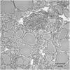
Thyroid morphology from thresher shark. This figure shows abundant extracellular colloidal protein, highly suggestive of Tg accumulation. [Reproduced from J. D. Borucinska and M. Tafur: Comparison of histological features, and description of histopathological lesions in thyroid glands from three species of free-ranging sharks from the northwestern Atlantic, the blue shark, Prionace glauca (L.), the shortfin mako, Isurus oxyrhinchus Rafinesque, and the thresher, Alopias vulpinus (Bonnaterre). J Fish Dis. 2009;32:785–793 (27), with permission. © John Wiley & Sons Ltd.]
Figure 5.
Figure 5.
Schematic of the regional primary structure of Tg. The 270-kB homo sapiens Tgn gene (GenBank accession no. NT_008046 [older] or NG_015832 [current]) located on chromosome 8q24.2–8q24.3 (28–33) contains 48 exons separated by introns of varying sizes up to 64 kB, ultimately encoding an 8.5-kB mRNA (also see Figure 13). Within the encoded polypeptide shown in the figure, the various domains, insertions, and connecting sequences are not drawn to scale. The 11 Tg type-1 domains are in dark gray with four nonhomologous insertions designated as white vertical bars. The three type-2 repeats are in white, and five type-3 repeats are in light gray. The ChEL domain follows, and Tg concludes with a short stretch of nondomain sequence. The numbers above are approximate positions based on numbering the mature Tg peptide as position 1. The numbers below indicate the number of Cys residues in each domain, and numbers below the arrows specify Cys residues contained within linker and hinge and in a segment connecting type-2 and type-3 repeats.
Figure 6.
Figure 6.
Modular architecture of vertebrate Tg type-1 domain-containing proteins. The various domains and connecting sequences are not to scale. Tg type-1 domains are in gray, with other domains in white. AC, testican acidic region; CL, invariant chain class II-associated invariant chain peptide (CLIP) fragment; EC, SMOC, and Testican, extracellular calcium-binding domains; EGF, epidermal growth factor–like domain; FS, follistatin-like domain; G1, nidogen G1 domain; G2, nidogen G2 domain; IC, invariant chain intracellular domain; IGFBP, IGF binding protein domain; LY, low-density lipoprotein receptor-like YWTD repeat; SMOC, SMOC-unique domain; Tg1, Tg type-1 repeat; Tg2, Tg type-2 repeat; Tg3, Tg type-3 repeat; TM, transmembrane region; TST, testican-unique domain.
Figure 7.
Figure 7.
Targeting Tg type-1 domains 1–10 with protease sensitivity, glycosylation, and iodination: insertions in the basic domain. A, Tg domains are aligned according to Ref. . Conserved residues are indicated when present at least five times (in bold), with the exception of Y130 and Y685; otherwise they are generically indicated as X. Numbers within the sequence indicate the length of segments without similarities between domains. The four large insertions in domains 1.3 (two), 1.7, and 1.8, and the linker (248 residues between domains 1.4 and 1.5) appear to be regions of preferential proteolytic sensitivity, glycosylation, and iodination. B, Spacing between cysteine residues in proteins possessing type-1 domains. Disulfide linkages and spacing between the cysteines for GA733–2 and IGFBP-1,6 are from Refs. and , respectively. In Tg, the spacings between linked cysteines in the basic domain common to GA733–2 and IGFBP-1,6 are variable as a result of four major (and some minor) insertions (see Figures 5 and 7A), whereas other spacings between adjacent disulfide linked pairs are highly conserved and equal to those of the GA733–2 and IGFBP-1,6.
Figure 8.
Figure 8.
The fold of Tg type-1 domain. Superimposition of three-dimensional structures of the Tg type-1 domain of IGFBP-6 (light gray) (46) and p41 fragment (dark gray) (58). The three disulfide bonds (shown as sticks) determine three loops, with the first containing an α-helix in its N-terminal part, and the second connecting an antiparallel β-sheet held in place by the second disulfide bond. Between various members of the family bearing this domain, including Tg itself, the domain structure tends to differ in the elaboration of sequences within loop I and loop II.
Figure 9.
Figure 9.
The Cholinesterase family fold. The ChEL domain of Tg shows homology with members of the α-β hydrolase fold superfamily (68, 69) including AChE (66, 70), whose structure is shown. Like ChEL, AChE forms a homodimer held together by a four-helix bundle composed of helices α-7/8 and α-10 from each subunit (gray cylindrical segments) (71–73).
Figure 10.
Figure 10.
Folding and secretion of truncated Tg segments. A, Autonomous folding and secretion of distinct Tg truncation mutations. From this we propose that folding the segment shown at bottom, including both linker and hinge regions, may be rate-limiting for Tg folding and export. See Section V for details. B, Intramolecular chaperone function of ChEL. ChEL rescues secretion of Tg region I-II-III. However, isolated II-III easily folds in the ER and is also rapidly secreted. Thus, the intramolecular rescue by ChEL appears to be directed mostly toward the improved folding stability of a portion of Tg region I. However, ChEL rescue of region I folding/stability requires the presence of II-III. See Section V for details.
Figure 11.
Figure 11.
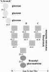
Structure of N-linked core oligosaccharide added to the polypeptide chains.
Figure 12.
Figure 12.
Acceptor and donor splice site mutations. Acceptor and donor splice site mutations produce exon skipping, when alternative natural splice sites are used (A); or altered exons, when cryptic splice sites are used (B). In exon skipping, if a donor splice site is mutated, skipping of the upstream exon may occur (A.i); or if an acceptor splice site is mutated, skipping of the downstream exon may occur (A.ii). In altered exons, if the cryptic splice site is located in an exon, part of a normal exon may be excluded (B.i); or, if the cryptic splice site is located in an intron, it may result in exonization of intronic sequences (B.ii). Both exon skipping and cryptic splice site activation may result in either a frameshift and downstream generation of a premature stop codon (the mRNA is then often subjected to nonsense-mediated mRNA decay), or preservation of the reading frame (the mRNA is normally translated, and a protein carrying an internal deletion is synthesized).
Figure 13.
Figure 13.
Alignment of Tgn exons with Tg repeat domains. Amino acid numbering is indicated both above the Tg protein structure and below the Tgn exons. The sequences are not drawn to scale. At the exon/exon boundary, a single number indicates that the nucleotide coding triplet is split by the boundary, whereas two numbers indicate that the adjacent nucleotides coding triplets are not split by the exon/exon boundary. A general exon/domain correspondence is notable in most type-1 repeats involving one exon for one repeat domain (repeats 1–2, 1–4, 1–7, and 1–10) or one exon for two repeats (1–5 plus 1–6). However, even when a repeat is interrupted by introns, the repeat boundaries correspond to exon boundaries, with the exception of repeat 1–1.

Similar articles

Cited by

References

    1. Pharoah PO, Buttfield IH, Hetzel BS. Neurological damage to the fetus resulting from severe iodine deficiency during pregnancy. Lancet. 1971;1:308–310. - PubMed
    1. Frati L, Bilstad J, Edelhoch H, Rall JE, Salvatore G. Biosynthesis of the 27 S thyroid iodoprotein. Arch Biochem Biophys. 1974;162:126–134. - PubMed
    1. Lee J, Di Jeso B, Arvan P. The cholinesterase-like domain of thyroglobulin functions as an intramolecular chaperone. J Clin Invest. 2008;118:2950–2958. - PMC - PubMed
    1. Novinec M, Kordis D, Turk V, Lenarcic B. Diversity and evolution of the thyroglobulin type-1 domain superfamily. Mol Biol Evol. 2006;23:744–755. - PubMed
    1. Takagi Y, Omura T, Go M. Evolutionary origin of thyroglobulin by duplication of esterase gene. FEBS Lett. 1991;282:17–22. - PubMed

Publication types

MeSH terms