. 2016 Jan;18(1):87-99.
doi: 10.1038/ncb3274. Epub 2015 Nov 23.

Conserved molecular interactions in centriole-to-centrosome conversion

Affiliations

Conserved molecular interactions in centriole-to-centrosome conversion

Jingyan Fu et al. Nat Cell Biol. 2016 Jan.

Abstract

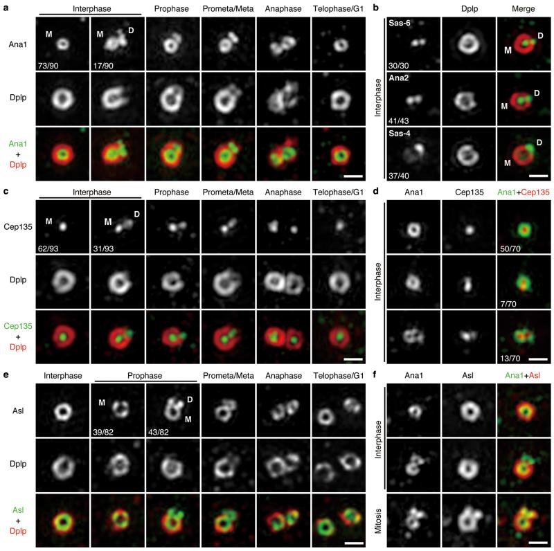
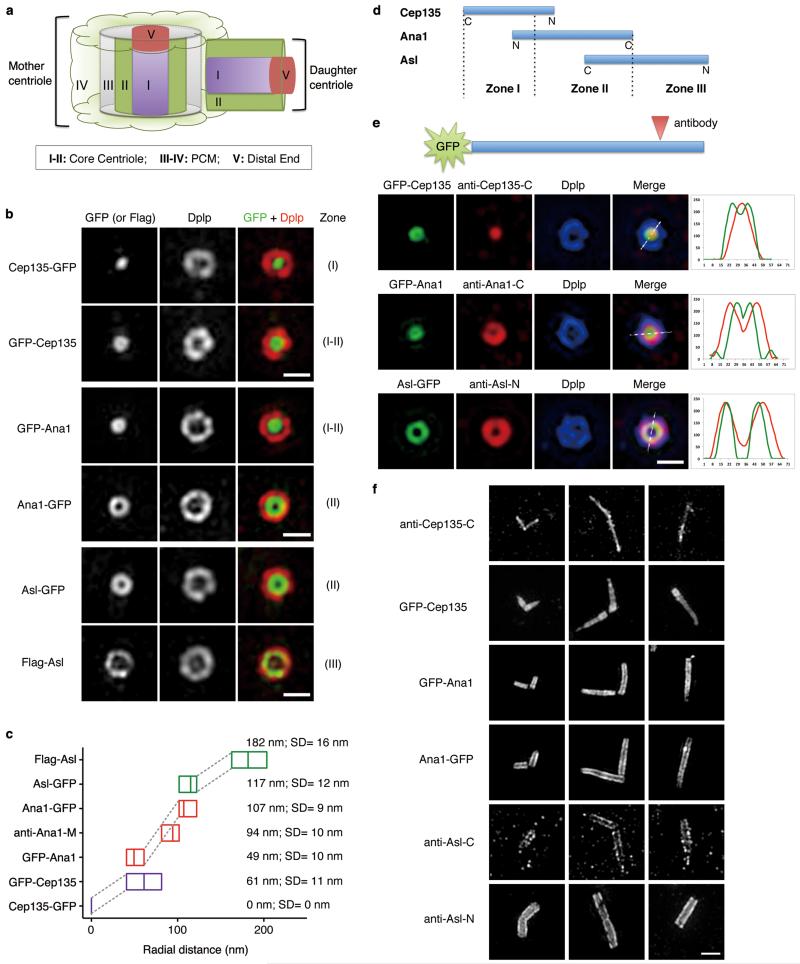
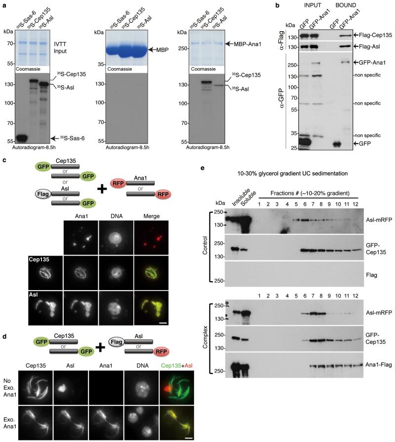
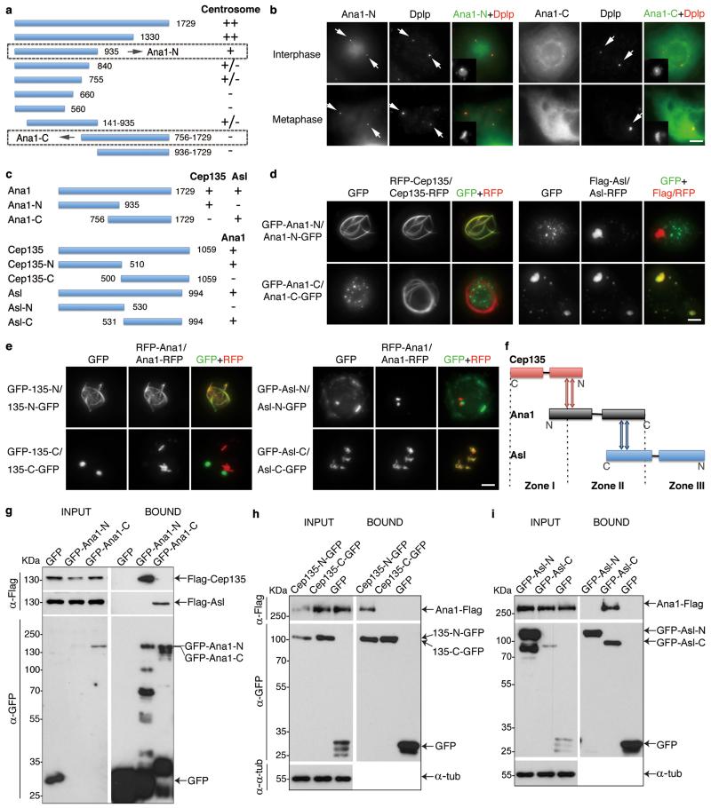
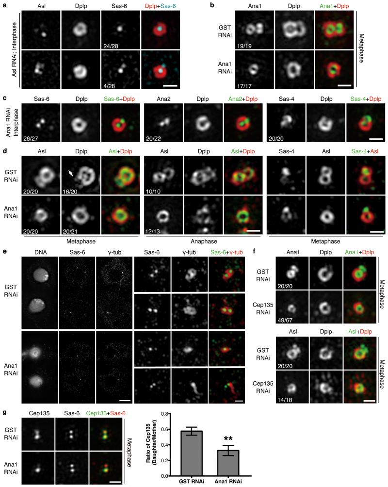
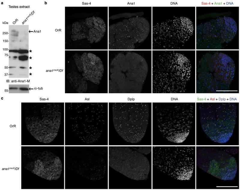
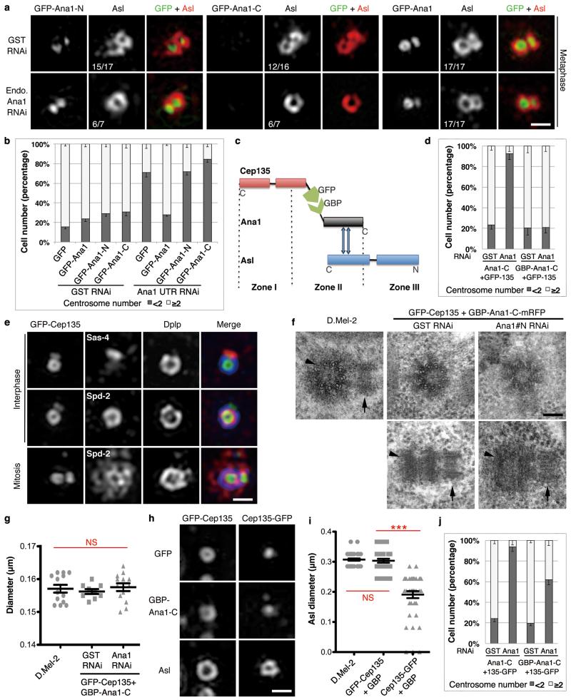
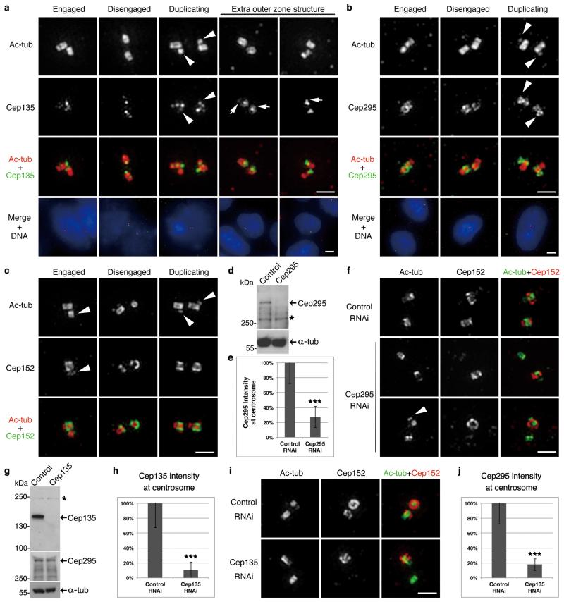
Centrioles are required to assemble centrosomes for cell division and cilia for motility and signalling. New centrioles assemble perpendicularly to pre-existing ones in G1-S and elongate throughout S and G2. Fully elongated daughter centrioles are converted into centrosomes during mitosis to be able to duplicate and organize pericentriolar material in the next cell cycle. Here we show that centriole-to-centrosome conversion requires sequential loading of Cep135, Ana1 (Cep295) and Asterless (Cep152) onto daughter centrioles during mitotic progression in both Drosophila melanogaster and human. This generates a molecular network spanning from the inner- to outermost parts of the centriole. Ana1 forms a molecular strut within the network, and its essential role can be substituted by an engineered fragment providing an alternative linkage between Asterless and Cep135. This conserved architectural framework is essential for loading Asterless or Cep152, the partner of the master regulator of centriole duplication, Plk4. Our study thus uncovers the molecular basis for centriole-to-centrosome conversion that renders daughter centrioles competent for motherhood.


Figures

Figure 1
Figure 1. Sequential loading of Cep135, Ana1 and Asl during centriole-to-centrosome conversion
(a) Localization of endogenous Ana1 throughout cell cycle. D.Mel-2 cells were immunostained to reveal Ana1, Dplp (Zone III marker) and DNA (to distinguish cell cycle stages, see Supplementary Fig. 1a), and analyzed by 3D-SIM. Interphase, prophase, prometaphase-metaphase, anaphase and telophase-G1 centrioles were shown. Only 19% (n=90) of interphase centrosomes have Ana1 signal on daughter centriole, suggesting Ana1 is recruited to daughter in late interphase. M, mother centriole; D, daughter. (b) Centrioles immunostained to reveal Dplp (red), DNA (not shown) and Sas-6, Ana2 or Sas-4 (green). Sas-6, Ana2 and Sas-4 appear on both mother and daughter centrioles in majority of interphase centrosomes (100%, 95% and 92%, respectively), indicating their early loading onto the daughters. n=30, 43 and 40 centrosomes. (c) Localization of endogenous Cep135 throughout cell cycle. 33% (n=93) of interphase centrosomes have Cep135 signal on the daughter. See Supplementary Fig. 1b for DNA staining. (d) Centrioles immunostained to reveal Ana1 (green), Cep135 (red) and DNA (not shown). Only 10% (n=70) of interphase centrosomes show earlier (less distinct) loading of Cep135 to the daughter than Ana1, indicating a close temporal recruitment of these two. (e) Localization of endogenous Asl throughout cell cycle. Asl starts to load on the daughter from prophase, when 52% (n=82) of centrosomes show Asl signal on the daughter. See Supplementary Fig. 1c for DNA staining. (f) Centrioles immunostained to reveal Ana1 (green), Asl (red) and DNA (not shown). Recruitment of Asl to the daughter happens later than Ana1. All scale bars, 500 nm.
Figure 2
Figure 2. Cep135, Ana1 and Asl are extended molecules that span from inner to outer centriole
(a) Scheme showing different Zones in Drosophila centrosome (modified from ref. 9). (b) D.Mel-2 cells constitutively expressing GFP-tagged Cep135, Ana1 or Asl were immunostained to reveal Dplp and DNA (not shown). Cells expressing Flag-Asl were immunostained with additional anti-Flag antibody (bottom panel). The signal of Cep135, Ana1 or Asl appears in different Zones at centriole when the fluorophore-epitope is at different end of the protein. Scale bars, 500 nm. (c) Average radial distance of different regions of Cep135, Ana1 or Asl. Low-high bar (horizontal) shows the range of radius and vertical line indicates mean. Cep135 in purple; Ana1, red; Asl, green. SD, Standard Deviation. From bottom up, n=35, 22, 14, 22, 15, 32 and 15 centrosomes, respectively. (d) Map showing the relative positions of Cep135, Ana1 and Asl within single centriole. N and C indicate protein orientation. (e) D.Mel-2 cells constitutively expressing GFP-tagged Cep135, Ana1 or Asl were immunostained for the respective antibodies recognizing the opposite end of the proteins and Dplp. Line scans reveal the stretched structure of exogenous Cep135, Ana1 and Asl at centriole. Scale bar, 500 nm. (f) Representative images of centrioles in successive developmental stages of fly spermatocytes. Immunofluorescence signals are seen at positions of successively increasing diameter extending from (inner-most) C-terminus of Cep135; N-terminus of Cep135; N-terminus of Ana1; C-terminus of Ana1; C-terminus of Asl; to (outer-most) N-terminus of Asl. Scale bar, 1 μm.
Figure 3
Figure 3. Ana1 provides a molecular linkage between Cep135 and Asl
(a) Recombinant MBP or MBP-Ana1 was immobilized on amylose resin and incubated with 35S-Met-labelled Cep135, Asl or Sas-6 (negative control) synthesized by coupled in vitro transcription-translation (IVTT). Inputs (9%, left panel) and affinity-purified complexes (middle and right panels) were subjected to SDS-PAGE, stained (Coomassie, upper panels), and dried for autoradiography (lower panels). Note: MBP-Ana1 directly and specifically binds to Cep135 and Asl. Uncropped scans are in Supplementary Fig. 6. (b) GFP or GFP-Ana1 was transiently co-expressed with Flag-Cep135 or Flag-Asl in D.Mel-2 cells for GFP-Trap pull-downs. Inputs and bound proteins were analyzed by Western blotting. GFP-Ana1 specifically co-purifies with Flag-Cep135 and Flag-Asl. Uncropped scans are in Supplementary Fig. 6. (c) Cep135 or Asl was transiently co-expressed with Ana1. Proteins were tagged either at the N- or C-terminus as indicated (cartoon, upper part). Representative images show both Cep135 (lower part, middle panel) and Asl (lower part, bottom panel) co-localize with Ana1 in cytoplasm, observed by GFP (green) or mRFP (red) signal. Flag-Asl was detected with anti-Flag antibody (green). (d) Cep135 and Asl were transiently co-expressed in cells, with or without additional Ana1 construct. Cep135 visualized by GFP fluorescence; Asl and Ana1, by mRFP fluorescence or Flag immunostaining as appropriate (cartoon, upper part). Cep135 and Asl form independent assemblies in cytoplasm in the absence of exogenous Ana1 (lower part, upper panel) but colocalize in the presence of exogenous Ana1 (lower part, lower panel). Scale bars in c and d, 5 μm. (e) Glycerol gradient sedimentation. D.Mel-2 cells were transiently co-transfected with GFP-Cep135 and Asl-mRFP (Control) or Ana1-Flag, GFP-Cep135 and Asl-mRFP (Complex), lysed and sedimented on 10-30% linear glycerol gradients and collected as 34 fractions. In the presence of Ana1-Flag, GFP-Cep135 and Asl-mRFP perfectly co-sediment (lower panel); when Ana1-Flag is absent, they only partially co-migrate with distinct main peaks (upper panel).
Figure 4
Figure 4. Cep135, Ana1 and Asl interact via adjacent regions
(a) Scheme to identify functional regions of Ana1. Fragments of Ana1 were tagged with GFP, transiently expressed in D.Mel-2 cells and analyzed for their localization using fluorescence microscopy. The N-terminal half (Ana1-N, 1-935aa) is necessary and sufficient for centrosome targeting whereas the C-terminal half (Ana1-C, 756-1729aa) is not. (b) Representative images of cells transiently expressing N- or C-terminally GFP-tagged Ana1-N or -C and immunostained to reveal Dplp and DNA. Arrows indicate centrosomes. (c) Summary of the interaction regions between Cep135, Ana1 and Asl. (d) Cep135 or Asl was transiently co-expressed with N- or C-terminal regions of Ana1 in cells, proteins tagged as indicated. Ana1-N strongly co-localizes with Cep135 (left panel) whereas Ana1-C with Asl (right panel) in cytoplasm, regardless of the positions of the tags. (e) Fragments of Cep135 or Asl were transiently co-expressed with Ana1 in cells, proteins tagged as indicated. N-terminal half of Cep135 (Cep135-N, 1-510aa) and C-terminal half of Asl (Asl-C, 531-994aa) strongly co-localize with Ana1 in cytoplasm, whereas C-terminal half of Cep135 (Cep135-C, 500-1059aa) and N-terminal half of Asl (Asl-N, 1-530aa) do not. (f) Summary of interaction surfaces between Cep135, Ana1 and Asl within a single centriole. (g-i) D.Mel-2 cells were transiently co-transfected with (g) GFP-tagged Ana1 fragments and Flag-tagged Cep135 or Asl, (h) GFP-tagged Cep135 fragments and Ana1-Flag, or (i) GFP-tagged Asl fragments and Ana1-Flag. Extracts were then subjected to GFP-Trap purification, and input and purified proteins (bound) were analyzed by Western blotting. α-tubulin (α-tub) served as loading control in (h) and (i). Note: Ana1 specifically binds Cep135-N via its N-terminal part, and Asl-C via its C-terminal part. Uncropped scans are in Supplementary Fig. 6. All scale bars, 5 μm.
Figure 5
Figure 5. Ana1 loads Asl to daughter centriole for centriole-to-centrosome conversion in cultured Drosophila cells
(a) D.Mel-2 cells were depleted of endogenous Asl and immunostained to reveal Asl, Dplp, Sas-6 and DNA. Note: Asl loses its ring structure whereas Dplp remains intact. 86% (n=28) of interphase centrosomes fail to recruit Sas-6 to a site for pro-centriole formation. (b) D.Mel-2 cells were depleted of GST (control) or Ana1 and immunostained to reveal Ana1, Dplp and DNA. Cells with single centrosome were selected indicating compromised duplication. Depletion of Ana1 does not affect existing centrioles but prevents Ana1 recruitment onto newly formed daughters. (c) Ana1-depleted cells were immunostained to reveal Dplp, DNA and Sas-6, Ana2 or Sas-4. Recruitment of Sas-6, Ana2 and Sas-4 to interphase daughter centrioles is not affected. (d) Control or Ana1-depleted cells were immunostained to reveal Asl, Dplp and DNA or Sas-4, Asl and DNA. Depletion of Ana1 leads to failure of Asl recruitment to daughters in metaphase and anaphase. Recruitment of Dplp is similarly impaired in metaphase. Sas-4 is seen on mitotic mother and daughter whereas Asl only on mother in the absence of Ana1. Arrow indicates Dplp on daughter centriole. (e) D.Mel-2 cells were depleted of GST or Ana1 and immunostained to reveal Sas-6, γ-tubulin and DNA. Note: in control telophase cells, mother and daughter (new mother-to-be) centrioles are both surrounded by γ-tubulin (upper panel, 30 cases out of 30); daughter centrioles in Ana1-depleted cells fail to recruit γ-tubulin by this stage (lower panel, 8 cases out of 8). (f) D.Mel-2 cells were depleted of GST or Cep135 and immunostained to reveal Dplp, DNA and Ana1 or Asl. Note: 73% (n=67) and 78% (n=18) of metaphase centrosomes fail to load Ana1 and Asl to daughter centrioles, respectively. (g) D.Mel-2 cells were depleted of GST or Ana1 and immunostained to reveal Cep135, Sas-6 and DNA. Loading of Cep135 to the daughter is impaired by Ana1 depletion. Error bars, SEM (Standard Error of the Mean); **, p<0.005 (two-tailed student’s t-test); n=20 and 21 centrosomes pooled across 3 independent experiments for GST and Ana1 RNAi, respectively. Scale bars, 5 μm for complete images of cells and 500 nm for magnified centrioles.
Figure 6
Figure 6. Ana1 in centriole-to-centrosome conversion in Drosophila testes
(a) Western blot analysis of testes extract from ana1mecB/Df(3R)Exel7357 males reveals loss of full-length Ana1. Control, wild type OregonR (OrR) testes. * nonspecific band. (b) Immunofluorescence showing absence of Ana1 at testes tips in ana1mecB/Df(3R)Exel7357 mutant flies. Sas-4 marks centrosomes in spermatogonia in either presence or absence of Ana1. (c) Immunofluorescence showing that recruitment of Asl and Dplp at the centrosomes in spermatogonia is greatly reduced in the absence of Ana1. Scale bars, 100 μm.
Figure 7
Figure 7. Cep135-Ana1-Asl interactions enable centriole-to-centrosome conversion
(a) D.Mel-2 cells constitutively expressing GFP-tagged Ana1-N, -C or full length were depleted of endogenous Ana1 and stained to reveal Asl and DNA. Only full-length Ana1 can rescue Asl recruitment. Scale bar, 500 nm. (b) Cells stably expressing GFP, GFP-tagged Ana1-N, -C or full length were depleted of endogenous Ana1 and immunostained to reveal Dplp. Only GFP-Ana1 can support centriole duplication following endogenous Ana1 depletion. n=3 independent experiments each scoring 500 cells; error bars, mean +/− SD. (c) Schematic of GBP-Ana1-C chimera binding GFP-tagged Cep135 and endogenous Asl. (d) Cells co-expressing GFP-Cep135 (constitutively) with Ana1-C-mRFP or GBP-Ana1-C-mRFP (inducible) were depleted of endogenous Ana1 (Ana1#N dsRNA, 4 days) and immunostained to reveal Dplp. GBP-Ana1-C-mRFP complements loss of endogenous Ana1 in presence of GFP-Cep135 to permit centriole duplication whereas Ana1-C-mRFP cannot. n=3 independent experiments each scoring 200 cells; error bars, mean +/− SD. (e) Cells co-expressing GFP-Cep135 (green) and GBP-Ana1-C-mRFP were depleted of endogenous Ana1 (Ana1#N, 5 days) and immunostained to reveal Dplp (blue), DNA and Sas-4 or Spd-2 (red). Rescued centrioles are positive for Sas-4, Spd-2, Dplp and capable of duplicating and recruiting PCM. Scale bar, 500 nm. (f, g) EM of cells co-expressing GFP-Cep135 and GBP-Ana1-C-mRFP with or without endogenous Ana1 (Ana1#N, 5 days). (f) Engineered centrioles maintain nine-fold symmetry and duplicate. Arrowheads, mothers; arrows, daughters. Scale bar, 100 nm. (g) Engineered centrioles are similar in diameter to controls. Error bars, SEM; NS, not significant (two-tailed student’s t-test p>0.1); from left to right, n=13, 9 and 13 centrosomes. (h, i) Cells co-expressing GFP-Cep135 or Cep135-GFP with GBP-Ana1-C-mRFP were depleted of endogenous Ana1 (Ana1#N, 5 days) and immunostained for Asl. GFP-Cep135 and GBP-Ana1-C-mRFP recruits Asl to similar radial position as in control cells; Cep135-GFP and GBP-Ana1-C-mRFP recruit Asl more interiorly. Scale bar, 500 nm. Error bars, SEM; NS, not significant (two-tailed student’s t-test p>0.1); ***, p<0.001; from left to right, n=40, 33 and 42 centrosomes. (j) Cells co-expressing Cep135-GFP with Ana1-C-mRFP or GBP-Ana1-C-mRFP were depleted of endogenous Ana1 (Ana1#N, 4 days) and immunostained to reveal Dplp. Note limited rescue of centriole duplication by GBP-Ana1-C-mRFP and no rescue by Ana1-C-mRFP. n=3 independent experiments each scoring 200 cells. Error bars, mean +/− SD.
Figure 8
Figure 8. Mechanism for centriole-to-centrosome conversion is conserved in human cells
(a-c) U2OS cells immunostained to reveal acetylated tubulin (ac-tub), DNA (by DAPI), and Cep135 (a), Cep295 (b) or Cep152 (c, antibody against C-terminus). Centrioles from different stages of centrosome cycle (engaged, disengaged and undergoing duplication) are shown. Cep135 and Cep295 are recruited onto daughter centrioles (arrowheads) early in centriole assembly; Cep152 is only fully loaded when daughter disengages from mother. Cep135 strongly localizes to the proximal end of centriole within microtubule wall, and in some cases at the base of the wall (arrows). Cep295 closely surrounds proximal part of microtubule wall. (d, e) U2OS cells were transfected with control or Cep295 siRNA, synchronized to G2 and subjected to Western blotting (d) or immunostaining for Cep295 and γ-tubulin (e; γ-tub as centrosome marker). Overall Cep295 is reduced by 96% (d, * nonspecific band), and at centrosome by 73% (e, n=30 centrosomes each; error bars, mean +/− SD; ***, p<0.001 (two-tailed student’s t-test)). (f) U2OS cells were depleted of Cep295, synchronized to G2 and immunostained to reveal ac-tub, Cep152 (antibody to N-terminus) and DNA. Note: newly disengaged daughter fails to load Cep152 when Cep295 is depleted (middle panel), correlating with failure of duplication (lower panel; arrowhead, daughter centriole.) (g, h) U2OS cells were transfected with control or Cep135 siRNA, synchronized to G2 and subjected to Western blotting (g) or immunostaining to reveal Cep135 and γ-tubulin (h). Overall Cep135 level is reduced by 99% (g, * nonspecific band), and at centrosome by 89% (h, n=30 centrosomes each; error bars, mean +/− SD; ***, p<0.001 (two-tailed student’s t-test)). Overall Cep295 level is not affected by Cep135 depletion (g). (i) U2OS cells were depleted of Cep135, synchronized to G2 and immunostained to reveal ac-tub, Cep152 (antibody to N-terminus) and DNA. Newly disengaged daughter fails to load Cep152 when Cep135 is depleted. (j) U2OS cells treated as in (i) were immunostained to reveal Cep295 and γ-tubulin. Note: intensity of Cep295 at centrosome is reduced by 82% following Cep135 depletion (n=30 centrosomes each; error bars, mean +/− SD; ***, p<0.001 (two-tailed student’s t-test)). Scale bars, 5 μm for complete images and 1 μm for magnified centrioles.

Comment in

Similar articles

Cited by

References

    1. Bornens M, Gonczy P. Centrosomes back in the limelight. Philos. Trans. R. Soc. Lond. B Biol. Sci. 2014;369 - PMC - PubMed
    1. Fu J, Hagan IM, Glover DM. The Centrosome and Its Duplication Cycle. Cold Spring Harb. Perspect. Biol. 2015;7 - PMC - PubMed
    1. Wang WJ, Soni RK, Uryu K, Tsou MF. The conversion of centrioles to centrosomes: essential coupling of duplication with segregation. J. Cell Biol. 2011;193:727–739. - PMC - PubMed
    1. Izquierdo D, Wang WJ, Uryu K, Tsou MF. Stabilization of cartwheel-less centrioles for duplication requires CEP295-mediated centriole-to-centrosome conversion. Cell Rep. 2014;8:957–965. - PMC - PubMed
    1. Sillibourne JE, et al. Assessing the localization of centrosomal proteins by PALM/STORM nanoscopy. Cytoskeleton. 2011;68:619–627. - PubMed

Publication types

MeSH terms

Substances

LinkOut - more resources