Review
. 2015 Oct 24;386(10004):1672-82.
doi: 10.1016/S0140-6736(15)00461-4.

Frontotemporal dementia

Affiliations
Review

Frontotemporal dementia

Jee Bang et al. Lancet. .

Abstract

Frontotemporal dementia is an umbrella clinical term that encompasses a group of neurodegenerative diseases characterised by progressive deficits in behaviour, executive function, or language. Frontotemporal dementia is a common type of dementia, particularly in patients younger than 65 years. The disease can mimic many psychiatric disorders because of the prominent behavioural features. Various underlying neuropathological entities lead to the frontotemporal dementia clinical phenotype, all of which are characterised by the selective degeneration of the frontal and temporal cortices. Genetics is an important risk factor for frontotemporal dementia. Advances in clinical, imaging, and molecular characterisation have increased the accuracy of frontotemporal dementia diagnosis, thus allowing for the accurate differentiation of these syndromes from psychiatric disorders. As the understanding of the molecular basis for frontotemporal dementia improves, rational therapies are beginning to emerge.


Conflict of interest statement

Declaration of interests

We declare no competing interests.

Figures
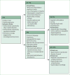
Figure 1
Figure 1. Diagnostic criteria for clinical diagnosis of FTD
These criteria focus mostly on symptoms that are present early in the illness, which tend to more clearly delineate the three variants of FTD. FTD=frontotemporal dementia. BV=behavioural variant. SV=semantic variant. NFV=non-fluent variant. PPA=primary progressive aphasia.
Figure 2
Figure 2. Neuropathology in FTLD-tau and FTLD-TDP
FTLD-tau (A) Pick bodies in Pick’s disease; (B) a tufted astrocyte in progressive supranuclear palsy; (C) an astrocytic plaque in corticobasal degeneration; FTLD-TDP (E) small compact or crescentic neuronal cytoplasmic inclusions and short, then neuropil threads in FTLD-TDP type A; (F) diffuse or granular neuronal cytoplasmic inclusions (with a relative paucity of neuropil threads) in FTLD-TDP type B; and (G) long, tortuous dystrophic neurites in FTLD-TDP type C.TDP can be seen within the nucleus in neurons lacking inclusions but mislocalises to the cytoplasm and forms inclusions in FTLD-TDP. The remaining FTLD cases are characterised by FUS-immunoreactive inclusions that stain negatively for tau and TDP-43; a vermiform neuronal nuclear inclusion in a dentate gyrus granule cell is shown (D); this neuron contains an ovoid cytoplasmic inclusion. In patients with hexanucleotide expansions in C9orf72, small juxtanuclear ubiquitin-positive, TDP-negative inclusions (H) are pathognomonic for the disorder. These inclusions contain dipeptide repeat proteins translated from the GGGGCC repeat in one of six reading frames. lmmunostains are 3-repeat tau (A), phospho-tau (B and C), FUS (D), TDP-43 (E–G) and ubiquitin (H). Sections are counterstained with haematoxylin. Scale bar applies to all panels and represents 50 µm in A, B, C, and H; 12 µm in D; and 100 µm in E and G. FTLD=frontotemporal lobar degeneration. TDP=TAR DNA-binding protein. FUS=fused-in-sarcoma.
Figure 3
Figure 3. Patterns of brain atrophy in FTLD pathologies
(A) Pick’s disease; (B) progressive supranuclear palsy; (C) corticobasal degeneration; (D) FTLD-TDP type A; (E) FTLD-TDP type B; (F) FTLD-TDP type C. FTLD-TDP type A is associated with an asymmetrical dorsal pattern of atrophy that includes the frontal lobe and temporal lobe (anterior, medial, and posterior regions), orbitofrontal cortex, anterior cingulate gyrus, inferior parietal lobe, striatum, and thalamus. FTLD-TDP type B is associated with a more medial pattern of atrophy mainly involving the medial and polar temporal lobe, anterior insular, cingulate and medial prefrontal cortices, and the orbitofrontal cortex. The frontal lobe is more severely affected in the posterior areas. FTLD-TDP type C is associated with either right-predominant or left-predominant anterior temporal lobe atrophy, with additional involvement of the amygdala, hippocampus, orbitofrontal cortex, and insular cortex. FTLD=frontotemporal lobar degeneration. TDP=TAR DN A-binding protein 43.

Similar articles

Cited by

References

    1. Vieira RT, Caixeta L, Machado S, et al. Epidemiology of early-onset dementia: a review of the literature. Clin Pract Epidemiol Ment Health. 2013;9:88–95. - PMC - PubMed
    1. Pick A. Über die Beziehungen der senilen Hirnatrophie zur Aphasie. Prager Med Wochenschr. 1892;17:165–67.
    1. Alzheimer A. Über eigenartige Krankheitsfälle der späteren Alters. Z Gesamte Neurol Psychiatr. 1911;4:356–85.
    1. Mesulam MM. Primary progressive aphasia. Ann Neurol. 2001;49:425–32. - PubMed
    1. Rascovsky K, Hodges JR, Knopman D, et al. Sensitivity of revised diagnostic criteria for the behavioural variant of frontotemporal dementia. Brain. 2011;134:2456–77. - PMC - PubMed

Publication types