. 2016 Jan;126(1):50-67.
doi: 10.1172/JCI80761. Epub 2015 Nov 23.

Immune activation caused by vascular oxidation promotes fibrosis and hypertension

Immune activation caused by vascular oxidation promotes fibrosis and hypertension

Jing Wu et al. J Clin Invest. 2016 Jan.

Erratum in

  • Immune activation caused by vascular oxidation promotes fibrosis and hypertension.
    Wu J, Saleh MA, Kirabo A, Itani HA, Montaniel KR, Xiao L, Chen W, Mernaugh RL, Cai H, Bernstein KE, Goronzy JJ, Weyand CM, Curci JA, Barbaro NR, Moreno H, Davies SS, Roberts LJ 2nd, Madhur MS, Harrison DG. Wu J, et al. J Clin Invest. 2016 Apr 1;126(4):1607. doi: 10.1172/JCI87425. Epub 2016 Apr 1. J Clin Invest. 2016. PMID: 27035819 Free PMC article. No abstract available.

Abstract

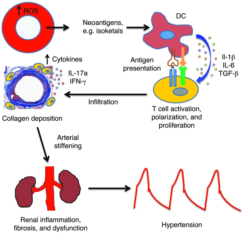
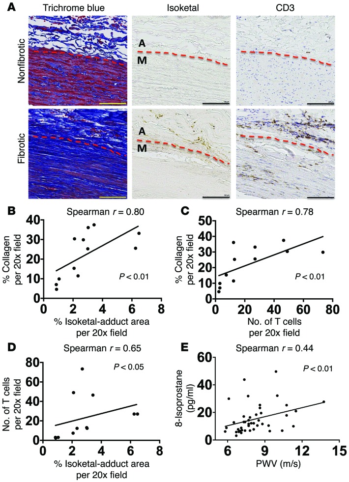
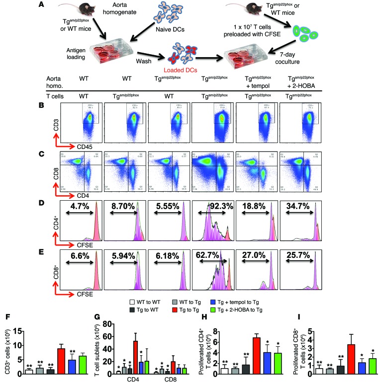
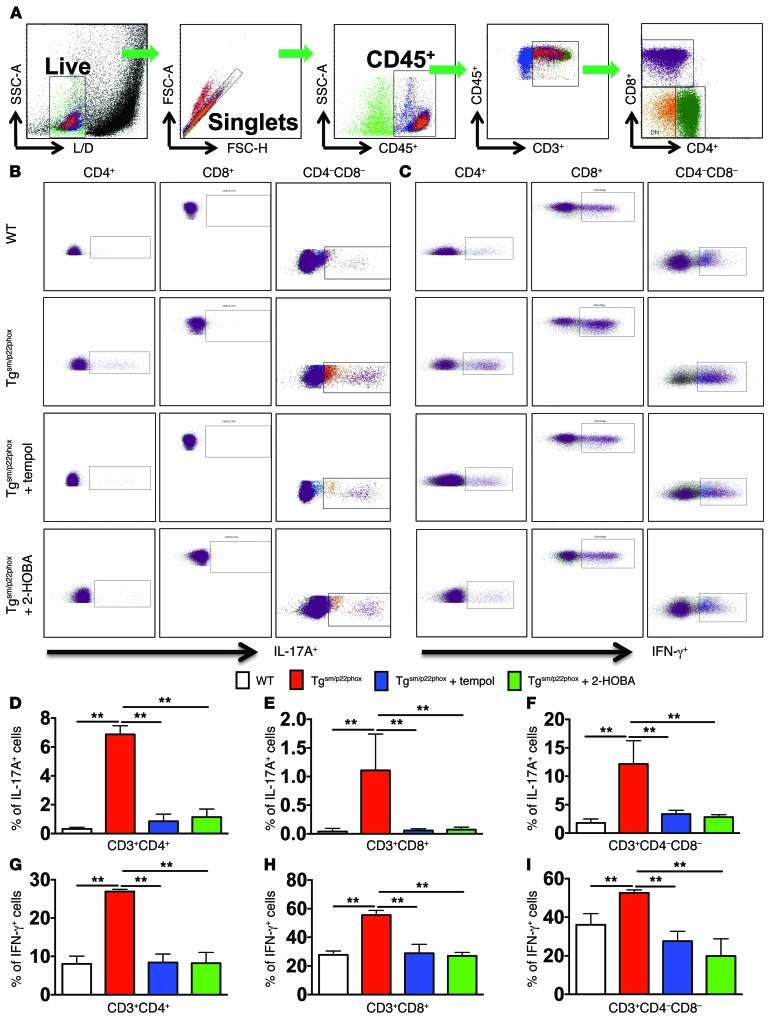
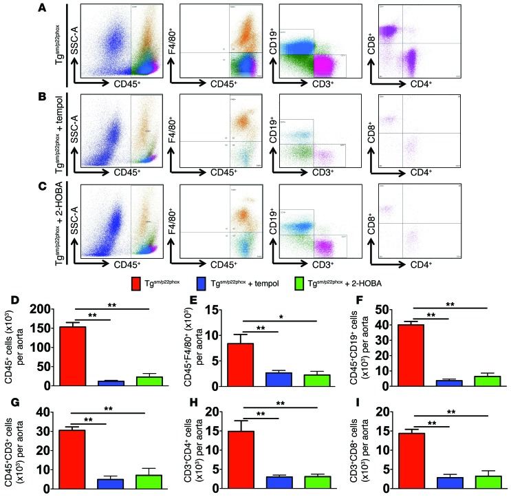
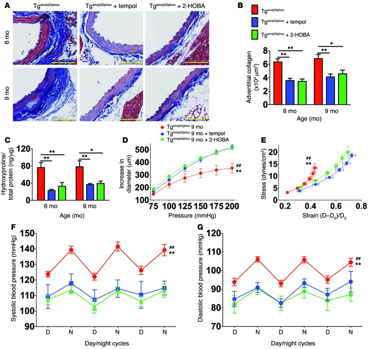
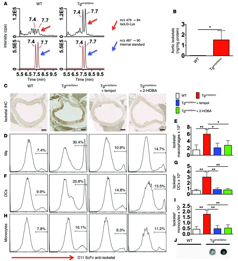
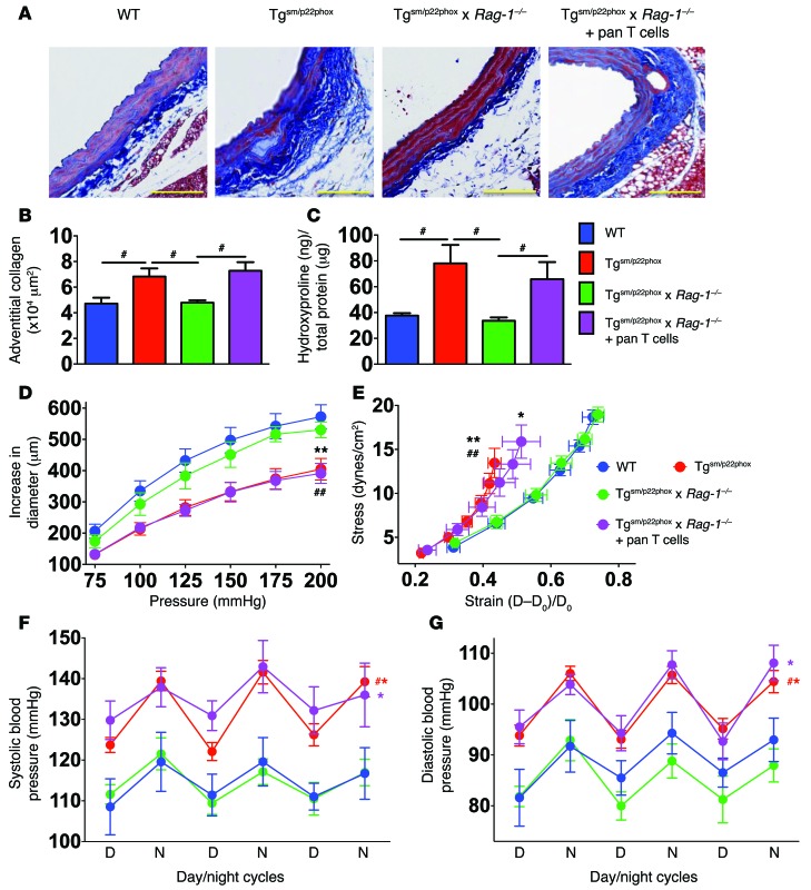
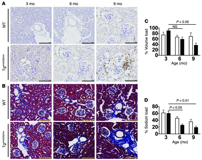
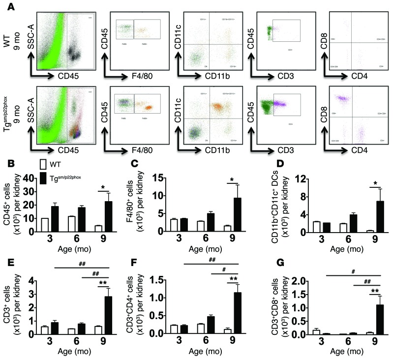
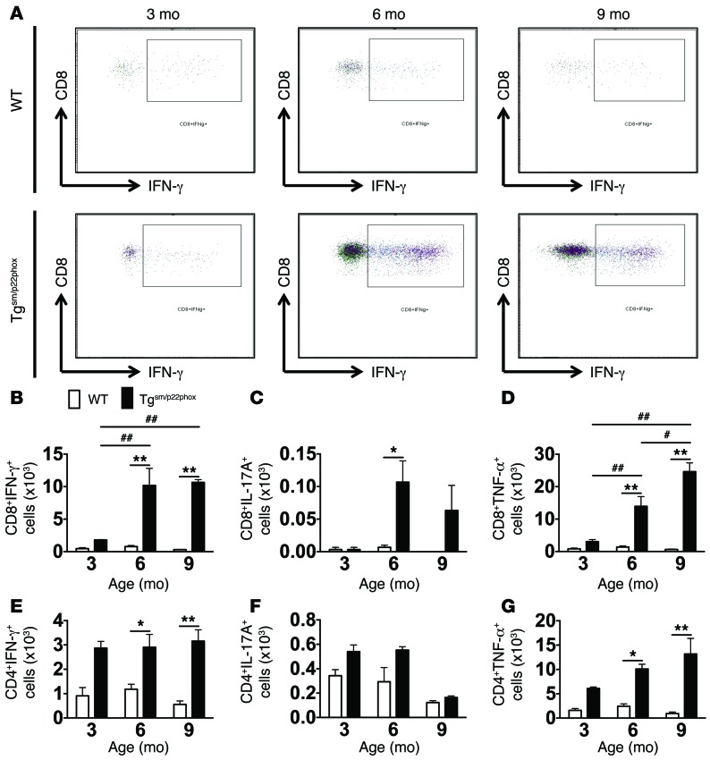
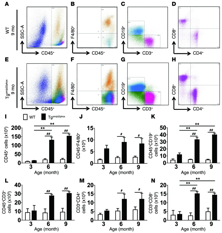
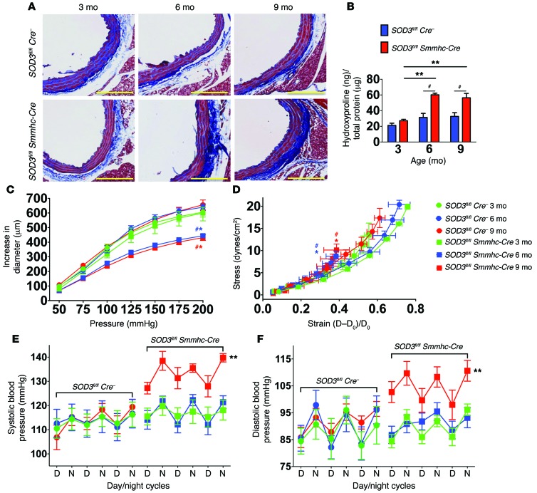
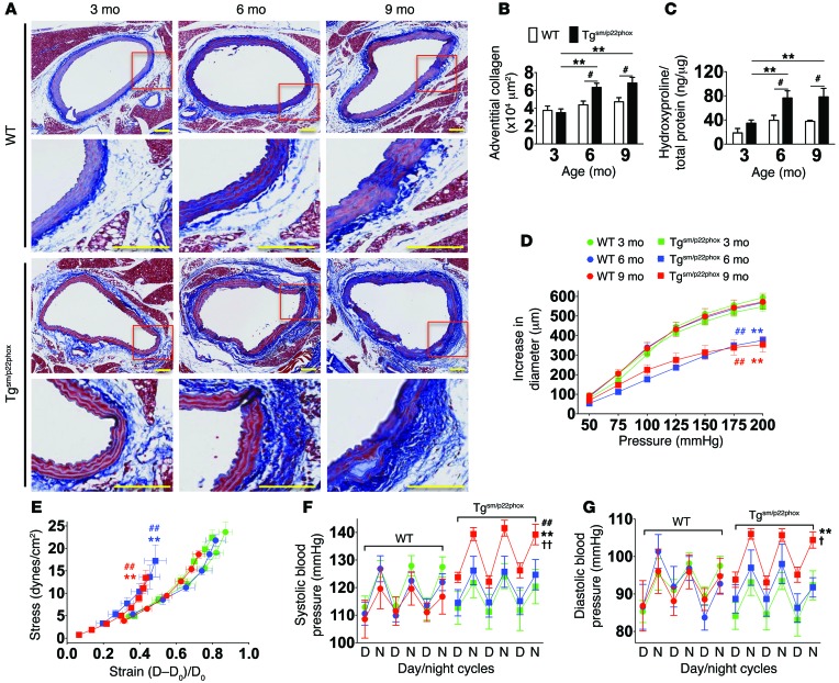
Vascular oxidative injury accompanies many common conditions associated with hypertension. In the present study, we employed mouse models with excessive vascular production of ROS (tg(sm/p22phox) mice, which overexpress the NADPH oxidase subunit p22(phox) in smooth muscle, and mice with vascular-specific deletion of extracellular SOD) and have shown that these animals develop vascular collagen deposition, aortic stiffening, renal dysfunction, and hypertension with age. T cells from tg(sm/p22phox) mice produced high levels of IL-17A and IFN-γ. Crossing tg(sm/p22phox) mice with lymphocyte-deficient Rag1(-/-) mice eliminated vascular inflammation, aortic stiffening, renal dysfunction, and hypertension; however, adoptive transfer of T cells restored these processes. Isoketal-protein adducts, which are immunogenic, were increased in aortas, DCs, and macrophages of tg(sm/p22phox) mice. Autologous pulsing with tg(sm/p22phox) aortic homogenates promoted DCs of tg(sm/p22phox) mice to stimulate T cell proliferation and production of IFN-γ, IL-17A, and TNF-α. Treatment with the superoxide scavenger tempol or the isoketal scavenger 2-hydroxybenzylamine (2-HOBA) normalized blood pressure; prevented vascular inflammation, aortic stiffening, and hypertension; and prevented DC and T cell activation. Moreover, in human aortas, the aortic content of isoketal adducts correlated with fibrosis and inflammation severity. Together, these results define a pathway linking vascular oxidant stress to immune activation and aortic stiffening and provide insight into the systemic inflammation encountered in common vascular diseases.


Figures

Figure 14
Figure 14. Proposed pathway.
Chronic vascular oxidative stress leads to formation of immunogenic isoketal-protein adducts, which accumulate in DCs and promote T cell activation. Activated T cells accumulate in perivascular tissues and release cytokines that enhance collagen deposition and aortic stiffening. This enhances pulse wave velocity. Renal inflammation and fibrosis follows these events, leading to overt hypertension.
Figure 13
Figure 13. Role of isoketal and oxidation in aortic collagen deposition and stiffening in humans.
(AD) Data from human aortas. (E) Comparison of PWV and isoprostane content in patients. Consecutive 6 μm sections were stained with anti-CD3, D-11 ScFv, and Masson’s trichrome blue (A). Images are ×20 magnification. Scale bars: 100 μm. Image J was used to quantify the area of D-11 staining for isoketal adducts and the area of blue in the Masson’s trichrome stains. Human T cells were quantified by counting positively stained cells per ×20 field (B and C). Spearman’s correlations comparing PWV and 8-isoprostanes in normotensive and treated hypertensive humans (E). M, media; A, adventitia.
Figure 12
Figure 12. Tgsm/p22phox aortic homogenates prime DCs to promote T cell proliferation and activation.
(A) Experimental design. Aortic homogenates from WT or tgsm/p22phox mice were used to prime naive DCs, which were then cocultured with T cells from these mice in various combinations. (B, C, F, and G) Flow cytometry and bar graphs identifying live CD3+ T cells and CD4+/CD8+ T cell subsets from cocultured DCs and T cells. (D, E, H, and I) Representative flow cytometry images and bar graphs showing percentages and numbers of proliferating CD4+ (D and H) and CD8+ (E and I) T cells, as determined using CSFE dilution (n = 6, 1-way ANOVA). Homo, homogenates. Black lines indicate original data, orange peaks indicate initial population, and purple peaks indicate generations of cells undergoing division as simulated by FlowJo 7.6.4 software. *P < 0.05 compared with Tgsm/p22phox homogenate + Tgsm/p22phox T cells; **P < 0.01 Tgsm/p22phox homogenate + Tgsm/p22phox T cells.
Figure 11
Figure 11. Cytokine production of T cells in the spleen of mice with chronic vascular oxidative stress.
(A) Gating strategy for identifying T cell populations among total spleenocytes. Single cell suspensions were prepared from freshly isolated mouse spleens via enzymatic digestion and mechanical dissociation. (BI) Intracellular staining indicating IL-17A (B, DF) and IFN-γ (C, GI) production in T cell subsets in WT and Tgsm/p22phox mice. Bar graphs were analyzed using 1-way ANOVA (n = 5–7). **P < 0.01.
Figure 10
Figure 10. Role of ROS and isoketals in vascular inflammation.
Tgsm/p22phox mice were treated with tempol or 2-HOBA from 4–9 months of age. (AC) Flow cytometric examples for leukocytes measured in single cells suspensions of aortas. (DI) Mean data analyzed using 1-way ANOVA (n = 6–8). *P < 0.05; **P < 0.01.
Figure 9
Figure 9. Scavenging superoxide or isoketals prevents aortic collagen deposition, aortic stiffening and hypertension in tgsm/p22phox mice.
Tgsm/p22phox mice received either no treatment, tempol, or 2-HOBA in the drinking water from 3–9 months of age. (A) Masson’s trichrome staining at ×20 magnification. Scale bars: 100 μm. (B and C) Mean data for quantification of collagen staining (B) and hydroxyproline content (C). *P < 0.05; **P < 0.01. (D and E) Pressure diameter and stress strain relationships for isolated aortic segments studied as in Figure 1. ##P < 0.01 vs. WT; **P < 0.01 vs. 2-HOBA. (F and G) Effect of antioxidant treatment on blood pressure in aged tgsm/p22phox mice. Collagen deposition was quantified using 1-way ANOVA. Aortic stiffness and blood pressure were analyzed with 1-way ANOVA with repeated measurements (n = 6–8).
Figure 8
Figure 8. Chronic vascular oxidative stress promotes isoketal-adduct formation.
(A and B) Stable isotope dilution multiple reactions monitoring MS analysis of isoketal-lysine-lactam (IsoK-Lys) adducts in aortas of 9-month-old animals. Representative LC/MS chromatographs from pooled samples for each group (A). The left panel shows chromatographs for IsoK-Lys in samples, while the right panel shows chromatographs for [13C615N2] internal standards in the same samples. Mann-Whitney tests were employed for analysis. The bar graph in B shows mean data. (C) Fix-perfused mouse aortas were embedded in paraffin and subjected to IHC for isoketal staining. Images are ×20 magnification. Scale bars: 100 μm. (DI) Intracellular staining for isoketals in spleen macrophages (D and E), DCs (F and G), and monocytes (H and I). These data were analyzed using 1-way ANOVA, n = 6. *P < 0.05; **P < 0.01. (J) DCs isolated from 9-month-old WT and tgsm/p22phox mice were exposed to weak acid to elute peptides presented on MHC-I complexes. The presence of isoketal-adducted peptides in the eluent was visualized by dot blot. Isoketal adducts were present in 3 of 4 tgsm/p22phox mice but not in WT mice (see Supplemental Figure 5C for full uncut gel).
Figure 7
Figure 7. T cells mediate age-related aortic collagen deposition, aortic stiffening, and elevation of blood pressure in tgsm/p22phox mice.
Pan T cells were isolated from the spleen of 3-month-old WT mice and adoptively transferred to age-matched tgsm/p22phox × Rag-1–/– mice to reconstitute T cell population. Mice were subsequently studied at 9 months of age. (A) Examples of aortic Masson’s trichrome blue staining. (B) Quantification of collagen staining by planimetry. (C) Quantification of aortic hydroxyproline levels. #P < 0.05. (D and E) Pressure diameter curves and stress strain relationships obtained using isolated aortic segments studied as in Figure 1. *P < 0.05 vs. Tgsm/p22phox × Rag-1–/–; **P < 0.01 vs. Tgsm/p22phox × Rag-1–/–; ##P < 0.01 vs. WT. (F and G) Telemetry recordings of blood pressures over 3 days. #P < 0.05 vs. WT. Collagen deposition was quantified using 1-way ANOVA. Aortic stiffness and blood pressure were analyzed using 1-way ANOVA with repeated measures (n = 6–8).
Figure 6
Figure 6. Development of age-related renal inflammation, fibrosis, and dysfunction in tgsm/p22phox mice.
(A and B) Consecutive 6-micron sections were obtained from paraffin-embedded mouse kidneys and were stained with anti-CD3 and Masson’s trichrome blue. Images are ×20 magnification. Scale bars: 100 μm. (C and D) Mice received a single i.p. injection of normal saline equal to 10% of body weight, and urine/sodium excretion in the subsequent 4 hours were monitored. These data were analyzed with 2-way ANOVA (n = 6–8).
Figure 5
Figure 5. Flow cytometry analysis of renal leukocytes.
Single cell suspensions were prepared from freshly isolated mouse kidneys via enzymatic digestion and mechanical dissociation. Live cell singlets were analyzed. (A) CD45+ total leukocytes, F4/80+ macrophages, CD11b+CD11c+ DCs, CD3+ T lymphocytes, and CD4+/CD8+ T cell subsets were identified in the aorta of 9-month-old WT and tgsm/p22phox mice. (BG) Quantification of infiltrating leukocyte subsets using 2-way ANOVA (n = 6–8). *P < 0.05; **P <0.01; #P < 0.05; ##P < 0.01.
Figure 4
Figure 4. Production of cytokines by aortic T cells using intracellular staining.
One million splenocytes were stimulated with ionomycin and phorbol myristate acetate and brefeldin A at 37°C for 5 hours. Intracellular staining was then performed with the Cytofix/Cytoperm Plus fixation and permeabilization solution kit using anti–IL-17A and anti–IFN-γ antibodies. (A) Representative images of IFN-γ–producing CD8+ T cells of WT and tgsm/p22phox mice at 3, 6, and 9 months of age. (BD) Quantification of CD8+ T cells producing IFN-γ, IL-17A, and TNF-α. (EG) Quantification of CD4+ T cells producing IFN-γ, IL-17A, and TNF-α. *P < 0.01 vs. WT; **P < 0.01 vs. WT; #P < 0.05 vs. 6 months; ##P <0.01 vs. 3 months. These data were analyzed using 2-way ANOVA.
Figure 3
Figure 3. Flow cytometry analysis of inflammatory cells in the aorta of WT and tgsm/p22phox mice.
Single cell suspensions were prepared from freshly isolated mouse aortas via enzymatic digestion and mechanical dissociation. Live cell singlets were analyzed for vascular inflammatory cells. (AH) CD45+ total leukocytes, F4/80+ macrophages, CD19+ B lymphocytes, CD3+ T lymphocytes, and CD4+/CD8+T cell subsets were identified in the aorta of 9-month-old WT (AD) and tgsm/p22phox mice (EH). (IN) Quantification of infiltrating leukocyte subsets using 2-way ANOVA (n = 6–8). **P < 0.01 vs. Tgsm/p22phox; #P < 0.05; ##P < 0.01.
Figure 2
Figure 2. Vascular oxidative stress induced by deletion of smooth muscle Sod3 promotes age-related aortic stiffening and hypertension.
(A) Masson’s trichrome blue staining in 3-, 6-, and 9-month-old Sod3fl/fl Cre and Sod3fl/fl Smmhc-Cre mice. Tamoxifen was administered at 3 mg/20 kg in corn oil via i.p. injection at 10 weeks of age. Images are ×20 magnification. Scale bars: 100 μm. (B) Aortic hydroxyproline content. (C and D) Pressure diameter relationship and stress-strain relationships. (E and F) Blood pressure measured by radiotelemetry (n = 6–8). #P < 0.05 vs. Cre–/–; *P < 0.05 vs. Sod3fl/fl Smmhc Cre 3 month; **P < 0.05 vs. Sod3fl/fl Smmhc-Cre 3 month. Aortic hydroxyproline content was analyzed using 2-way ANOVA. Pressure diameter relationships, stress-strain relationships, and blood pressures were analyzed using 1-way ANOVA with repeated measures.
Figure 1
Figure 1. Tgsm/p22phox mice develop age-related aortic stiffening and hypertension.
(A) Effects of aging on vascular collagen deposition in WT and tgsm/p22phox mice. Perfusion-fixed sections of the thoracic aortas were sectioned (6 μm) and stained with Masson’s trichome to highlight collagen (blue). Areas within the red boxes were magnified (×20) and shown below the original images (×6). Scale bars: 100 μm. (B) Adventitial collagen area was quantified by planimetry. #P < 0.05 vs. WT; **P < 0.01 vs. 3 months. (C) Aortic collagen quantification by hydroxyproline assay. (D and E) Freshly isolated aortas were mounted on a myograph system in Ca2+-free buffer to determine pressure-diameter relationships. Stress-strain relationships were constructed from intraluminal pressure, wall thickness, and inner and outer diameters. These parameters were measured at 25 mmHg step changes in pressure from 0–200 mmHg. **P < 0.01 vs. Tg 3 months; ##P < 0.01 vs. WT. (F and G) Telemetry blood pressure of WT and tgsm/p22phox mice at 3, 6, and 9 months of age. P < 0.05 vs. Tg 6 months; ††P <0.01 vs. Tg 6 months. Data were analyzed using 2-way ANOVA; n = 6–9.

Comment in

Similar articles

Cited by

References

    1. Meaume S, Benetos A, Henry OF, Rudnichi A, Safar ME. Aortic pulse wave velocity predicts cardiovascular mortality in subjects > 70 years of age. Arterioscler Thromb Vasc Biol. 2001;21(12):2046–2050. doi: 10.1161/hq1201.100226. - DOI - PubMed
    1. Laurent S, et al. Aortic stiffness is an independent predictor of fatal stroke in essential hypertension. Stroke. 2003;34(5):1203–1206. doi: 10.1161/01.STR.0000065428.03209.64. - DOI - PubMed
    1. Kaess BM, et al. Aortic stiffness, blood pressure progression, and incident hypertension. JAMA. 2012;308(9):875–881. doi: 10.1001/2012.jama.10503. - DOI - PMC - PubMed
    1. Cooper LL, et al. Components of hemodynamic load and cardiovascular events: the Framingham Heart Study. Circulation. 2015;131(4):354–361. doi: 10.1161/CIRCULATIONAHA.114.011357. - DOI - PMC - PubMed
    1. Kals J, et al. Inflammation and oxidative stress are associated differently with endothelial function and arterial stiffness in healthy subjects and in patients with atherosclerosis. Scand J Clin Lab Invest. 2008;68(7):594–601. doi: 10.1080/00365510801930626. - DOI - PubMed

Publication types