. 2016 Jan;126(1):99-111.
doi: 10.1172/JCI83312. Epub 2015 Nov 23.

Ultrasound ablation enhances drug accumulation and survival in mammary carcinoma models

Ultrasound ablation enhances drug accumulation and survival in mammary carcinoma models

Andrew W Wong et al. J Clin Invest. 2016 Jan.

Abstract

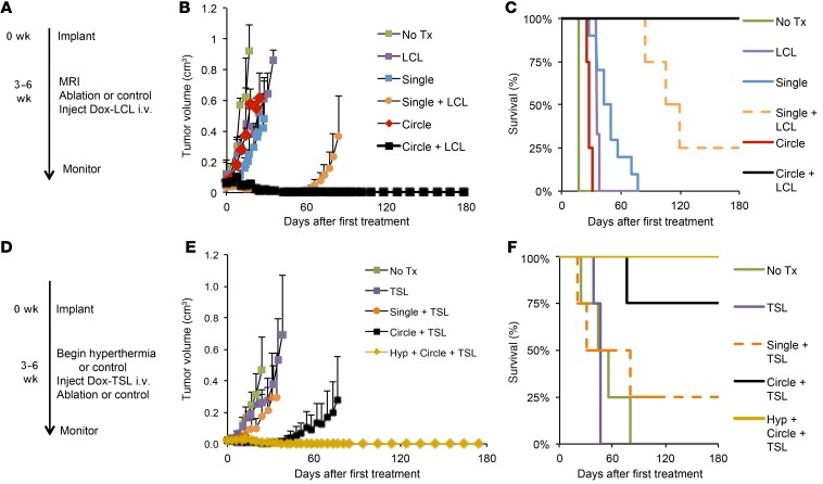
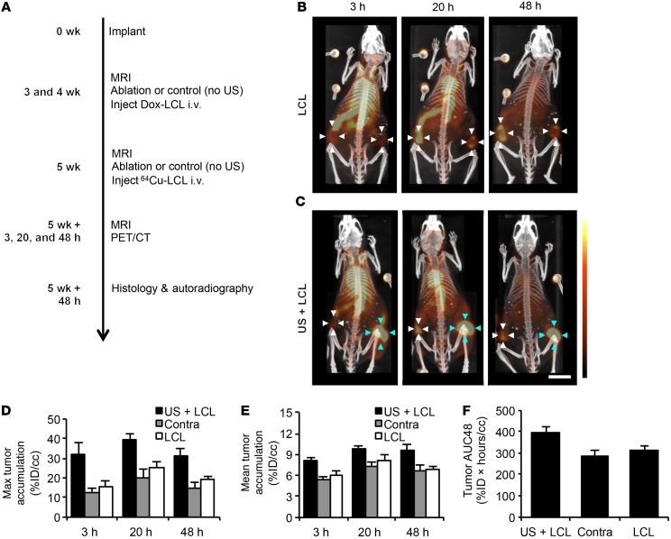
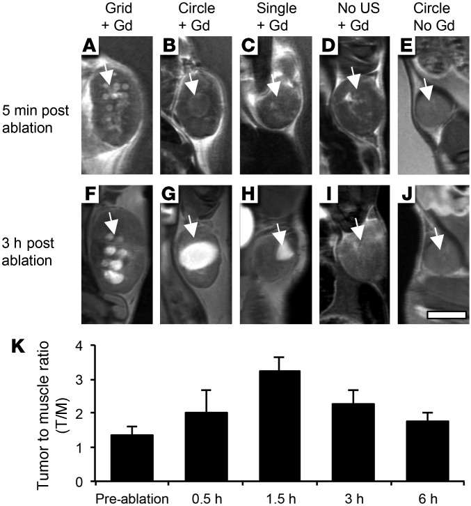
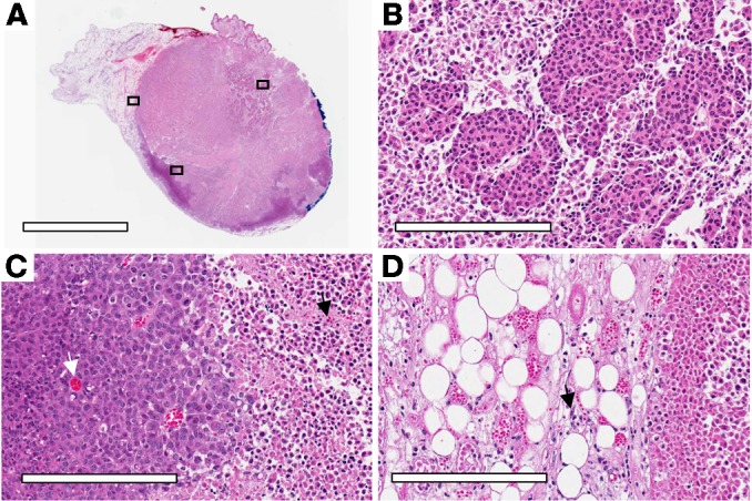
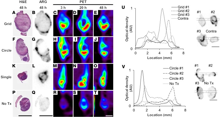
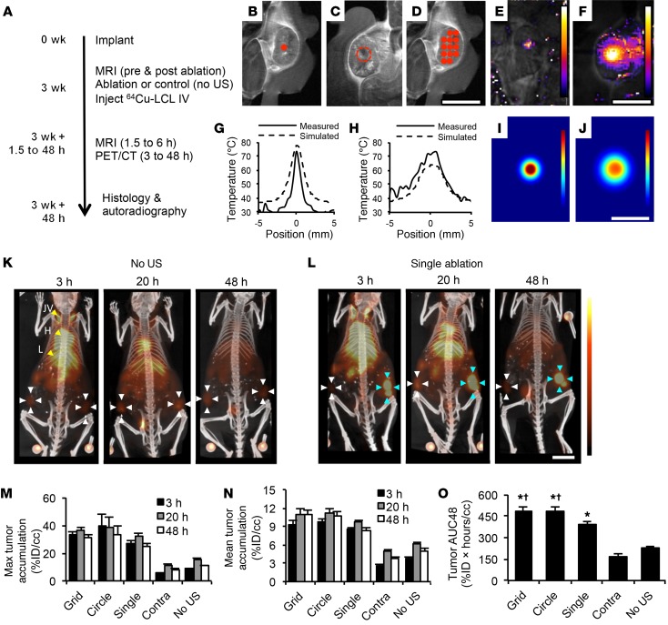
Magnetic resonance-guided focused ultrasound (MRgFUS) facilitates noninvasive image-guided conformal thermal therapy of cancer. Yet in many scenarios, the sensitive tissues surrounding the tumor constrain the margins of ablation; therefore, augmentation of MRgFUS with chemotherapy may be required to destroy remaining tumor. Here, we used 64Cu-PET-CT, MRI, autoradiography, and fluorescence imaging to track the kinetics of long-circulating liposomes in immunocompetent mammary carcinoma-bearing FVB/n and BALB/c mice. We observed a 5-fold and 50-fold enhancement of liposome and drug concentration, respectively, within MRgFUS thermal ablation-treated tumors along with dense accumulation within the surrounding tissue rim. Ultrasound-enhanced drug accumulation was rapid and durable and greatly increased total tumor drug exposure over time. In addition, we found that the small molecule gadoteridol accumulates around and within ablated tissue. We further demonstrated that dilated vasculature, loss of vascular integrity resulting in extravasation of blood cells, stromal inflammation, and loss of cell-cell adhesion and tissue architecture all contribute to the enhanced accumulation of the liposomes and small molecule probe. The locally enhanced liposome accumulation was preserved even after a multiweek protocol of doxorubicin-loaded liposomes and partial ablation. Finally, by supplementing ablation with concurrent liposomal drug therapy, a complete and durable response was obtained using protocols for which a sub-mm rim of tumor remained after ablation.


Figures

Figure 6
Figure 6. Combination of ablation and doxorubicin encapsulated in LCL (Dox-LCL) or TSL (Dox-TSL) can produce complete local regression in the NDL tumor model.
(AC) Treatments included weekly single-point ablation (single, n = 8), circle pattern ablation (circle, n = 4), single-point ablation plus Dox-LCL (single + LCL, n = 4), circle ablation plus Dox-LCL (circle + LCL, n = 4), no ultrasound and no drug (no tx, n = 8), or Dox-LCL only (LCL, n = 4). Dox-LCL (or control saline) was injected biweekly (4 ablations and 8 injections total). (B) At 180 days after the start of treatment/152 days after completion (study end point), tumors could not be detected in mice treated with circle ablation plus Dox-LCL. In all other groups, tumor growth was delayed, but a complete response was not typically achieved. (C) At study end point, all (n = 4) mice treated with circle ablation plus Dox-LCL and 25% (1 of 4) of mice treated with single-point ablation plus Dox-LCL survived. (DF) TSL therapy combined with ablation. (D) Treatments included weekly: single-point ablation plus Dox-TSL (single + TSL, n = 4), circle ablation plus Dox-TSL (circle + TSL, n = 4), circle ablation preceded by hyperthermia and Dox-TSL (hyp + circle + TSL, n = 4), no ultrasound and no drug (no tx, n = 4), or Dox-TSL only (n = 4) for a period of 4 weeks. (E and F) At study end point, tumors could not be detected in all mice treated with circle ablation plus Dox-TSL combined with hyperthermia. In all other groups, tumor growth was delayed, and a complete response was also achieved in 75% of mice treated with circle ablation plus Dox-TSL and 25% of mice treated with single-point ablation plus Dox-TSL. Data are pooled from 6 experiments.
Figure 5
Figure 5. Single-point MRgFUS ablation continues to enhance tumor accumulation of liposomes in the NDL tumor model after treatment for 2 weeks with MRgFUS ablation and therapeutic doxorubicin liposomes (Dox-LCL).
(A) Mice were treated with weekly ablation (single-point protocol) or control (no ultrasound) and biweekly Dox-LCL (i.v., 6 mg/kg) for 2 weeks, then treated with ablation or control (no ultrasound) and injected with 64Cu-LCL. (B) PET-CT MIP images of mice treated only with repeated Dox-LCL (LCL) demonstrate heterogeneous tumor accumulation of 64Cu-LCL (white arrows). (C) PET-CT MIP images of mice treated with ablation plus Dox-LCL (US + LCL) reveal enhanced accumulation in ultrasound-treated tumors (blue arrows). PET image–derived tumor activity of 64Cu-LCL was obtained by manual segmentation with data plotted as (D) maximum pixel intensity within volume of interest, (E) activity averaged over volume of interest, and (F) AUC48. n = 3 each for US + LCL, contralateral tumors, and LCL. Scale bar: 1 cm. PET color bar ranges from 25%ID/cc to 0%ID/cc.
Figure 4
Figure 4. As compared with controls, grid and circular ablation protocols alter the tumor distribution of a single injection of gadoteridol contrast agent in the NDL tumor model.
Results were similar whether gadoteridol was injected before or after ablation. Columns represent the same tumor imaged at 5 minutes (AE) and 3 hours (FJ) after ablation with the following protocols: (A and F) grid, (B and G) circle, (C and H) single point, (D and I) no ultrasound, gadoteridol before ablation, (E and J) circle ablation with no gadoteridol. (K) For gadoteridol injection after ablation, ratio of intensity in ablation region and quadriceps muscle reaches 3.2 ± 0.4 at 1.5 hours, n = 4. (AJ) Representative images with n = 4 each for grid, single-point ablation, no ultrasound plus gadoteridol (Gd), and circle no gadoteridol, n = 12 for circle plus gadoteridol. Scale bar: 5 mm. Data are pooled from 2 experiments.
Figure 3
Figure 3. Mechanisms for enhanced accumulation after ablation at the boundary between viable and nonviable tissue stained with H&E in the NDL tumor model.
(A) Representative tumor following circle protocol ablation with orientation dye at boundary and black boxes at locations of insets. (B) View of heat-fixed tissue central to beam with surrounding loss of cell-cell adhesion. Necrotic tissue displays loss of glandular tumor architecture visible in heat-fixed and viable tumor. (C) View at border of viable (left) and necrotic (right) tumor and (D) view at border of mammary fat pad with light inflammatory reaction in stroma (black arrow in D) and necrotic tissue (right). (C and D) Viable fat pad and tumor tissue demonstrate dilated blood vessels (white arrow in C) and extravasation of blood cells (black arrow in C), indicating disruption of vascular integrity. Histology was included in all (n = 117) studies. Scale bars: 4 mm (A); 200 μm (BD).
Figure 2
Figure 2. Ablation greatly enhances 64Cu-LCL accumulation in tumor rim and remaining viable tumor in the NDL tumor model.
(AT) Comparison of H&E-stained frozen sections (A, F, K, and P), autoradiography (B, G, L, and Q), and PET image slices (CE, HJ, MO, RT) acquired at 3 hours, 20 hours, and 48 hours from tumors sectioned 48 hours after MRgFUS treatment with grid, circle, single-point ablation, or no ultrasound. Grid and circle MRgFUS exposures increased accumulation of 64Cu-LCL in rims surrounding ablation (dark purple rim on A and F corresponds with black rim on B and G). Limited resolution of PET images blurs rim in CE and HJ, but also shows blood and lymphatic radioactivity in CE, HJ, and MO. Single-point ablation also increased accumulation of liposomes (LO). Accumulation in untreated tumor was homogeneous and low (QT). (U and V) Region of interest analysis of autoradiography (rectangular black box was plotted as line profile) demonstrating that central accumulation in tumors treated with (U) grid protocol and (V) circle protocol is comparable to that in untreated tumors in 5 of 6 tumors. n = 4, 3, 12, 19, and 6 mice for grid, circle, single point, contralateral tumors, and no treatment. Scale bars: 5 mm. Color bar scaled from 25%ID/cc to 0%ID/cc. Data are representative of 5 experiments.
Figure 1
Figure 1. MRgFUS ablation enhanced accumulation of 64Cu-LCL in NDL mammary carcinoma.
(A) Three weeks after tumor implantation, tumors were ablated and imaged with pre- and postablation T1w-MRI. Mice were then injected with i.v. 64Cu-LCL and imaged with PET-CT at 3, 20, and 48 hours after treatment. Control treatment was no ultrasound (no US). (BJ) MRgFUS protocols and temperature profiles. T1w images localize ultrasound for (B) single-point, (C) circle, and (D) grid protocols. MR thermometry images following (E) 20 seconds single-point or (F) 60 seconds circle protocol MRgFUS. Temperature of measured and simulated ultrasound focus is plotted after (G) 20 seconds single-point or (H) 60 seconds circle protocol. Temperature distribution was simulated following (I) 20 seconds single-point or (J) 60 seconds circle protocol. Grid protocol shown in D and MO is equivalent to E and G repeated in grid pattern, due to sequential application of ablation points and cool-down period. (K) PET-CT maximum intensity projection images of 64Cu-LCL without ablation. White arrows indicate tumors; yellow arrowheads indicate jugular vein (JV), heart (H), and liver (L). (L) At 3, 20, and 48 hours following single-point ablation (blue arrows), liposome accumulation is enhanced as compared with that in contralateral tumors (white arrows) in PET-CT images. 64Cu-LCL PET image–derived volume of interest analysis plotted as (M) maximum voxel intensity, (N) average activity, and (O) AUC48. Significance is analyzed with 1-way ANOVA followed by Tukey’s test. For MO, n = 4, 3, 12, 19, and 6 mice for grid, circle, single point, contralateral tumors (contra), and no treatment. Scale bars: 1 cm. Thermometry color bars range from 70°C to 40°C; PET color bar ranges from 25%ID/cc to 0%ID/cc. *P < 1 × 10⁻⁴ vs. untreated tumors; P < 0.05 vs. single-point ablation. Data are pooled from 5 experiments.

References

    1. Alm El-Din MA, Taghian AG. Breast conservation therapy for patients with locally advanced breast cancer. Semin Radiat Oncol. 2009;19(4):229–235. doi: 10.1016/j.semradonc.2009.05.005. - DOI - PubMed
    1. Gralow JR, et al. Preoperative therapy in invasive breast cancer: pathologic assessment and systemic therapy issues in operable disease. J Clin Oncol. 2008;26(5):814–819. doi: 10.1200/JCO.2007.15.3510. - DOI - PubMed
    1. Weinberg BD, Krupka TM, Haaga JR, Exner AA. Combination of sensitizing pretreatment and radiofrequency tumor ablation: evaluation in rat model. Radiology. 2008;246(3):796–803. doi: 10.1148/radiol.2463070228. - DOI - PubMed
    1. Ahmed M, et al. Combination radiofrequency ablation with intratumoral liposomal doxorubicin: effect on drug accumulation and coagulation in multiple tissues and tumor types in animals. Radiology. 2005;235(2):469–477. doi: 10.1148/radiol.2352031856. - DOI - PubMed
    1. Ahmed M, et al. Radiofrequency thermal ablation sharply increases intratumoral liposomal doxorubicin accumulation and tumor coagulation. Cancer Res. 2003;63(19):6327–6333. - PubMed

Publication types

MeSH terms