. 2015 Nov 24:5:17016.
doi: 10.1038/srep17016.

Kill-painting of hypoxic tumours in charged particle therapy

Affiliations

Kill-painting of hypoxic tumours in charged particle therapy

Walter Tinganelli et al. Sci Rep. .

Abstract

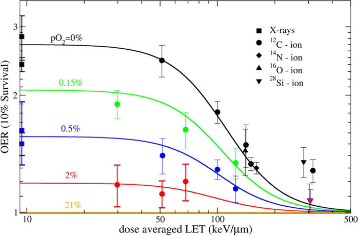
Solid tumours often present regions with severe oxygen deprivation (hypoxia), which are resistant to both chemotherapy and radiotherapy. Increased radiosensitivity as a function of the oxygen concentration is well described for X-rays. It has also been demonstrated that radioresistance in anoxia is reduced using high-LET radiation rather than conventional X-rays. However, the dependence of the oxygen enhancement ratio (OER) on radiation quality in the regions of intermediate oxygen concentrations, those normally found in tumours, had never been measured and biophysical models were based on extrapolations. Here we present a complete survival dataset of mammalian cells exposed to different ions in oxygen concentration ranging from normoxia (21%) to anoxia (0%). The data were used to generate a model of the dependence of the OER on oxygen concentration and particle energy. The model was implemented in the ion beam treatment planning system to prescribe uniform cell killing across volumes with heterogeneous radiosensitivity. The adaptive treatment plans have been validated in two different accelerator facilities, using a biological phantom where cells can be irradiated simultaneously at three different oxygen concentrations. We thus realized a hypoxia-adapted treatment plan, which will be used for painting by voxel of hypoxic tumours visualized by functional imaging.


Figures

Figure 1
Figure 1. Oxic and hypoxic survival rates at different LET.
A subset of the measured survival curves used to extract OER points, for different LET and pO2 values, including the corresponding (contemporary) oxic measurement. Each curve is obtained by fitting three independent measurements (different symbol colors), except the hypoxic curve in panel d, where only two measurements were considered.
Figure 2
Figure 2. OER model.
Two-dimensional semiempirical description of the OER dependence on LET and pO2 proposed in this work.
Figure 3
Figure 3. OER model verification.
Collection of all OER measurements compared to model surface cuts.
Figure 4
Figure 4. Kill-painting.
Planned survival rates (values in legend) on the differently oxygenated target shown on panel (a), without (b) and with (c) considering the inhomogeneous oxygen concentration in the optimization.
Figure 5
Figure 5. Experimental verification setup.
Single (a) and triple (b) hypoxic chambers used for experiments in Germany (GSI and HIT); chamber set used at NIRS for survival curve experiments (c), holding several specific Petri dishes (d); “hypoxic phantom” (e) used for all the extended target measurements. The GSI triple chambers (b) were used both in Japan and Germany, and additional measurement points in normoxic regions were collected with normal tissue culture flasks (TCF).
Figure 6
Figure 6. Non-optimized plan verification
Extended target survival measurements performed at HIMAC compared with the calculation by our new TPS version. A beam of 290 MeV/u 12C-ions was used from both sides, passively modulated in a SOBP of 6 cm, on a phantom of 18 cm. Dose in the target was recalculated from the oxic control as 9.5 Gy (RBE) (see M&M).
Figure 7
Figure 7. OER optimized plan verification.
Comparison of expected survival in an OER optimized plan with experimental results, performed at GSI. An actively scanned 12C ion beam, composed of 17 monoenergetic slices ranging from 234.64 to 155.26 MeV/u was used from both sides. The target length was 6 cm on a phantom of 16 cm. The beam was optimized with a prescribed survival level in the target of 0.1, corresponding to a RBE-weighted dose of 6.5 Gy (RBE) in normoxia. RBE-weighted dose in the entrance is 2.8 Gy(RBE). The dashed curve represents the expected survival across the phantom, when a normoxic plan is applied (similar to previous figure). Absolute measured and calculated data are shown, with no recalculation adjustment applied.
Figure 8
Figure 8. Kill-painting therapeutic improvement.
Tumour control probability computed as a function of a biologically isoeffective dose (BED) for the two different plans. The BED is computed in the entrance channel at 25 mm depth, in order to indicate a similar damage to the normal tissue, after application of several fractions of each plan to the target.

Similar articles

Cited by

References

    1. Hanahan D. & Weinberg R. A. Hallmarks of cancer: the next generation. Cell 144, 646–674 (2011). - PubMed
    1. Wilson W. R. & Hay M. P. Targeting hypoxia in cancer therapy. Nat. Rev. Cancer 11, 393–410 (2011). - PubMed
    1. Brown J. M. & Wilson W. R. Exploiting tumour hypoxia in cancer treatment. Nat. Rev. Cancer 4, 437–447 (2004). - PubMed
    1. Schardt D., Elsässer T. & Schulz-Ertner D. Heavy-ion tumor therapy: Physical and radiobiological benefits. Rev. Mod. Phys. 82, 383–425 (2010).
    1. Kamada T. et al.. Carbon ion radiotherapy in Japan: an assessment of 20 years of clinical experience. Lancet Oncol 16, e93–e100 (2015). - PubMed

Publication types

LinkOut - more resources