. 2015 Nov 24:5:17107.
doi: 10.1038/srep17107.

p53 dynamics upon response element recognition explored by molecular simulations

Affiliations

p53 dynamics upon response element recognition explored by molecular simulations

Tsuyoshi Terakawa et al. Sci Rep. .

Abstract

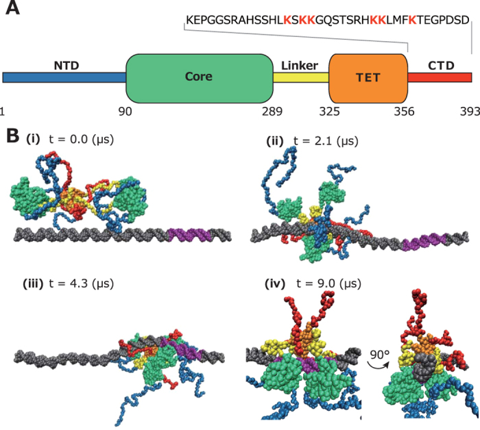
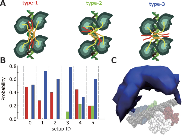
p53 is a representative transcription factor that activates multiple target genes. To realize stimulus-dependent specificities, p53 has to recognize targets with structural variety, of which molecular mechanisms are largely unknown. Here, we conducted a series of long-time scale (totally more than 100-ms) coarse-grained molecular dynamics simulations, uncovering structure and dynamics of full-length p53 tetramer that recognizes its response element (RE). We obtained structures of a full-length p53 tetramer that binds to the RE, which is strikingly different from the structure of p53 at search. These structures are not only consistent with previous low-resolution or partial structural information, but also give access to previously unreachable detail, such as the preferential distribution of intrinsically disordered regions, the contacts between core domains, the DNA bending, and the connectivity of linker regions. We also explored how the RE variation affects the structure of the p53-RE complex. Further analysis of simulation trajectories revealed how p53 finds out the RE and how post-translational modifications affect the search mechanism.


Figures

Figure 1
Figure 1. p53 domain structures and simulation snapshots.
(A) The domain map of p53. The bars represent intrinsically disordered regions and the rounded rectangles represent folded domains. The numbers represent residue IDs. The amino acid sequence of the CTD is shown with acetylation sites colored red. (B) Representative snapshots taken from the coarse-grained molecular dynamics simulation trajectories. p53 slid along dsDNA (ii). After one of the Cores bound to the RE (iii), the other three Cores bound one by one, forming p53-RE complex (iv). dsDNA is colored grey and the RE is colored purple. The color-coding of p53 is the same as (A). In (iv), p53 in the recognition mode is viewed from two different orientations.
Figure 2
Figure 2. The p53-RE complex with varying spacer lengths.
(A–C) Representative structures of the p53-RE complex in which the REs contain 1- (A), 2- (B), and 10- (C) bps spacers. (D) Probability distributions and time trajectories (inset) of Q-score of the inter-Core contacts of longitudinally aligned two Cores in which the RE contains 0- (red), 1- (green), 2- (blue), and 10-bps (magenta) spacers. (E) DNA bending scores vs base-pair IDs. The color assignment is the same as that of (D). We also plotted the scores of naked dsDNA (grey) and dsDNA to which p53 non-specifically binds (black).
Figure 3
Figure 3. Conformations of full-length p53 bound on the RE.
(A) A definition of the three types in which the four Cores are tethered to the TET. The color-coding of p53 is the same as Fig. 1A. The DNA is colored light green. (B) Probabilities of the three types (type-1 in red, type-2 in green, and type-3 in blue) from the six sets of the simulations with different setups (Table 1). (C) An iso-surface (0.001) of a spatial probability distribution of the center of the Core (blue) that forms dimers with the fixed one (white). The Cores that bind to the RE are depicted by four different colors (white, red, blue, and green). The dsDNA is colored grey.
Figure 4
Figure 4. Effects of charge neutralization in CTD.
(A) The time trajectories of nearest distance between p53 and dsDNA in the simulations in which the charges of CTD is not neutralized (p530; left) and is neutralized (p53n; right). The black circles indicate the time when the first core recognized the RE. The roman numerals represent the snapshot IDs in Fig. 1B (left) and Fig. 4B (right). (B) Representative snapshots taken from the trajectories for p53n. p53 diffused around dsDNA (ii). After one of the Cores bound to the RE (iii), the other three Cores bound one by one, forming p53-RE complex (iv).
Figure 5
Figure 5. The analysis of the sequential binding of the Core to the RE.
(A) The scheme of the four Cores binding to the RE with rate constants (p530 in red and p53n in blue). (B) The fractions of the 1- (left), 2- (center), and 3- (right) Core bound states against duration. (C) The approximate free energy along the distance between the RE and the unbound Core that is the nearest from the RE in the 1- (red), 2- (blue), and 3- (green) Core bound states. (D) Free energy profiles along the reaction coordinate. The solid and dashed lines represent free energy profiles from the two independent simulations with the same setup. The similarity of these lines shows the convergence of the simulation.

References

    1. Funk W. D., Pak D. T., Karas R. H., Wright W. E. & Shay J. W. A transcriptionally active DNA-binding site for human p53 protein complexes. Mol. Cell. Biol. 12, 2866–2871 (1992). - PMC - PubMed
    1. Ginsberg D., Mechta F., Yaniv M. & Oren M. Wild-type p53 can down-modulate the activity of various promoters. Proc. Natl. Acad. Sci. USA 88, 9979–9983 (1991). - PMC - PubMed
    1. Levine A. J. & Oren M. The first 30 years of p53: growing ever more complex. Nat. Rev. Cancer 9, 749–758 (2009). - PMC - PubMed
    1. Riley T., Sontag E., Chen P. & Levine A. Transcriptional control of human p53-regulated genes. Nat. Rev. Mol. Cell Biol. 9, 402–412 (2008). - PubMed
    1. Weinberg R. L., Veprintsev D. B., Bycroft M. & Fersht A. R. Comparative Binding of p53 to its Promoter and DNA Recognition Elements. J. Mol. Biol. 348, 589–596 (2005). - PubMed

Publication types

Substances