Review
. 2016 Mar 18;11(3):554-63.
doi: 10.1021/acschembio.5b00830. Epub 2015 Dec 7.

Reading between the Lines: "ADD"-ing Histone and DNA Methylation Marks toward a New Epigenetic "Sum"

Affiliations
Review

Reading between the Lines: "ADD"-ing Histone and DNA Methylation Marks toward a New Epigenetic "Sum"

Kyung-Min Noh et al. ACS Chem Biol. .

Erratum in

Abstract

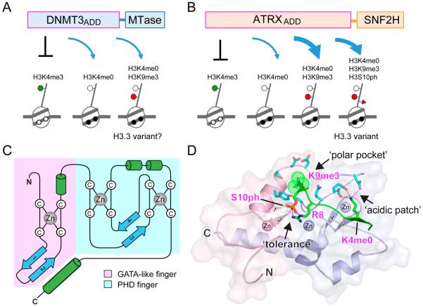
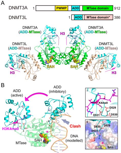
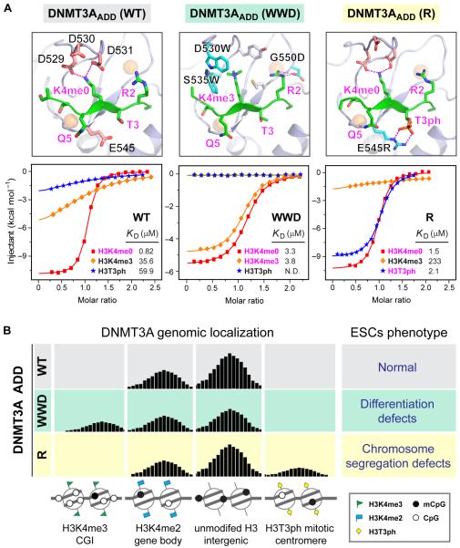
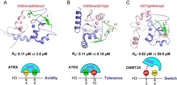
Covalent modifications of both DNA and histones act in concert to define the landscape of our epigenome. In this review, we explore the interconnections between histone and DNA modifications by focusing on a conserved chromatin-binding regulatory domain, the ATRX-DNMT3-DNMT3L (ADD) domain. New studies show that the ADD domain is capable of sensing, and therefore integrating, the status of multiple histone modifications. This in turn dictates the in vivo localization or allosteric regulation of the full-length ADD-containing protein and its ability to function in downstream chromatin remodeling events. Strategies to re-engineer the ADD "reader pocket" in the de novo DNA methyltransferase DNMT3A such that it redirects this "writer" to new genomic loci proved useful in understanding important biological downstream consequences of mis-targeting of DNA methylation via altered reading of histone marks. Combined with genome-editing tools, this approach stands as a poof-of-principle and will be broadly applicable to the elucidation of epigenetic networks that have been altered by "reader" mutations, either artificially or as naturally occurs in some human diseases.


Figures

Figure 1
Figure 1
Linking histone modification, DNA methylation, and chromatin remodeling through ADD domain. (A) DNMT3 family ADD domain binds to unmodified H3K4 N-tails regardless of H3K9 methylation. DNMT3 proteins function broadly throughout the genome although their link to H3.3 variant is unclear. MTase, DNA methyltransferase domain. (B) ATRX ADD domain binds to H3K9 methylation in conjunction with unmodified H3K4 which limits its location largely to the repeat elements. ATRX deposits H3.3 variant in the repeat elements. ATRX ADD also tolerates H3S10 phosphorylation. SNF2H, SWI/SNF homology ATPase domain that has chromatin remodeling activity. (C) Topology of the ADD fold. ADD domain consists of a GATA-like zinc finger, a PHD finger and a C-terminal α-helix. Blue arrow, β-strand; green cylinder, α-helix. Tight association of the GATA-like finger and the PHD finger is notably stabilized by an extended C-terminal α-helical segment. (D) A structural illustration of the ATRX ADD fold. The GATA-like finger is colored light pink, and the PHD finger is colored light blue. Three zinc ions are depicted as spheres. A semitransparent surface of ATRX ADD is presented to highlight tight integration of the GATA-like and PHD fingers as well as the reader pocket. ATRX ADD contains an acidic surface patch and a polar pocket to recognize unmodified H3K4 (H3K4me0) and trimethylated H3K9 (H3K9me3, dotted spheres), respectively. Histone H3 peptide (green) forms an antiparallel β-sheet within the PHD finger subdomain around H3K4. Recognition of H3K4me0 is conserved among all ADDs. However, H3K9me3 readout and H3S10ph tolerance is unique to ATRX due to lack of functional pocket in DNMT3 ADDs. S10ph tolerance is partly contributed by intramolecular hydrogen bonding with H3R8. PDB entry 4W5A was used for figure preparation.
Figure 2
Figure 2
Complex structure, allosteric regulation, and autoinhibition of de novo DNA methyltransferases. (A) Top, domain architecture of mammalian de novo DNA methyltransferases DNMT3A and its cofactor DNMT3L. “*” denotes that the methyltransferase (MTase) domain of DNMT3L is catalytically inactive. Bottom, structural model of DNMT3L–DNMT3A–DNMT3A–DNMT3L tetramer bound to histone H3 peptides and S-adenosyl-l-homocysteine (SAH). The model was built by combining experimental structures of a minimal DNMT3A (ADD-MTase)-DNMT3L (MTase) tetramer bound to histone H3 peptide (PDB code: 4U7T) and full length DNMT3L (ADD-MTase) bound to histone H3 (PDB code: 2PVC). The protein structure is shown in ribbon representation, and domains of DNMT3A and 3L are color-coded as defined in domain architecture. The cofactor SAH is shown in space-filling spheres. Three zinc ions are depicted as light blue spheres. (B) Superimposed structures of H3K4me0-bound (PDB code: 4U7T) and peptide free (PDB code: 4U7P) DNMT3A ADD-MTase domains. MTase domain (green) was superimposed for structural alignment. Note the large domain movement of ADD from an inhibitory state to an active state upon H3 peptide (magenta) binding. Without histone H3, DNMT3A ADD adopts an autoinhibitory conformation and interacts with the MTase domain to block DNA substrate (light orange) binding. The modeled DNA was introduced by structural alignment with a bacterial CpG-specific DNA methyltransferase M. MpeI (PDB code: 4DKJ). Magenta star denotes the active site where the methyl transfer reaction occurs. Acidic residues that participate in autoinhibition and H3K4me0 recognition are shown as salmon sticks, with close-up views in (i) for H3K4me0 recognition and (ii) for autoinhibition near the active pocket. The surface of the active pocket is colored by its electrostatic potential with blue for positive charge and red for negative charge.
Figure 3
Figure 3
Structure-based reader engineering of DNMT3A ADD and its functional outcomes. (A) Structures (top) and calorimetric titration fitting curves (bottom) of wild type (WT) (PDB code: 4QBQ), WWD mutant (modeled based on a G550D structure, PDB code: 4QBR), and R mutant DNMT3A ADDs (PDB code: 4QBS). Histone peptides are shown as green ribbons. Mutant residues are shown as cyan sticks. Binding KD's are listed below the fitting curve with strong binding highlighted in magenta. (B) Diagram of genome-wide distribution of wild type and re-engineered DNMT3A and its phenotypic outcomes in embryonic stem cells (ESCs).
Figure 4
Figure 4
Neighboring histone modification cross-talk centered on ADD domains. (A) ATRX ADD domain recognizes a combinatorial methylation pattern of unmodified H3K4 (H3K4me0; KD = 3.0 μM) and trimethylated H3K9 (H3K9me3; KD = 0.11 μM) with enhanced binding affinity. (B) ATRX ADD domain recognition of H3K9me3 (KD = 0.11 μM) tolerates H3S10 phosphorylation (KD = 0.19 μM) with minimally affected binding affinity. (C) The binding of DNMT3A ADD domain to H3K4me0 (KD = 0.82 μM) can be disrupted by phosphorylation at H3T3 (H3T3ph; KD = 59.9 μM) through a binary switch mechanism. Histone peptides are depicted as green ribbons. The ADD domains are in ribbon representation with pink for the GATA-like finger and blue for the PHD finger. Zinc ions are shown as spheres. Binding KD's are taken from refs 60 and .

References

    1. Allis CD, Caparros M-L, Jenuwein T, Reinberg D. Epigenetics. 2nd ed. Chapter 3. Cold Spring Harbor Laboratory Press, Cold Spring Harbor; New York: 2015. Overview and Concepts.
    1. Jackson JP, Lindroth AM, Cao X, Jacobsen SE. Control of CpNpG DNA methylation by the KRYPTONITE histone H3 methyltransferase. Nature. 2002;416:556–560. - PubMed
    1. Tamaru H, Selker EU. A histone H3 methyltransferase controls DNA methylation in Neurospora crassa. Nature. 2001;414:277–283. - PubMed
    1. Tamaru H, Zhang X, McMillen D, Singh PB, Nakayama J, Grewal SI, Allis CD, Cheng X, Selker EU. Trimethylated lysine 9 of histone H3 is a mark for DNA methylation in Neurospora crassa. Nat. Genet. 2003;34:75–79. - PubMed
    1. Jones PA. Functions of DNA methylation: islands, start sites, gene bodies and beyond. Nat. Rev. Genet. 2012;13:484–492. - PubMed

Publication types