Comparative Study
. 2015 Nov 23:17:101.
doi: 10.1186/s12968-015-0205-2.

Breath-hold imaging of the coronary arteries using Quiescent-Interval Slice-Selective (QISS) magnetic resonance angiography: pilot study at 1.5 Tesla and 3 Tesla

Affiliations
Comparative Study

Breath-hold imaging of the coronary arteries using Quiescent-Interval Slice-Selective (QISS) magnetic resonance angiography: pilot study at 1.5 Tesla and 3 Tesla

Robert R Edelman et al. J Cardiovasc Magn Reson. .

Abstract

Background: Coronary magnetic resonance angiography (MRA) is usually obtained with a free-breathing navigator-gated 3D acquisition. Our aim was to develop an alternative breath-hold approach that would allow the coronary arteries to be evaluated in a much shorter time and without risk of degradation by respiratory motion artifacts. For this purpose, we implemented a breath-hold, non-contrast-enhanced, quiescent-interval slice-selective (QISS) 2D technique. Sequence performance was compared at 1.5 and 3 Tesla using both radial and Cartesian k-space trajectories.

Methods: The left coronary circulation was imaged in six healthy subjects and two patients with coronary artery disease. Breath-hold QISS was compared with T2-prepared 2D balanced steady-state free-precession (bSSFP) and free-breathing, navigator-gated 3D bSSFP.

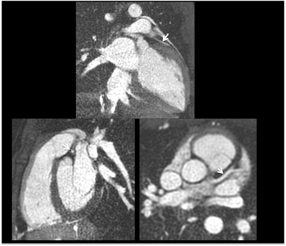
Results: Approximately 10 2.1-mm thick slices were acquired in a single ~20-s breath-hold using two-shot QISS. QISS contrast-to-noise ratio (CNR) was 1.5-fold higher at 3 Tesla than at 1.5 Tesla. Cartesian QISS provided the best coronary-to-myocardium CNR, whereas radial QISS provided the sharpest coronary images. QISS image quality exceeded that of free-breathing 3D coronary MRA with few artifacts at either field strength. Compared with T2-prepared 2D bSSFP, multi-slice capability was not restricted by the specific absorption rate at 3 Tesla and pericardial fluid signal was better suppressed. In addition to depicting the coronary arteries, QISS could image intra-cardiac structures, pericardium, and the aortic root in arbitrary slice orientations.

Conclusions: Breath-hold QISS is a simple, versatile, and time-efficient method for coronary MRA that provides excellent image quality at both 1.5 and 3 Tesla. Image quality exceeded that of free-breathing, navigator-gated 3D MRA in a much shorter scan time. QISS also allowed rapid multi-slice bright-blood, diastolic phase imaging of the heart, which may have complementary value to multi-phase cine imaging. We conclude that, with further clinical validation, QISS might provide an efficient alternative to commonly used free-breathing coronary MRA techniques.


Figures

Fig. 1
Fig. 1
Pulse sequence diagrams for radial QISS (a) and 2D T2-prepared bSSFP (b). QISS (flow dependent) applies a slice-selective FOCI pulse for inversion of in-plane spins, followed by a quiescent interval (QI) to allow for replenishment of in-plane arterial spins. With T2-prepared bSSFP (flow independent), a spatially non-selective T2 preparation is applied
Fig. 2
Fig. 2
Examples of thin (4 to 10-mm) maximum intensity projections reconstructed from single breath hold, radial QISS (10–12 slices per breath hold, slice thickness = 2.1-mm with 20-50 % slice overlap, in-plane spatial resolution of 0.4-mm to 0.5-mm after interpolation). Images were acquired at 3 Tesla using various scan orientations. a Aorta and left coronary circulation. LM = left main; LAD = left anterior descending; D1 = first diagonal branch; D2 = second diagonal branch; LCx = left circumflex; OM = obtuse marginal branch. b Right coronary circulation. RCA = right coronary artery; AM = acute marginal branch
Fig. 3
Fig. 3
39-year-old male evaluated for chest pain. Left: 5-mm MIP of coronary CT angiography shows mild narrowing of the proximal LAD and a punctate coronary calcification (arrow). Middle: MIP of breath-hold radial QISS MRA obtained at 1.5 Tesla demonstrates the left main and LAD coronary arteries, including the D1 and D2 branches. The appearances are similar to the coronary CT angiogram except that the wall calcification is not visible. Right: MIP from navigator-gated 3D bSSFP also demonstrates the left coronary anatomy comparably to the coronary CTA. Compared with QISS, there is increased pericardial fluid signal (arrows)
Fig. 4
Fig. 4
Radial QISS images acquired in three orthogonal planes within a single breath-hold show the LAD (arrow) in long and short axes. Left and right ventricular myocardium, pulmonary veins, and mitral valve leaflets are also well depicted
Fig. 5
Fig. 5
Montage of radial QISS images that were oriented orthogonally to the long axis of the LAD. Images were acquired at 1.5 Tesla in two breath-holds (14 images shown out of 18 acquired). The LAD and LCx, including their takeoffs from the left main coronary artery, are well seen. Magnified view (inset) shows the LAD, left circumflex, and posterior descending branch of the right coronary artery (PDA)
Fig. 6
Fig. 6
Comparison of source images from breath-hold radial QISS (left) and T2-prepared radial 2D bSSFP (right) in a healthy volunteer at 1.5 Tesla. Compared with QISS, pericardial fluid signal (arrows) is substantially increased with T2-prepared bSSFP
Fig. 7
Fig. 7
57-year-old patient with hyperlipidemia and chest pain. Breath-hold 2D T2-prepared bSSFP and free-breathing 3D T2-prepared bSSFP showed similar findings to the coronary CTA, which was prospectively interpreted as showing a severe LAD stenosis (arrow). However, radial QISS indicated an LAD occlusion, which was confirmed by subsequent x-ray coronary catheterization

References

    1. Pennell DJ. Cardiovascular magnetic resonance. Circulation. 2010;121:692–705. doi: 10.1161/CIRCULATIONAHA.108.811547. - DOI - PubMed
    1. Kato S, Kitagawa K, Ishida N, et al. Assessment of coronary artery disease using magnetic resonance coronary angiography: a national multicenter trial. J Am Coll Cardiol. 2010;56(12):983–91. doi: 10.1016/j.jacc.2010.01.071. - DOI - PubMed
    1. In the original publication (Magn Reson Med 2010;63:951–958), the QISS acronym stood for “quiescent-interval single shot”. However, we have modified the acronym to stand for “quiescent-interval slice-selective” in order to more flexibly encompass more recent implementations of the technique for carotid and coronary MRA.
    1. Hodnett PA, Koktzoglou I, Davarpanah AH, et al. Evaluation of peripheral arterial disease with nonenhanced quiescent-interval single-shot MR angiography. Radiology. 2011;260(1):282–93. doi: 10.1148/radiol.11101336. - DOI - PMC - PubMed
    1. Oshinski JN, Delfino JG, Sharma P, Gharib AM, Pettigrew RI. Cardiovascular magnetic resonance at 3.0 T: current state of the art. J Cardiovasc Magn Reson. 2010;12:55. doi: 10.1186/1532-429X-12-55. - DOI - PMC - PubMed

Publication types

MeSH terms