

Unique expression of cytoskeletal proteins in human soft palate muscles
- PMID: 26597319
- PMCID: PMC5341545
- DOI: 10.1111/joa.12417
Unique expression of cytoskeletal proteins in human soft palate muscles
Abstract
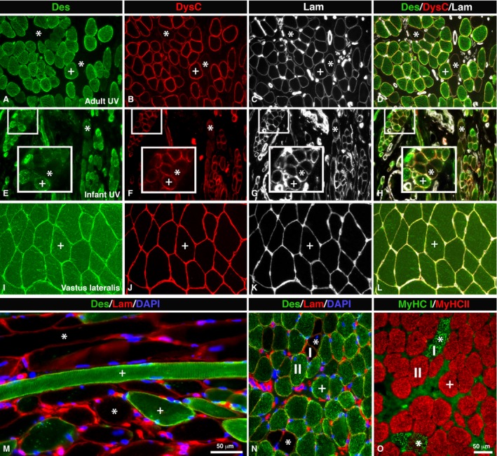
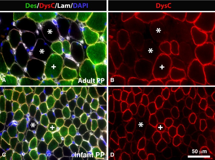
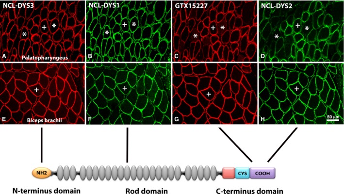
The human oropharyngeal muscles have a unique anatomy with diverse and intricate functions. To investigate if this specialization is also reflected in the cytoarchitecture of muscle fibers, intermediate filament proteins and the dystrophin-associated protein complex have been analyzed in two human palate muscles, musculus uvula (UV) and musculus palatopharyngeus (PP), with immunohistochenmical and morphological techniques. Human limb muscles were used as reference. The findings show that the soft palate muscle fibers have a cytoskeletal architecture that differs from the limb muscles. While all limb muscles showed immunoreaction for a panel of antibodies directed against different domains of cytoskeletal proteins desmin and dystrophin, a subpopulation of palate muscle fibers lacked or had a faint immunoreaction for desmin (UV 11.7% and PP 9.8%) and the C-terminal of the dystrophin molecule (UV 4.2% and PP 6.4%). The vast majority of these fibers expressed slow contractile protein myosin heavy chain I. Furthermore, an unusual staining pattern was also observed in these fibers for β-dystroglycan, caveolin-3 and neuronal nitric oxide synthase nNOS, which are all membrane-linking proteins associated with the dystrophin C-terminus. While the immunoreaction for nNOS was generally weak or absent, β-dystroglycan and caveolin-3 showed a stronger immunostaining. The absence or a low expression of cytoskeletal proteins otherwise considered ubiquitous and important for integration and contraction of muscle cells indicate a unique cytoarchitecture designed to meet the intricate demands of the upper airway muscles. It can be concluded that a subgroup of muscle fibers in the human soft palate appears to have special biomechanical properties, and their unique cytoarchitecture must be taken into account while assessing function and pathology in oropharyngeal muscles.
Keywords: cytoskeleton; desmin; dystrophin; muscle fiber; palatopharyngeus; sleep apnea; soft palate; uvula.
© 2015 Anatomical Society.
Figures
References
-
- Blake DJ, Tinsley JM, Davies KE (1996) Utrophin: a structural and functional comparison to dystrophin. Brain Pathol 6, 37–47. - PubMed
-
- Campbell KP (1995) Three muscular dystrophies: loss of cytoskeleton‐extracellular matrix linkage. Cell 80, 675–679. - PubMed
-
- Capetanaki Y, Bloch R, Kouloumenta A, et al. (2007) Muscle intermediate filaments and their links to membranes and membranous organelles. Exp Cell Res 313, 2063–2076. - PubMed
-
- Davies KE, Nowak KJ (2006) Molecular mechanisms of muscular dystrophies: old and new players. Nat Rev Mol Cell Biol 7, 762–773. - PubMed
Publication types
MeSH terms
LinkOut - more resources
Full Text Sources
Other Literature Sources
Miscellaneous
