. 2015 Nov 24:5:17185.
doi: 10.1038/srep17185.

Characterization of stem/progenitor cell cycle using murine circumvallate papilla taste bud organoid

Affiliations

Characterization of stem/progenitor cell cycle using murine circumvallate papilla taste bud organoid

Eitaro Aihara et al. Sci Rep. .

Abstract

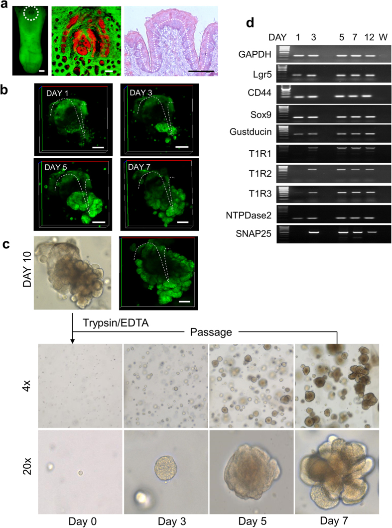
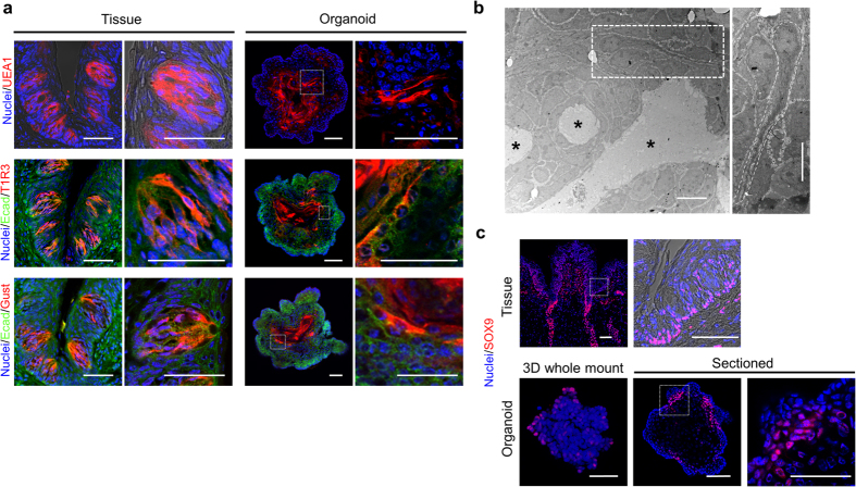
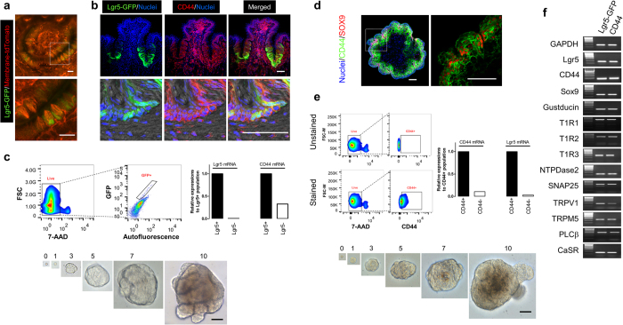
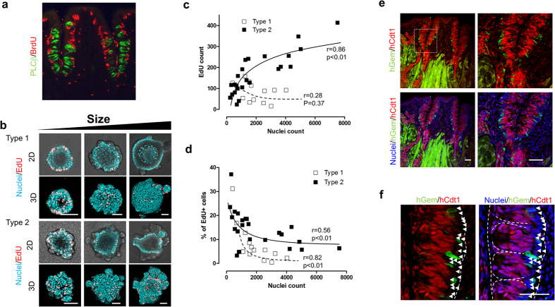
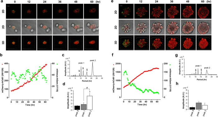
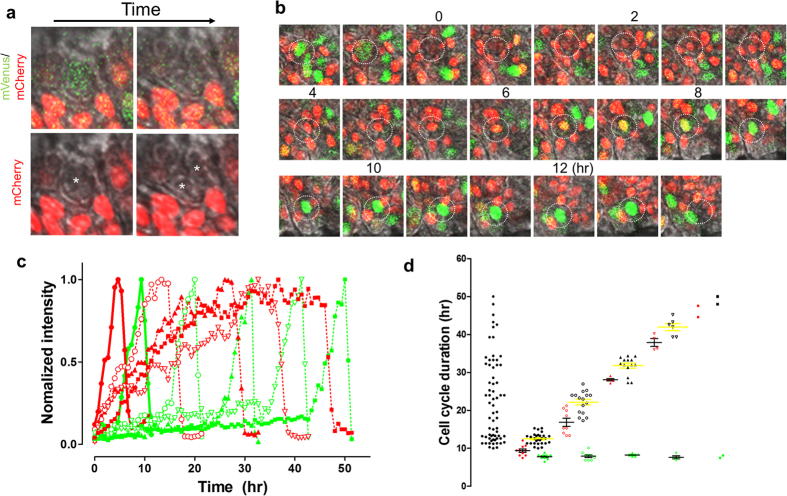
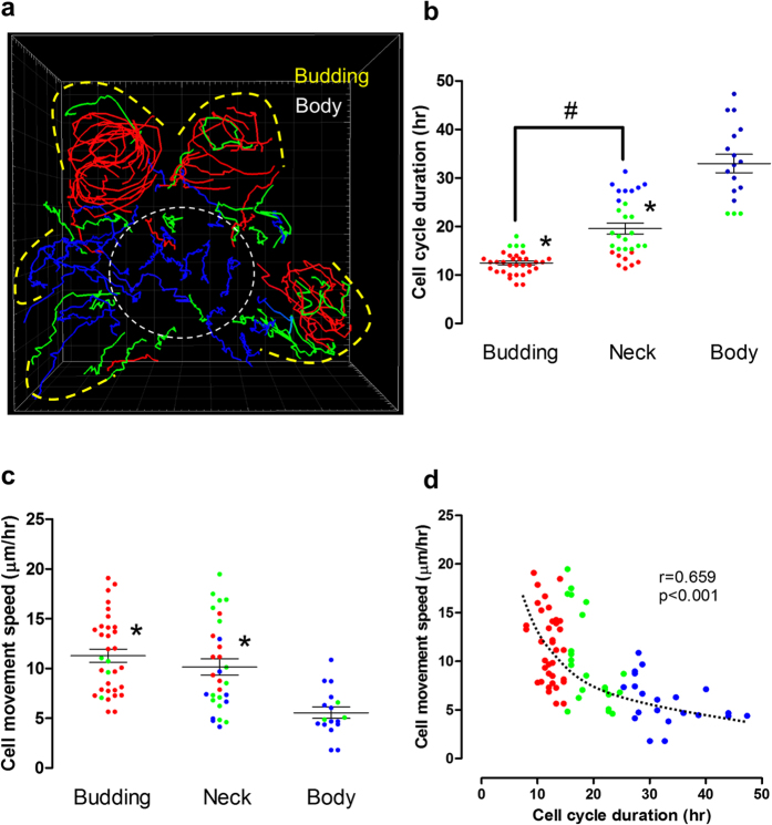
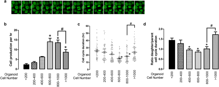
Leucine-rich repeat-containing G-protein coupled receptor 5-expressing (Lgr5(+)) cells have been identified as stem/progenitor cells in the circumvallate papillae, and single cultured Lgr5(+) cells give rise to taste cells. Here we use circumvallate papilla tissue to establish a three-dimensional culture system (taste bud organoids) that develops phenotypic characteristics similar to native tissue, including a multilayered epithelium containing stem/progenitor in the outer layers and taste cells in the inner layers. Furthermore, characterization of the cell cycle of the taste bud progenitor niche reveals striking dynamics of taste bud development and regeneration. Using this taste bud organoid culture system and FUCCI2 transgenic mice, we identify the stem/progenitor cells have at least 5 distinct cell cycle populations by tracking within 24-hour synchronized oscillations of proliferation. Additionally, we demonstrate that stem/progenitor cells have motility to form taste bud organoids. Taste bud organoids provides a system for elucidating mechanisms of taste signaling, disease modeling, and taste tissue regeneration.


Figures

Figure 1
Figure 1. Development of taste bud organoids from CV tissue.
(a) Isolated tongue from YC mouse (white circle area: circumvallate papilla, GFP filter, Scale bars = 1000 μm), confocal image and H&E staining of CV (white dotted line: epithelium, Scale bars = 200 μm). Nuclei stained with Hoechst 33342 (Red). (b) 3D structure of taste bud organoid grows from isolated CV (white circle area in (a) from YC mouse. 3D YFP reconstructed from 1, 3, 5 or 7 day cultures. The white dotted line shows morphology of isolated CV epithelium. Scale bars = 100 μm (c) After 10 days in culture, taste bud organoids were broken up to single cells by trypsin/EDTA and re-embedded in Matrigel. Images were taken 0, 3, 5 or 7 days after passage. (d) Stem/progenitor cell or taste bud lineage marker mRNA was determined 1, 3, 5, 7, or 12 days after passage. W: water.
Figure 2
Figure 2. Expression of taste bud cell lineage markers in the organoid.
(a) Sectioned CV tissue or organoid were stained with UEA1 (red), E-cadherin (green) and T1R3 (red) or E-cadherin (green) and gustducin (red), respectively. Cell nucleus (blue) was stained by Hoechst 33342. Scale bars = 50 μm. (b) Transmission electron microscopy images shows the middle of the organoid with low resolution, and high resolutions of taste cell (white rectangle). Asterisks indicate the lumen. Scale bars = 10 μm. (c) Sectioned CV tissue (low magnification and high magnification of white rectangle), 3D whole mount organoid, or sectioned organoid (low magnification and high magnification of white rectangle) were stained with SOX9 (red). Nuclei (blue) was stained with Hoechst 33342. Scale bars = 50 μm.
Figure 3
Figure 3. Lgr5+ or CD44+ cell-induced taste bud organoids.
(a) 3D images CV region of tongue freshly isolated from Lgr5-GFP/membrane-tdTomato mouse with low magnification and high magnification of white rectangle indicated in top panel. Scale bars = 50 μm. (b) Sectioned Lgr5-GFP mouse CV tissue was immunostained for GFP (green), CD44 (red) and nuclei (Hoechst 33342: blue). Scale bars = 50 μm. (c) Representative histograms of Lgr5-GFP stem cell sorting from isolated CV tissue, and expression of Lgr5 or CD44 mRNA were determined. Images of taste bud organoid growth from sorted Lgr5-GFP stem cell, at indicated days after cell plating. (d) Sectioned organoid was stained with CD44 (green), SOX9 (red) and nuclei (blue). Scale bars = 50 μm. (e) Representative histograms of CD44+ cell sorting from taste bud organoids, and expression of Lgr5 or CD44 mRNA were determined. Images of taste bud organoid growth from sorted CD44+ cell. (f) After 12 days culture of Lgr5-GFP cell or CD44+ cell-derived taste bud organoid, stem/progenitor cell or taste bud lineage markers mRNA was determined by RT-PCR.
Figure 4
Figure 4. Proliferation zone within taste bud organoids.
Representative images of BrdU (red) staining in the CV tissue (PLCβ:green) (a), 2D (top) and 3D (bottom) EdU (red) staining of different sizes of type 1 (sphere) or type 2 (with budding) taste bud organoid (b). (c) Numbers of EdU positive cells were counted in the different sizes of type 1 (sphere: open rectangle) or type 2 (with budding: closed rectangle) taste bud organoid. (d) % EdU positive cells versus nuclei were calculated from different sizes of type 1 (sphere: open rectangle) or type 2 (with budding: closed rectangle) taste bud organoid shown in (c). (e) Representative images of expression of mVenus (green) and mCherry (red) in the CV tissue. (f) High magnification image of mVenus (green) and mCherry (red) in the CV tissue of white rectangle indicated in (e). Symbol ▵ shows cells that both mVenus (green) and mCherry (red) did not detect. Nuclei (blue) was stained with Hoechst 33342. The white dotted line shows morphology of taste buds. Scale bars = 50 μm (b,e,f).
Figure 5
Figure 5. Monitoring the cell cycle using FUCCI2 taste bud organoids.
Taste bud organoids were created from FUCCI2 transgenic mice and organoid growth was monitored from 3–6 days (a) or 9–12 days (e) on the confocal microscope. Scale bars = 50 μm. Numbers of mCherry (red) or mVenus (green) positive cells in 4D were counted using Imaris software (b or f). (c or g) Representative FFT analysis data of time courses of number of mVenus-hGeminin positive cells from organoids monitored from 3–6 days (a) or 9–12 days (e), respectively. (d or h) The average amplitudes of peak 1 and peak 2 were calculated from 3 different taste bud organoids. The average amplitude of peak $ was calculated from detected peaks other than peak 1 and peak 2. *p < 0.05 vs. peak $, #p < 0.05 vs. peak 1.
Figure 6
Figure 6. Diversity of cell cycle duration in the taste bud organoid.
Taste bud organoids were created from FUCCI2 transgenic mice and growth was monitored by confocal microscope. (a) High magnification image of mVenus/mCherry (top), or mCherry (bottom) with differential interference contrast (DIC) superimposed on the fluorescence. Note that cell division (white asterisk) occurs following the disappearance of mVenus fluorescence. (b) Representative images of 12 hr cell cycle determined by tracking FUCCI2 fluorescence appearance in the taste bud organoid. (c) Changes of mVenus or mCherry fluorescence intensity during cell cycle, calculated from b (●), Supplementary Fig. 7a (○), 7b (▲), 7c (▽) or from 52 hr cycling cells (■). (d) Duration of the cell cycle from 68 cells tracked (left: black circle), and divided into 5 groups based on clustering of cell cycle duration (Supplementary Fig. 6). Each group shows duration of mCherry (red), mVenus (green) (black line) and mCherry + mVenus (black: yellow line).
Figure 7
Figure 7. Tracking individual cells in the taste bud organoid.
Manual cell tracking was performed using Imaris 7.7 on FUCCI2 organoids showed in Fig. 5e and Supplementary video 4. (a) Image showed summary of proliferative cell tracking, while tracking 3D movie showed in Supplementary video 5. The line colors were separated by cell cycle duration (Red: <15 hr, Green: 15–25 hr, Blue >25). The cell cycle durations (b) or cell movement speeds (c) are shown at different positions in the organoid (In (a), Budding: yellow outlined, Neck: between yellow and body, Sphere Body: while outlined). Additionally, correlation between cell movement speed and cell cycle duration was shown in d. *p < 0.05 vs. body. #p < 0.05 vs. neck.
Figure 8
Figure 8. Tracking daughter cells from parent cells in taste bud organoids.
Taste bud organoids were created from H2B-EGFP transgenic mice and growth monitored on the confocal microscope. (a) Representative image of cell division of the taste bud organoid. (b) H2B-EGFP was counted in 4D using Imaris software, and calculated rate of cell number changes over time (cell production/hr) from size grouping of organoids as a <200, 200–400, 400–600, 600–800, 800–1000, and >1000. (c) Time between cell divisions (431 cells) was tracked from 11 different taste bud organoids. Cell cycle duration was separated based on cell numbers in the organoid. (d) Tracking daughter cell cycle duration from parent cell (raw data shown in Supplementary Fig. 8a), and data shows the ratio of daughter cell to parent cell duration based on cell numbers in the organoid. *p < 0.05 vs. <200. #p < 0.05 vs. 800–1000.

Similar articles

Cited by

References

    1. Chaudhari N. & Roper S. D. The cell biology of taste. J Cell Biol 190, 285–296 (2010). - PMC - PubMed
    1. Barlow L. A. & Klein O. D. Developing and regenerating a sense of taste. Curr Top Dev Biol 111, 401–419 (2015). - PMC - PubMed
    1. Ozdener H. et al. Characterization and long-term maintenance of rat taste cells in culture. Chem Senses 31, 279–290 (2006). - PubMed
    1. Ozdener M. H. & Rawson N. E. Primary culture of mammalian taste epithelium. Methods Mol Biol 945, 95–107 (2013). - PMC - PubMed
    1. Kishi M., Emori Y., Tsukamoto Y. & Abe K. Primary culture of rat taste bud cells that retain molecular markers for taste buds and permit functional expression of foreign genes. Neuroscience 106, 217–225 (2001). - PubMed

Publication types