. 2015 Nov 23;84(2):467-79.
doi: 10.1128/IAI.01171-15. Print 2016 Feb.

The Chromosome-Encoded Hypothetical Protein TC0668 Is an Upper Genital Tract Pathogenicity Factor of Chlamydia muridarum

Affiliations

The Chromosome-Encoded Hypothetical Protein TC0668 Is an Upper Genital Tract Pathogenicity Factor of Chlamydia muridarum

Turner Allen Conrad et al. Infect Immun. .

Abstract

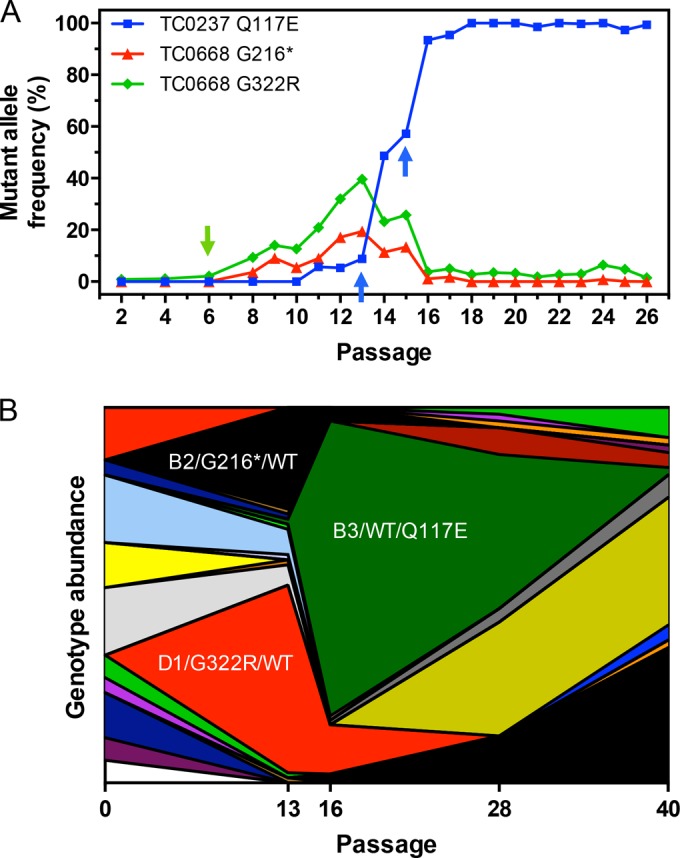
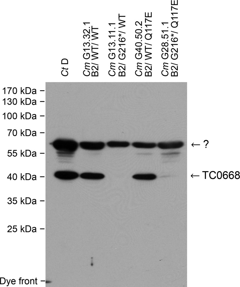
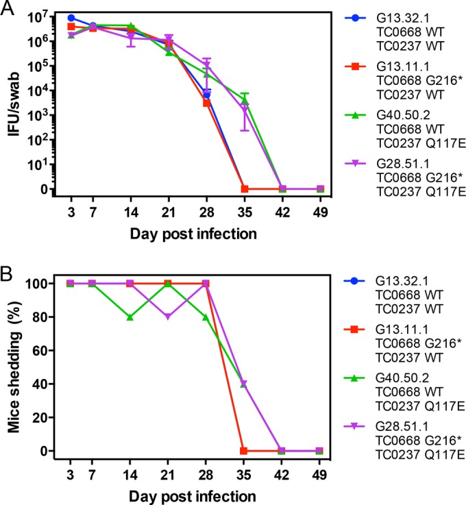
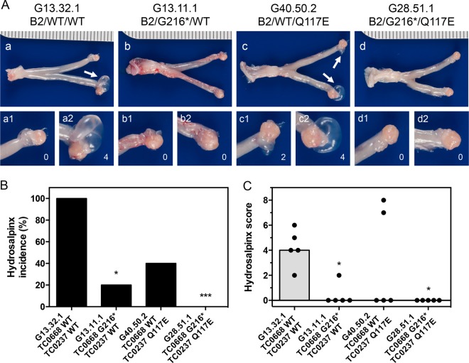
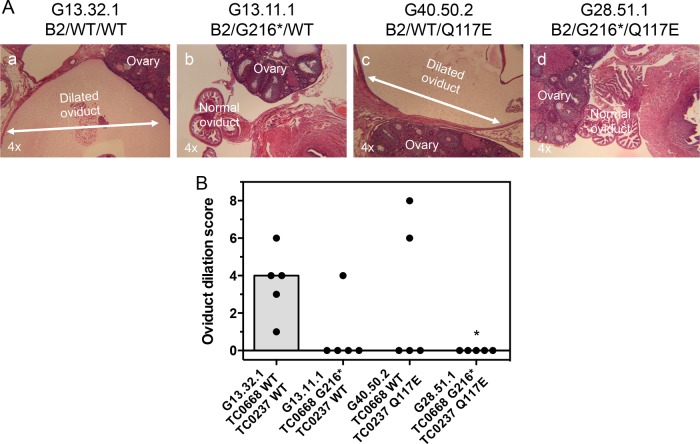
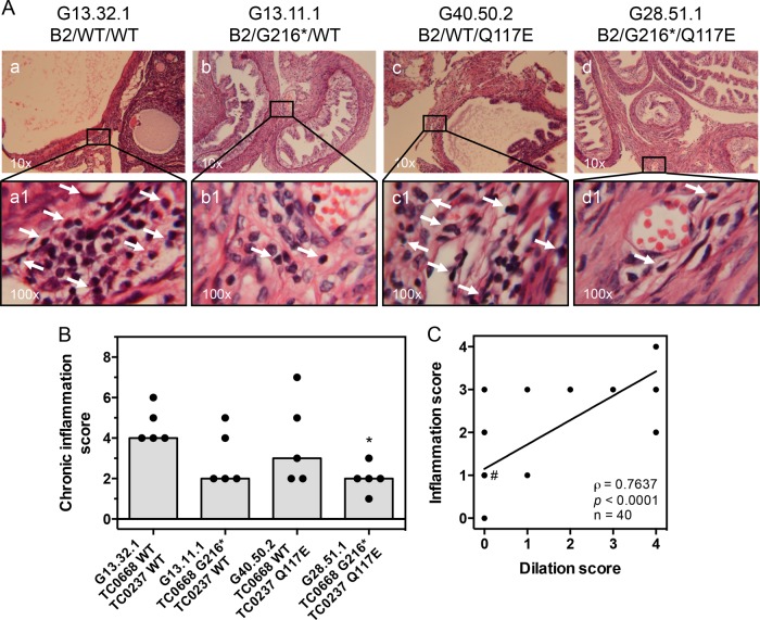
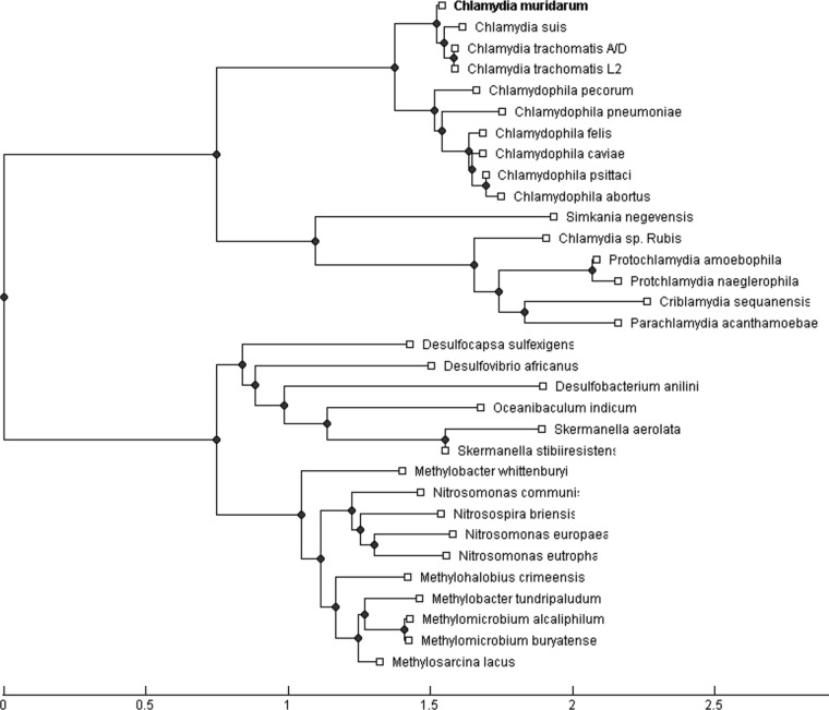
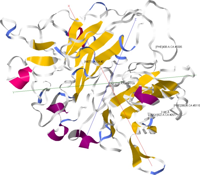
We previously associated a missense mutation of the tc0668 gene of serial in vitro-passaged Chlamydia muridarum, a murine model of human urogenital C. trachomatis, with severely attenuated disease development in the upper genital tract of female mice. Since these mutants also contained a TC0237 Q117E missense mutation that enhances their in vitro infectivity, an effort was made here to isolate and characterize a tc0668 single mutant to determine its individual contribution to urogenital pathogenicity. Detailed genetic analysis of C. muridarum passages revealed a truncated variant with a G216* nonsense mutation of the 408-amino-acid TC0668 protein that does not produce a detectable product. Intracellular growth and infectivity of C. muridarum in vitro remain unaffected in the absence of TC0668. Intravaginal inoculation of the TC0668 null mutant into C3H/HeJ mice results in a typical course of lower genital tract infection but, unlike a pathogenic isogenic control, is unable to elicit significant chronic inflammation of the oviduct and fails to induce hydrosalpinx. Thus, TC0668 is demonstrated as an important chromosome-encoded urogenital pathogenicity factor of C. muridarum and the first with these characteristics to be discovered for a Chlamydia pathogen.


Figures

FIG 1
FIG 1
Population dynamics of mutations arising from C. muridarum Nigg3 passage. (A) Mutant allele frequencies of tc0237 and tc0668 variants across early C. muridarum Nigg3 passage were calculated from the ratio of mutant to wild-type allele relative fluorescence signals in Sanger sequencing chromatograms. Source DNA was purified from passaged populations and PCR amplified prior to sequencing. The green arrow indicates the first appearance of tc0668 mutation (passage 6), and blue arrows indicate selective sweep of TC0237 Q117E (unassisted passages 13 and 15). (B) Combined TC0412, TC0237, and TC0668 genotypes of Nigg3 plaque clones as determined by Sanger sequencing. Each continuous area represents a unique genotype encompassing all three genes. For genotype B2/G216*/WT (black), B2 signifies the proteoform of TC0412, G216* represents the nonsense mutation of TC0668, and wild type (WT) indicates no mutation in TC0237. The D1/G322R/WT and B3/WT/Q117E genotypes are highlighted in red and dark green, respectively. Abundances are in relation to the number of plaques isolated from each Nigg3 passage shown, with a total of 326 clones across the five passages sampled. Discontinuity in unique genotypes is largely due to selective sweep of TC0237 Q117E.
FIG 2
FIG 2
Western blot detection of TC0668 protein in C. muridarum Nigg3 mutant clones. The CT389 and TC0668 proteins from 1 × 10⁷ heat-denatured C. trachomatis serovar D (Ct D) and C. muridarum (Cm) Nigg3 EBs were detected by a mouse polyclonal anti-CT389 antibody. Nigg3 EB organisms are labeled by their clone designations followed by their TC0412/TC0668/TC0237 proteoform/genotypes as indicated on top of the figure, including C. muridarum variant clones G13.31.1 with a genotype of B2/WT/WT, G13.11.1 with a genotype of B2/G216*/WT, G40.50.2 with a genotype of B2/WT/Q117E, and G28.51.1 with a genotype of B2/G216*/Q117E (see Table S2 in the supplemental material for details). TC0668 protein (∼47 kDa) is abundant in clones with the wild-type tc0668 sequence but not detectable in clones with the G216* mutation. An unknown (?) band of approximately ∼65 kDa is likely a secondary target of the polyclonal antibody and serves as a loading reference.
FIG 3
FIG 3
In vitro fitness phenotypes of C. muridarum Nigg3 mutant clones. (A) Clone dependence on inoculum centrifugation for infection. Fold increase of HeLa cell infection was calculated by dividing the titers of the centrifuged by those of the noncentrifuged samples derived from the same source material (P < 0.0001, by one-way ANOVA; **** P < 0.0001, by a Holm-Sidak multiple-comparison test against the control organism). Bars are representative of means of two to three experiments with standard errors of the means. (B) Intracellular growth and development of clones during logarithmic growth. The number of progeny IFU per input IFU was calculated from duplicate infections using the same source material; one infection was directly titrated, while another was harvested at 24 h postinfection (hpi) and titrated (P = 0.75, by one-way ANOVA). Bars represent means from four experiments with standard errors of the means.
FIG 4
FIG 4
Plaque sizes of C muridarum Nigg3 mutant clones. (A) Plaques formed by clones after 6 days of growth in confluent McCoy B monolayers. (B) Relative sizes of plaques in scanned-plate image pixels formed by the mutant clones (P < 0.0001 by one-way ANOVA; ****, P < 0.0001 by a Holm-Sidak multiple-comparison test against the control organism). Individual values are shown as gray points, and means with standard errors of the means are shown as black lines.
FIG 5
FIG 5
In vivo lower genital tract dynamics of C. muridarum Nigg3 mutant clones. (A) Lower genital tract shedding course of clones following intravaginal inoculation with 2 × 10⁵ IFU. Shedding course was titrated from cervicovaginal swabs collected on the indicated days postinfection (dpi). Points represent means of 5 mice per group (n = 5) with standard errors of the means. (B) Percentage of mice in each infection group with positive cervicovaginal shedding on the day postinfection indicated.
FIG 6
FIG 6
Gross pathology of murine genital tracts infected with C. muridarum Nigg3 mutant clones. (A) Representative gross pathology images of whole genital tracts harvested at 56 dpi. Left and right oviducts and ovaries are amplified for detail. White arrows indicate hydrosalpinges. (B) Incidence of hydrosalpinx at 56 dpi (*, P < 0.05; ***, P < 0.001, by Fisher's exact test against the results for the control organism; n = 5 per group). (C) Severity of hydrosalpinx at 56 dpi (P = 0.021, by a Kruskal-Wallis test; *, P < 0.05, by Dunn's multiple-comparison test against the results for the control organism). Both individual bilateral scores (black dots) and medians (gray bars) are shown.
FIG 7
FIG 7
Oviduct dilation of mice infected with C. muridarum Nigg3 mutant clones. (A) Representative micrographs of H&E-stained oviducts from infected mice at 56 dpi. Oviduct dilation is illustrated by white double-headed arrows. (B) Individual (black dots) and median (gray bars) bilateral oviduct dilation scores determined from H&E-stained tissue sections (P = 0.0363, by a Kruskal-Wallis test; *, P < 0.05, by Fisher's exact test against results for the control organism).
FIG 8
FIG 8
Chronic oviduct inflammation of mice infected with C. muridarum Nigg3 mutant clones. (A) Representative micrographs of H&E-stained oviducts from infected mice at 56 dpi. Top panels (a to d) present a broad view of tissue sections. Chronic inflammatory cell infiltrate foci are illustrated by white arrows in bottom panels (a1 to d1). (B) Individual (black dots) and median (gray bars) bilateral chronic inflammatory cell infiltrate scores determined from H&E-stained tissue sections (P = 0.0533, by a Kruskal-Wallis test; *, P < 0.05, by Dunn's multiple-comparison test against results for the control organism). (C) Unilateral oviduct dilation scores plotted against corresponding unilateral oviduct chronic inflammation scores. Deming linear regression (black line) illustrates an unbiased trend of the overlapping ordinal score pairs. A majority of the points (23 of 40) lie within the (0, 1) coordinate (#). Spearman rank correlation is highly significant at a P value of <0.0001 with a correlation coefficient of ρ = 0.7637.
FIG 9
FIG 9
Neighbor-joining tree of TC0668 homologs. Homologous protein sequences were discovered by a protein-protein BLAST search (38) against the NCBI nonredundant protein database. The neighbor-joining phylogenetic tree is rooted by the longest distance as calculated using the Grishin general method (39).
FIG 10
FIG 10
Predicted model of TC0668 protein. TC0668 tertiary protein structure was predicted using the I-TASSER suite (35–37). Residues are labeled at the upper right by position and include the N-terminal M1 and C-terminal F408, G216, G322, and F386 residues. The distance in angstroms between the G322 and F386 residues is indicated by a dashed line (7.96 Å). Colors distinguish secondary structure features. Model depictions were acquired using the Jmol plug-in within the MATLAB Bioinformatics Toolbox.

Similar articles

Cited by

References

    1. Nigg C. 1942. An unidentified virus which produces pneumonia and systemic infection in mice. Science 95:49–50. doi:10.1126/science.95.2454.49. - DOI - PubMed
    1. Nigg C, Eaton MD. 1944. Isolation from normal mice of a pneumotropic virus which forms elementary bodies. J Exp Med 79:497–510. doi:10.1084/jem.79.5.497. - DOI - PMC - PubMed
    1. Read TD, Brunham RC, Shen C, Gill SR, Heidelberg JF, White O, Hickey EK, Peterson J, Utterback T, Berry K, Bass S, Linher K, Weidman J, Khouri H, Craven B, Bowman C, Dodson R, Gwinn M, Nelson W, DeBoy R, Kolonay J, McClarty G, Salzberg SL, Eisen J, Fraser CM. 2000. Genome sequences of Chlamydia trachomatis MoPn and Chlamydia pneumoniae AR39. Nucleic Acids Res 28:1397–1406. doi:10.1093/nar/28.6.1397. - DOI - PMC - PubMed
    1. Horn M, Collingro A, Schmitz-Esser S, Beier CL, Purkhold U, Fartmann B, Brandt P, Nyakatura GJ, Droege M, Frishman D, Rattei T, Mewes H-W, Wagner M. 2004. Illuminating the evolutionary history of Chlamydiae. Science 304:728–730. doi:10.1126/science.1096330. - DOI - PubMed
    1. Torrone E, Papp J, Weinstock H, Centers for Disease Control and Prevention . 2014. Prevalence of Chlamydia trachomatis genital infection among persons aged 14–39 years—United States, 2007–2012. MMWR Morb Mortal Wkly Rep 63:834–838. - PMC - PubMed

Publication types

LinkOut - more resources