. 2015 Nov 24:6:10033.
doi: 10.1038/ncomms10033.

Gekko japonicus genome reveals evolution of adhesive toe pads and tail regeneration

Affiliations

Gekko japonicus genome reveals evolution of adhesive toe pads and tail regeneration

Yan Liu et al. Nat Commun. .

Abstract

Reptiles are the most morphologically and physiologically diverse tetrapods, and have undergone 300 million years of adaptive evolution. Within the reptilian tetrapods, geckos possess several interesting features, including the ability to regenerate autotomized tails and to climb on smooth surfaces. Here we sequence the genome of Gekko japonicus (Schlegel's Japanese Gecko) and investigate genetic elements related to its physiology. We obtain a draft G. japonicus genome sequence of 2.55 Gb and annotated 22,487 genes. Comparative genomic analysis reveals specific gene family expansions or reductions that are associated with the formation of adhesive setae, nocturnal vision and tail regeneration, as well as the diversification of olfactory sensation. The obtained genomic data provide robust genetic evidence of adaptive evolution in reptiles.


Figures

Figure 1
Figure 1. Phylogenetic analysis of the whole-genomes of 6 reptilian species and 10 additional vertebrate species.
The species in the phylogenetic tree include Danio rerio (D. rerio), Xenopus tropicalis (X. tropicalis), Chelonia mydas (C. mydas), Pelodiscus sinensis (P. sinensis), Alligator sinensis (A. sinensis), Python molurus bivittatus (P. bivittatus), Anolis carolinensis (A. carolinensis), Gekko japonicus (G. japonicus), Taeniopygia guttata (T. guttata), Gallus gallus (G. gallus), Ornithorhynchus anatinus (O. anatinus), Canis familiaris (C. familiaris), Mus musculus (M. musculus), Homo sapiens (H. sapiens), Oryzias latipes (O. latipes) and Meleagris gallopavo (M. gallopavo). Before the Permian period is represented in brown. The Permian period to the Triassic period is represented in green. The Triassic period to the Paleogene period is represented in purple. The Paleogene period to the present is represented in blue.
Figure 2
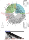
Figure 2. Phylogenetic tree of β-keratin families from G. japonicus, An. carolinensis and Al. sinensis.
(a) The β-keratins in black font belong to G. japonicus, those in red belong to An. carolinensis and those in blue belong to Al. sinensis. Gene copy number is listed in parentheses. The green background denotes β-keratins in scales and claws. The grey background denotes β-keratins in setae. The blue background denotes β-keratins in digital scales and pad lamella for supporting setae. A schematic diagram of toe of G. japonicus, An. carolinensis and Al. sinensis, which possess branched setae, unbranched setae and no setae are presented, respectively. The setae of gecko G. japonicus are ∼60 μm in length, and that in An. carolinensis is ∼25 μm. (b) Synteny diagram of β-keratin genes in A. carolinensis (upper line: GL343369, blue: 23 β-keratin genes) and G. japonicus (lower line: scaffold 426, red: 48 β-keratins).
Figure 3
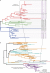
Figure 3. Evolutionary analysis of setae β-keratins.
(a) Phylogenetic tree of β-keratins from G. japonicus. The red branches represent β-keratins belonging to the primary components of setae, the blue branches represent the components in pad lamella for supporting setae and the green branches represent β-keratins in scales or claws. A total of 48 β-keratins (purple font) are clustered in scaffold 426, of these 46 have a single exon. The combinatorial numbers following the keratin names indicate the following protein characteristics: 1: S-core box (SEVTIQPPPCTVVVPGPVLA, sequence similarity ≥70%, 35 proteins); 2: cysteine-rich (Cys >10%, 19 proteins); 3: glycine-rich (Gly >15%, 36 proteins); 4: isoelectric point (pI >7, 34 proteins); 5: molecular weight (Wt <15,000, 58 proteins); 0: none of the above. β-keratins associated with clinging ability have undergone extensive expansion and have higher isoelectric points. (b) Calculation of the expansion period for primary gecko setae β-keratins using protein sequences from 71 β-keratins of G. japonicas (orange), 23 β-keratins of An. carolinensis (light blue), 2 β-keratins of Al. sinensis and 1 β-keratin-like of Crocodylus niloticus (pink), and 36 β-keratins associated with bird's claw, scale and feather (violet, including the following birds: Gallus gallus, Chlamydotis macqueenii, Opisthocomus hoazin, Mesitornis unicolor, Haliaeetus leucocephalus, Leptosomus discolor, Nestor notabilis, Chaetura pelagica, Pterocles gutturalis, Tinamus guttatus, Pygoscelis adeliae, Tauraco erythrolophus, Manacus vitellinus, Picoides pubescens, Mycteria americana, Cathartes aura and Eurypyga helias). The divergence of scale and claw keratins occurs in a birds ancestor ∼156 Myr ago, and the feather keratins expansion occurred in birds ∼66 Myr ago. β-keratins in setae of G. japonicus have undergone two expansion periods: one approximately 105–96 Myr ago and the other approximately 87–80 Myr ago.
Figure 4
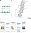
Figure 4. Opsins genes in the G. japonicus genome.
(a) Analysis of RH1 and SWS2 pseudogenes showed mutations in both initiation and termination codons. In addition, the exons of RH1 and SWS2 in G. japonicus were lost, incomplete or shifted in comparison with those in A. carolinensis. The green boxes indicate exons with similarity to those in A. carolinensis. The red boxes represent the lost exons. (b) Phylogenetic analysis of opsin genes from Drosophila melanogaster, Danio rerio, Oryzias latipes, Xenopus tropicalis, Chelonia mydas, Gallus gallus, Taeniopygia guttata, Anolis carolinensis, Gekko gecko and Homo sapiens. The pseudogenes in G. japonicus (P-RH1 and P-SWS2) are indicated in red. (c) Opsin evolution in G. japonicus at genomic level agrees with observed evolutionary variation of retinal cell type and visual sense.
Figure 5
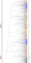
Figure 5. Phylogenetic tree of functional OR genes in seven species.
The Class I genes (including α, β, ɛ, ζ and δ ORs) encode proteins used for scent detection in water. The Class II gene (γ-OR) encode proteins used for scent detectionin air. G. japonicus has undergone extensive expansion in Class II genes. Red, G. japonicus; blue, X. tropicalis; green, D. rerio; purple, An. carolinensis; yellow, H. sapiens; pink, Al. sinensis; black, T. rubripes.
Figure 6
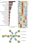
Figure 6. Positively selected genes (PSGs) related to tail regeneration in G. japonicus.
(a) Analysis of 155 PSGs in G. japonicus based on representative gene ontology biological processes. Categories with red bars such as cell cycle (GO:0007049), response to wounding (GO:0009611), wound healing (GO:0042060), tissue regeneration (GO:0042246), tissue remodelling (GO:0048771), blood coagulation (GO:0007596) and prostaglandin biosynthetic process (GO:0001516) are likely to be involved in tail regeneration after autotomy. (b) Heatmap of 107 PSGs in G. japonicus at different time points following tail autotomy. Approximately 70% of the PSGs were detected in transcriptome data. (c) Pathway of arachidonic acid (ARA) metabolism in G. japonicus. The circles with green backgrounds represent ARA and its derivatives. The boxes with orange backgrounds show the key synthases under positive selection in G. japonicus genome. The boxes with blue backgrounds show the normal synthases in G. japonicus genome.

References

    1. Wang D. Y., Kumar S. & Hedges S. B. Divergence time estimates for the early history of animal phyla and the origin of plants, animals and fungi. Proc. Biol. Sci. 266, 163–171 (1999) . - PMC - PubMed
    1. Peter U. The original descriptions of reptiles. Zootaxa 2334, 59–68 (2010) .
    1. Torstrom S. M., Pangle K. L. & Swanson B. J. Shedding subspecies: the influence of genetics on reptile subspecies taxonomy. Mol. Phylogenet. Evol. 76, 134–143 (2014) . - PubMed
    1. Vidal N. & Hedges S. B. The phylogeny of squamate reptiles (lizards, snakes, and amphisbaenians) inferred from nine nuclear protein-coding genes. C. R. Biol. 328, 1000–1008 (2005) . - PubMed
    1. Wever E. G. The lizard ear: Gekkonidae. J. Morphol. 143, 121–165 (1974) . - PubMed

Publication types

Associated data

LinkOut - more resources