. 2015 Dec;185(12):3141-51.
doi: 10.1016/j.ajpath.2015.08.020.

miR-223 Deficiency Protects against Fas-Induced Hepatocyte Apoptosis and Liver Injury through Targeting Insulin-Like Growth Factor 1 Receptor

Affiliations

miR-223 Deficiency Protects against Fas-Induced Hepatocyte Apoptosis and Liver Injury through Targeting Insulin-Like Growth Factor 1 Receptor

Ximena V Qadir et al. Am J Pathol. 2015 Dec.

Abstract

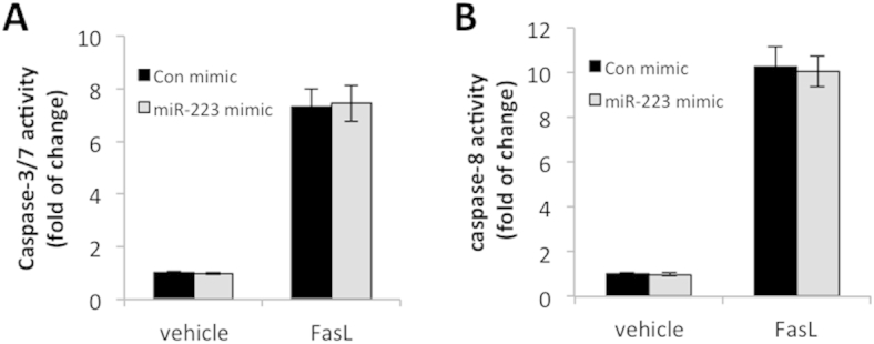
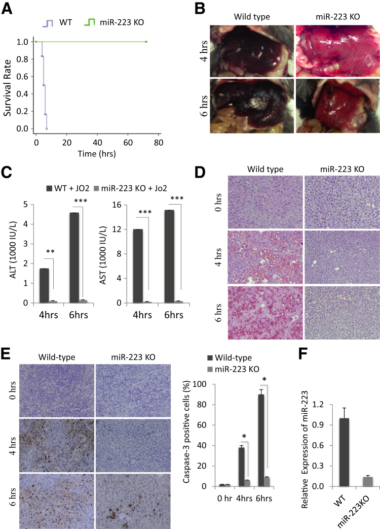
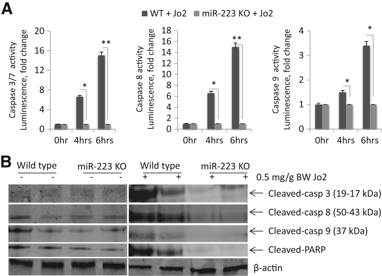
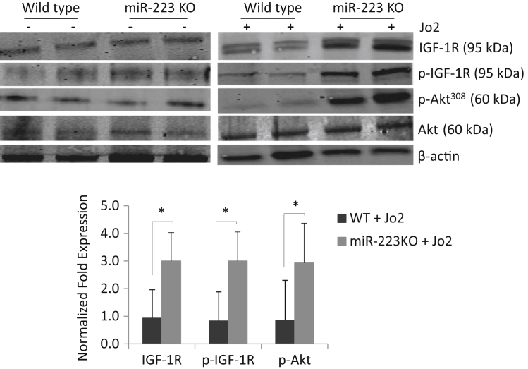
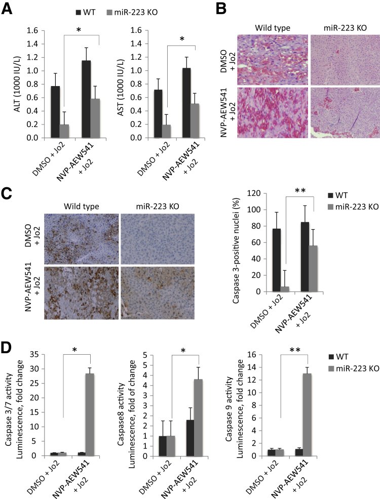
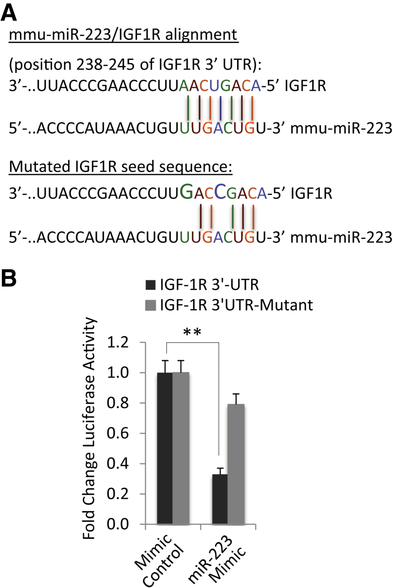
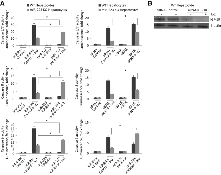
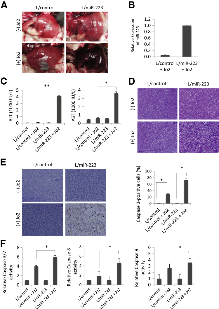
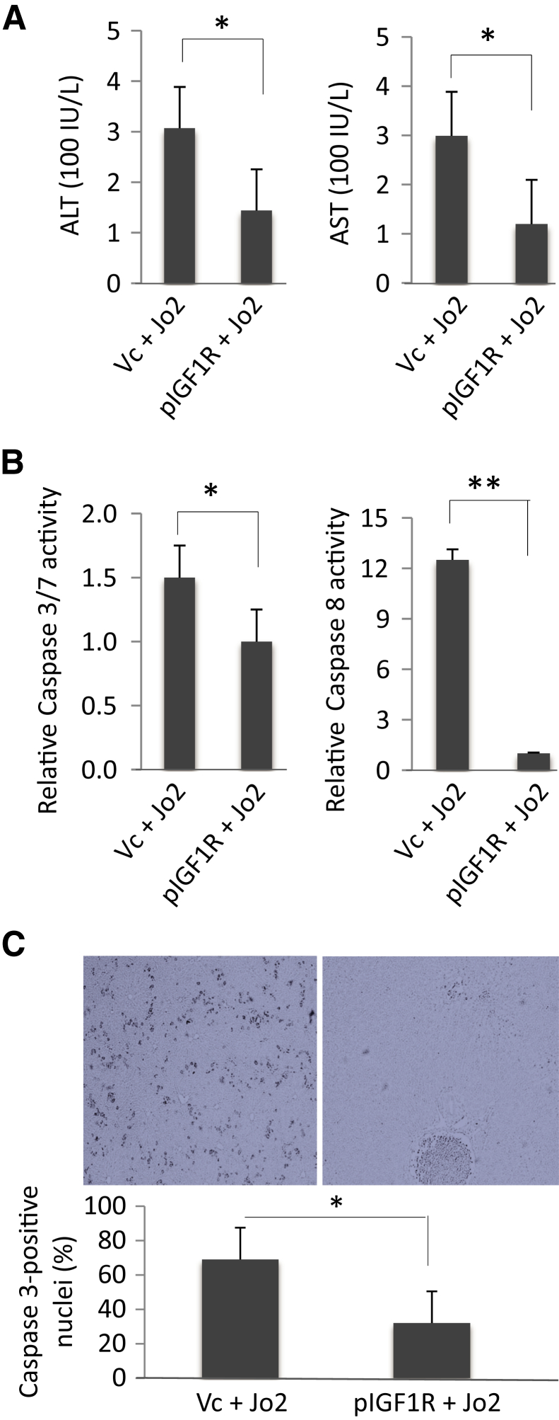
The biological functions and molecular mechanisms of miR-223 action in liver cells and liver diseases remain unclear. We therefore determined the effect and mechanism of action of miR-233 in Fas-induced hepatocyte apoptosis and liver injury. Wild-type (WT) and miR-223 knockout (KO) mice were treated i.p. with 0.5 μg/g body weight anti-Fas antibody Jo2, and the animals were monitored for survival and the extent of liver injury. Although WT mice died 4 to 6 hours after Jo2 injection (n = 6), all of the miR-223 KO mice (n = 6) survived. In comparison to WT mice, the miR-223 KO mice showed resistance to Fas-induced liver injury, as indicated by less tissue damage under histopathological examination, fewer apoptotic hepatocytes under caspase-3 immunostaining, and less elevation of serum transaminases. miR-223 KO livers showed less caspase-3, caspase-8, and caspase-9 activation and less poly (ADP-ribose) polymerase cleavage compared with WT livers (P < 0.05). Furthermore, tail vein injection of miR-223 lentiviral vector to miR-223 KO mice restored Jo2-induced liver injury. Transfection of miR-223 KO hepatocytes with miR-223 mimic enhanced Jo2-induced activation of caspase-3, caspase-8, and caspase-9, whereas transfection of WT hepatocytes with the miR-223 inhibitor attenuated Jo2-induced apoptosis. These findings demonstrate that miR-223 deficiency protects against Fas-induced hepatocyte apoptosis and liver injury. Further in vitro and in vivo data indicate that miR-223 regulates Fas-induced hepatocyte apoptosis and liver injury by targeting the insulin-like growth factor 1 receptor.


Figures

Supplemental Figure S1
The effect of miR-223 on Fas-induced apoptosis in HepG2 cells. HepG2 cells were transfected with synthetic human miR-223 mimic and control (Con) miRNA mimic and treated with 200 ng/mL recombinant human FasL for 24 hours. The activities of caspase-3/7 (A) and caspase-8 (B) were measured by the Caspase-Glo Assay Kit. The experiments were repeated three times. The data are expressed as means ± SD.
Figure 1
Figure 1
Deletion of miR-223 prevents Fas-induced liver injury. A: Wild-type (WT) and miR-223 knockout (KO) mice were i.p. injected with 0.5 μg/g Jo2 per 1 g body weight, and the animal survival was recorded at hourly intervals. B: Gross images of livers from WT and miR-223 KO mice 4 and 6 hours after Jo2 injection. C: Serum transaminase levels [alanine aminotransferase (ALT) and aspartate aminotransferase (AST)] from WT and miR-223 KO mice (4 and 6 hours after Jo2 treatment). Jo2-treated miR-223 KO mice show lower serum ALT and AST levels compared with Jo2-treated WT mice. D: Hematoxylin and eosin staining of liver tissues from WT and miR-223 KO mice (0, 4, and 6 hours after Jo2 injection). E: Caspase-3 immunostaining of liver tissues from WT and miR-223 KO mice (0, 4, and 6 hours after Jo2 injection) and the percentages of caspase-3–positive cells. F: Relative fold expression of miR-223 in WT and miR-223 KO livers, as determined by quantitative real-time PCR. Data represent means ± SD of fold changes (C). N = 6 per group (A). P < 0.05, ∗∗P < 0.01, and ∗∗∗P < 0.001. Original magnification, ×100 (D and E).
Figure 2
Figure 2
Deletion of miR-223 protects against Fas-induced caspase activation and poly (ADP-ribose) polymerase (PARP) cleavage. A: Wild-type (WT) and miR-223 KO mice were i.p. injected with 0.5 μg/g body weight Jo2. The liver tissues were harvested 0, 4, and 6 hours after Jo2 injection and processed for caspase (Casp) activity assays. The levels of caspase-3, caspase-8, and caspase-9 in miR-223 knockout (KO) livers are significantly lower compared with WT livers. B: Western blot analysis of liver tissue samples for caspase-3, caspase-8, and caspase-9 and PARP. Jo2 induces less cleavage of caspase-3, caspase-8, caspase-9, and PARP in miR-223 mice compared with WT mice. The result represents three individual experiments. The results represent means ± SD of fold changes (A). N = 6 per group (A). P < 0.05, ∗∗P < 0.01.
Figure 3
Figure 3
Deletion of miR-223 enhances insulin-like growth factor 1 receptor (IGF1R) signaling during Fas-induced liver injury. Wild-type (WT) and miR-223 knockout (KO) mice were i.p. injected with 0.5 μg/g body weight Jo2. In silico analysis for target gene prediction using TargetScan led to identification of Igf1r as a candidate gene of miR-223 target. Western blot analysis was performed to measure the levels of IGF1R and p-IGF1R as well as its downstream target Akt. The miR-223 KO livers show higher levels of IGF1R, p-IGF1R, and p-Akt compared with WT livers after Jo2 treatment. Representative Western blots and the average densitometry data are shown. Data indicate means ± SD. N = 6 per group. P < 0.05.
Figure 4
Figure 4
The effect of insulin-like growth factor 1 receptor (IGF1R) inhibitor on Fas-induced liver injury. Wild-type (WT) and miR-223 knockout (KO) mice were injected with 25 mg/kg of body weight IGF1R inhibitor (NVP-AEW541) or vehicle [dimethyl sulfoxide (DMSO)] 30 minutes before injection with 0.5 μg/g body weight Jo2 (the animals were sacrificed 4 hours after Jo2 injection. A: The levels of serum transaminases [alanine aminotransferase (ALT) and aspartate aminotransferase (AST)]. B: Representative hematoxylin and eosin staining of the liver tissues. C: Caspase-3 immunostaining of the liver tissues. The percentages of caspase-3–positive cells are shown. D: Caspase-3, caspase-8, and caspase-9 activities in the liver tissues. The data are expressed as means ± SD (A) or means ± SD of fold changes (D). N = 6 per group (A); N = 3 (D). P < 0.05, ∗∗P < 0.01. Original magnification, ×100 (B and C).
Figure 5
Figure 5
Insulin-like growth factor 1 receptor (IGF1R) is a direct target of miR-223. A: Putative miR-223 binding site in IGF1R–3′-untranslated region (UTR). The IGF1R–3′-UTR sequence that contains seven base nucleotides interacting with miR-223 and the sequence of IGF1R–3′-UTR-mutant generated in this study (with mutation of two nucleotides in the seed sequence) are shown. B: IGF1R 3′-UTR luciferase reporter activity assay. Primary hepatocytes isolated from wild-type (WT) mice were cotransfected with miR-223 mimic plus IGF1R 3′-UTR reporter plasmid (with or without miR-223 binding site mutation). miR-223 mimic decreases the IGF1R 3′-UTR luciferase activity when the cells were transfected with the WT IGF1R 3′-UTR reporter construct; this effect is abolished when the miR-223 binding site at the IGF1R 3′-UTR is mutated. ∗∗P < 0.01.
Figure 6
Figure 6
A: The effect of hepatocyte miR-223 on Jo2-induced apoptosis. Left panels: Primary hepatocytes isolated from wild type (WT) were transfected with miR-223 inhibitor or control miRNA inhibitor for 24 hours; the cells were then treated with 0.5 μg/mL Jo2 for 4 hours, and the cell lysates were analyzed for caspase-3, caspase-8, and caspase-9 activities. Right panels: miR-223 isolated from miR-223 knockout (KO) mice were transfected with miR-223 mimic (syn-mmu-miR-223-3p) or control miRNA mimic for 24 hours. The cells were then treated with 0.5 μg/mL Jo2 for 4 hours, and the cell lysates were analyzed for caspase-3, caspase-8, and caspase-9 activities. B: The effect of siRNA knockdown of insulin-like growth factor 1 receptor (IGF1R). Hepatocytes isolated from WT or miR-223 KO mice were transfected with IGF1R siRNA or control siRNA for 24 hours, followed by cells treated with 0.5 μg/mL Jo2 for 4 hours. The cell lysates were then obtained to determine caspase-3, caspase-8, and caspase-9 activities. The efficiency of IGF1R depletion by siRNA is shown verified by Western blot analysis. The data are expressed as means ± SD of fold changes (A and B). N = 3 (A and B). P < 0.05.
Figure 7
Figure 7
Restoration of miR-223 in miR-223 knockout (KO) mice enhances Fas-induced liver injury. miR-223 KO mice at 8 weeks of age were injected with lentiviral particles expressing pre–miR-223 (L/miR-223) or with lentiviral particles expressing control miRNA (L/control) via tail vein injections. Seven days after tail vein injections, the mice were treated with 0.5 μg/g body weight Jo2 for 4 hours; the animals were sacrificed and the livers were harvested for further analyses [hematoxylin and eosin (H&E) stain, caspase-3 immunostain, caspase activity assay, and Western blot analysis]. A: Gross images depicting greater Jo2-induced liver injury in L/miR-223–injected mice compared with L/control-injected animals. B: The levels of miR-223 in the livers of miR-223 KO mice receiving L/control or L/miR-223, as determined by quantitative real-time PCR analysis. C: The levels of serum transaminases [alanine aminotransferase (ALT) and aspartate aminotransferase (AST)] in the L/control and L/miR-223–injected mice (with or without Jo2 treatment). D: H&E staining of the liver tissues showing more prominent liver injury in L/miR-223–injected compared with L/control-injected mice after Jo2 treatment. E: Caspase-3 immunostaining of the liver tissues showing more prominent hepatocyte apoptosis in L/miR-223–injected compared with L/control-injected mice after Jo2 treatment. The percentages of caspase-3–positive cells are depicted in the bar graph. F: Caspase Glo assay shows increased caspase-3, caspase-8, and caspase-9 activities in L/miR-223–injected compared with L/control-injected mice after Jo2 treatment. The results are presented as means ± SD (C) or means ± SD of fold changes (F). N = 3 for each group (A and C–E). P < 0.05, ∗∗P < 0.01. Original magnification, ×100 (E).
Figure 8
Figure 8
Insulin-like growth factor 1 receptor (IGF1R) overexpression protects wild-type (WT) mice against Fas-induced liver injury. WT mice received 20 μg/mL of IGF1R expression plasmid (pBabe-bleo-IGF1R) or control plasmid via hydrodynamic tail vein injection. Forty-eight hours after tail vein injection, the mice were treated with 0.5 μg/g body weight of Jo2 via i.p. injection. A: Serum alanine aminotransferase (ALT) and aspartate aminotransferase (AST) levels. IGF1R overexpression attenuates Jo2-induced transaminase increase. B: Caspase-3 and caspase-8 activities in the liver tissues. IGF1R overexpression attenuates Jo2-induced activation of caspase-3 and caspase-8. C: Immunostaining for activated caspase-3 in the liver tissues. The bar graph shows percentages of caspase-3–positive cells. P < 0.05, ∗∗P < 0.01. Original magnification, ×100 (C). Vc, vector control plasmid.

References

    1. Johnnidis J.B., Harris M.H., Wheeler R.T., Stehling-Sun S., Lam M.H., Kirak O., Brummelkamp T.R., Fleming M.D., Camargo F.D. Regulation of progenitor cell proliferation and granulocyte function by microRNA-223. Nature. 2008;451:1125–1129. - PubMed
    1. Fukao T., Fukuda Y., Kiga K., Sharif J., Hino K., Enomoto Y., Kawamura A., Nakamura K., Takeuchi T., Tanabe M. An evolutionarily conserved mechanism for microRNA-223 expression revealed by microRNA gene profiling. Cell. 2007;129:617–631. - PubMed
    1. Fazi F., Rosa A., Fatica A., Gelmetti V., De Marchis M.L., Nervi C., Bozzoni I. A minicircuitry comprised of microRNA-223 and transcription factors NFI-A and C/EBPalpha regulates human granulopoiesis. Cell. 2005;123:819–831. - PubMed
    1. Taibi F., Metzinger-Le Meuth V., Massy Z.A., Metzinger L. miR-223: an inflammatory oncomiR enters the cardiovascular field. Biochim Biophys Acta. 2014;1842:1001–1009. - PubMed
    1. Haneklaus M., Gerlic M., O'Neill L.A., Masters S.L. miR-223: infection, inflammation and cancer. J Intern Med. 2013;274:215–226. - PMC - PubMed

Publication types

MeSH terms