. 2015 Nov 24:5:17212.
doi: 10.1038/srep17212.

The role, mechanism and potentially novel biomarker of microRNA-17-92 cluster in macrosomia

Affiliations

The role, mechanism and potentially novel biomarker of microRNA-17-92 cluster in macrosomia

Jing Li et al. Sci Rep. .

Abstract

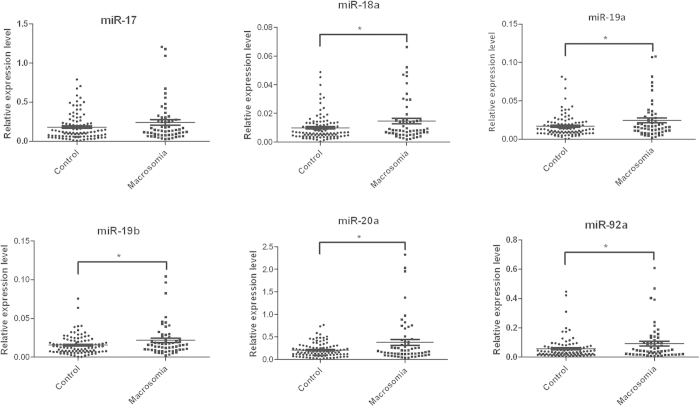
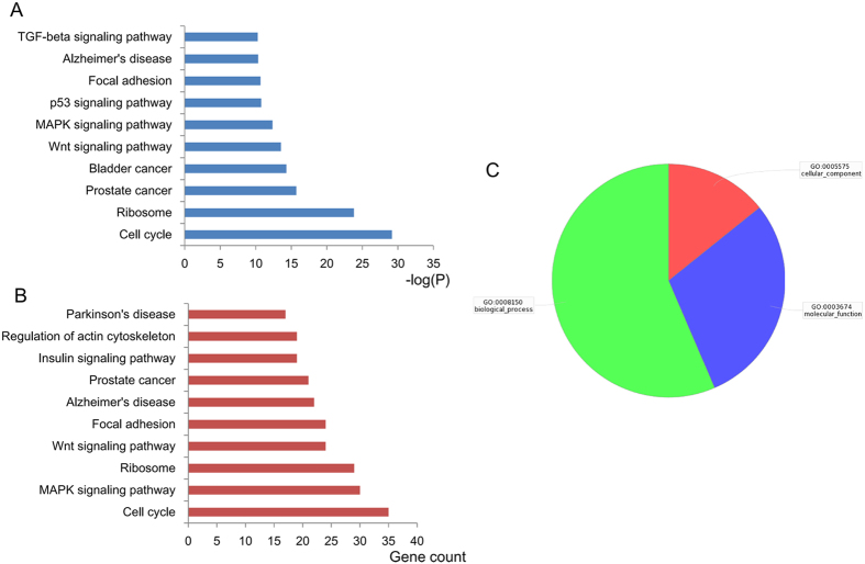
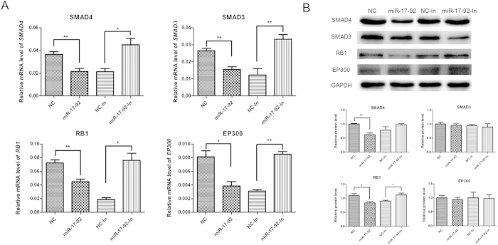
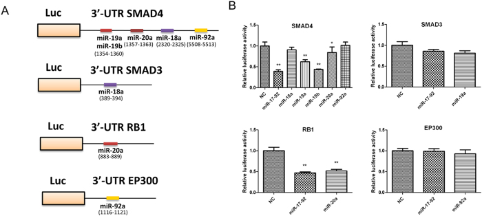
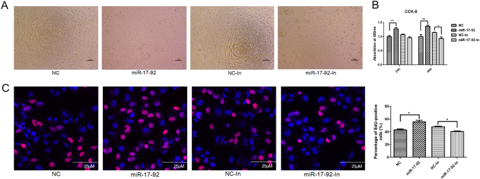
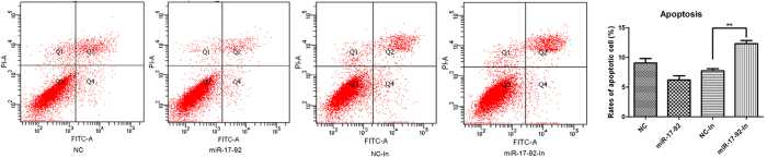
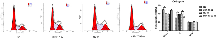
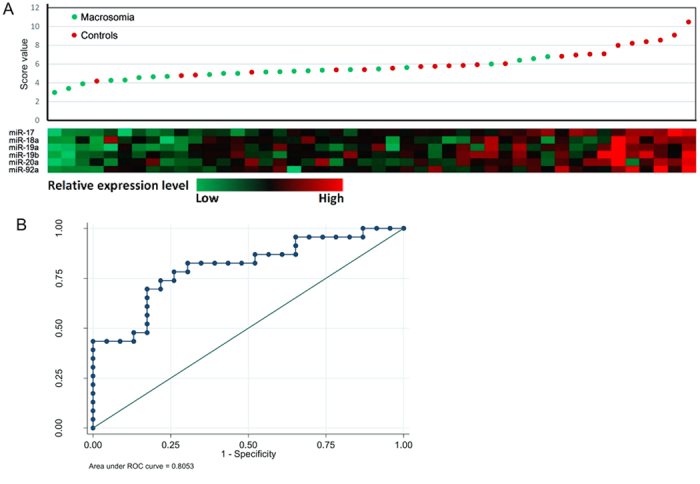
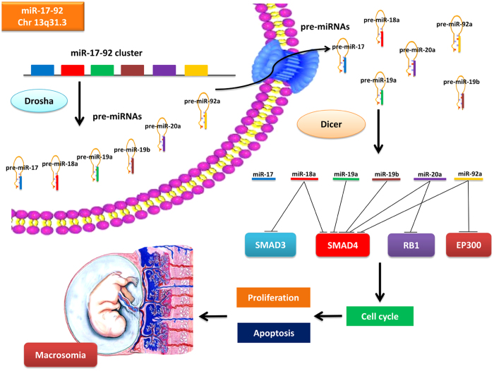
Macrosomia is one of the most common perinatal complications of pregnancy and has life-long health implications for the infant. microRNAs (miRNAs) have been identified to regulate placental development, yet the role of miRNAs in macrosomia remains poorly understood. Here we investigated the role of miR-17-92 cluster in macrosomia. The expression levels of five miRNAs in miR-17-92 cluster were significantly elevated in placentas of macrosomia, which may due to the up-regulation of miRNA-processing enzyme Drosha and Dicer. Cell cycle pathway was identified to be the most relevant pathways regulated by miR-17-92 cluster miRNAs. Importantly, miR-17-92 cluster increased proliferation, attenuated cell apoptosis and accelerated cells entering S phase by targeting SMAD4 and RB1 in HTR8/SVneo cells. Furthermore, we found that expression of miR-17-92 cluster in serum had a high diagnostic sensitivity and specificity for macrosomia (AUC: 80.53%; sensitivity: 82.61%; specificity: 69.57%). Our results suggested that miR-17-92 cluster contribute to macrosomia development by targeting regulators of cell cycle pathway. Our findings not only provide a novel insight into the molecular mechanisms of macrosomia, but also the clinical value of miR-17-92 cluster as a predictive biomarker for macrosomia.


Conflict of interest statement

The authors declare no competing financial interests.

Figures

Figure 1
Figure 1. miR-17-92 cluster miRNAs were up-regulated in placenta tissues of macrosomia.
Expression levels of miR-17, miR-18a, miR-19a, miR-19b, miR-20a and miR-92a were analyzed by qRT-PCR in placentas of macrosomia infants (n = 57) and controls (n = 100), and normalized to RNU6B. *P < 0.05; compared with the controls.
Figure 2
Figure 2. Target genes and pathways of the related miR-17-92 cluster miRNAs in macrosomia were enriched.
Top ten KEGG pathways regulated by miR-17-92 cluster are enriched according to P value (A) or gene count (B). (C) GO terms are grouped into three categories.
Figure 3
Figure 3. miR-17-92 cluster miRNAs inhibited expression of SMAD4 and RB1 in HTR8/SVneo cells.
(A) Detection of SMAD4, SMAD3, RB1, and EP300 mRNA expression in miR-17-92 mimics or miR-17-92 inhibitors-transfected HTR8/SVneo cells by qRT-PCR, and normalized to GAPDH. (B) Detection of SMAD4, SMAD3, RB1, and EP300 protein expression by Western-blot. Data were normalized to the level of GAPDH. *P < 0.05; **P < 0.01; compared with the control.
Figure 4
Figure 4. miR-17-92 cluster miRNAs targeted SMAD4 and RB1 in HTR8/SVneo cells.
(A) A schematic presentation of putative miR-17-92 cluster miRNAs-binding sites in the 3′-UTR regions of SMAD4, SMAD3, RB1 and EP300. (B) Luciferase assay in which the activities of 3′-UTRs of SMAD4, SMAD3, RB1 and EP300 fused to the luciferase gene constructs were measured in the presence of individually miR-17-92 cluster miRNAs or miR-17-92 cluster mix in HEK 293T cells. Data are expressed as the mean ± SEM (n = 3). *P < 0.05; **P < 0.01; compared with the control.
Figure 5
Figure 5. miR-17-92 cluster miRNAs enhanced the proliferation of HTR8/SVneo cells.
The effect of miR-17-92 cluster miRNAs on cell proliferation was detected using CCK-8 and EdU after transfection with miR-17-92 mimics or inhibitors. (A) Phase contrast microscopy of HTR8/SVneo cells treated with miR-17-92 mimics or inhibitors for 24h. (B) The effect of miR-17-92 cluster miRNAs on cell proliferation was detected at 24h and 48h using CCK-8. (C) Cell proliferation was determined by the EdU assay. Forty-eight hours after transfection, HTR8/SVneo cells were stained with EdU and DAPI. The percentage of EdU-positive HTR8/SVneo cells was quantified. Data are expressed as the mean ± SEM (n = 5). *P < 0.05; **P < 0.01; compared with the control.
Figure 6
Figure 6. miR-17-92 cluster miRNAs attenuated apoptosis of HTR8/SVneo cells.
Apoptosis was detected by flow cytometry with Annexin V-FITC staining in miR-17-92 mimics or miR-17-92 inhibitors-transfected HTR8/SVneo cells for 48h. Percentage of apoptotic cells were analyzed by FACS Calibur Flow Cytometer. Data are expressed as the mean ± SEM (n = 3). **P < 0.01; compared with the control.
Figure 7
Figure 7. miR-17-92 cluster miRNAs affected cell cycle in HTR8/SVneo cells.
The cell cycle was detected by flow cytometry with PI staining in HTR8/SVneo cells after transfection with miR-17-92 mimics or inhibitors for 48 h. The percentage of cells in G1, S, and G2 cell cycle phases were depicted in the bar graph. Data are expressed as the mean ± SEM (n = 3). *P < 0.05; compared with the control.
Figure 8
Figure 8. Risk-score and ROC curves for the ability of the maternal serum expression of six miRNAs in miR-17-92 cluster to differentiate the macrosomia from the controls.
(A) Risk-score distribution and color-gram of serum-miRNA expression profiles of macrosomia were performed. Rows represent miRNAs and columns represent subjects. Green denotes down-regulated expression and red denotes up-regulated expression compared with the mean. (B) ROC curve was shown for the ability of the six miRNAs to differentiate macrosomia from the controls.
Figure 9
Figure 9. A model was presented for miR-17-92 cluster mediated macrosomia.
The miR-17-92 cluster encodes for six distinct miRNAs. Drosha executes the initial step of miRNA processing in the nucleus, and the resultant pre-miRNAs are exported to the cytoplasm where they are cleaved by Dicer to generate the final mature miRNAs. Up-regulation of Drosha and Dicer causes miR-17-92 overexpression, which drives macrosomia by inhibiting multiple negative regulators of cell cycle pathway that mediated the pathology of macromosia.

Similar articles

Cited by

References

    1. Lu Y., Zhang J., Lu X., Xi W. & Li Z. Secular trends of macrosomia in southeast China, 1994–2005. BMC public health 11, 818, 10.1186/1471-2458-11-818 (2011). - DOI - PMC - PubMed
    1. Li G. et al. Prevalence of macrosomia and its risk factors in china: a multicentre survey based on birth data involving 101,723 singleton term infants. Paediatr. Perinat. Epidemiol. 28, 345–350, 10.1111/ppe.12133 (2014). - DOI - PubMed
    1. Zhang X., Decker A., Platt R. W. & Kramer M. S. How big is too big ? The perinatal consequences of fetal macrosomia. Am. J. Obstet. Gynecol. 198, 517 e511–516, 10.1016/j.ajog.2007.12.005 (2008). - DOI - PubMed
    1. Boney C. M., Verma A., Tucker R. & Vohr B. R. Metabolic syndrome in childhood: association with birth weight, maternal obesity, and gestational diabetes mellitus. Pediatrics 115, e290–296, 10.1542/peds.2004-1808 (2005). - DOI - PubMed
    1. Wei J. N. et al. Low birth weight and high birth weight infants are both at an increased risk to have type 2 diabetes among schoolchildren in taiwan. Diabetes care 26, 343–348 (2003). - PubMed

Publication types

MeSH terms

LinkOut - more resources