Review
. 2016 Mar;13(3):798-805.
doi: 10.1016/j.hrthm.2015.11.023. Epub 2015 Nov 17.

Cardiac output and vasodilation in the vasovagal response: An analysis of the classic papers

Affiliations
Review

Cardiac output and vasodilation in the vasovagal response: An analysis of the classic papers

Wouter Wieling et al. Heart Rhythm. 2016 Mar.

Abstract

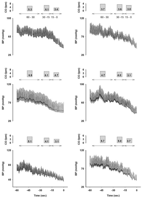
The simple faint is secondary to hypotension and bradycardia resulting in transient loss of consciousness. According to Ohm's law applied to the circulation, BP = SVR × CO, hypotension can result from a decrease in systemic vascular resistance (SVR), cardiac output (CO), or both. It is important to understand that when blood pressure (BP) is falling, SVR and CO do not change reciprocally as they do in the steady state. In 1932, Lewis, assuming that decreased SVR alone accounted for hypotension, defined "the vasovagal response" along pathophysiologic lines to denote the association of vasodilation with vagal-induced bradycardia in simple faint. Studies performed by Barcroft and Sharpey-Schafer between 1940 and 1950 used volume-based plethysmography to demonstrate major forearm vasodilation during extreme hypotension and concluded that the main mechanism for hypotension was vasodilation. Plethysmographic measurements were intermittent and not frequent enough to capture rapid changes in blood flow during progressive hypotension. However, later investigations by Weissler, Murray, and Stevens performed between 1950 and 1970 used invasive beat-to-beat BP measurements and more frequent measurements of CO using the Fick principle. They demonstrated that CO significantly fell before syncope, and little vasodilation occurred until very late in the vasovagal reaction Thus, since the 1970s, decreasing cardiac output rather than vasodilation has been regarded as the principal mechanism for the hypotension of vasovagal syncope.

Keywords: Blood pressure; Cardiac output; Central blood volume; Heart rate; Orthostasis; Stroke volume; Systemic vascular resistance; Vasovagal syncope; lower body negative pressure.


Figures

Figure 1
Figure 1
Ohm’s law: Mean arterial pressure = Cardiac output (CO) × Systemic Vascular Resistance (SVR). LA = left atrium; LV = left ventricle; RA = right atrium; RV = right ventricle; Venous pooling = accumulation of blood in the venous system.
Figure 2
Figure 2
Vasovagal faint induced by venesection 1020 cc. Steeps fall in blood pressure (BP) and heart rate (HR) are observed at the moment of the faint. See text for details. CO = cardiac output. (Reproduced with permission from Barcroft H, Edholm OG, McMichael J, Sharpey-Schafer EP. Posthaemorrhagic fainting. Study by cardiac output and forearm flow. Lancet 1944; I:489–491.) RAP = right atrial pressure; TPR = total peripheral resistance
Figure 3
Figure 3
Aborting a vasovagal faint after antigravity suit inflation to 60 mm Hg. Note progressive fall in intra-arterial pressure. Shaded section indicates the period of inflation. Central venous pressure increases instantaneously after inflation. The increase in blood pressure (BP) is delayed by about 3 secounds due to the transit time from the right to the left ventricle. The increase in BP was solely explained by the increase in cardiac output (increase by a factor of 1.4). Note that deep sighs during the fall in pressure increase BP for 3–4 beats (effect of the thoracoabdominal pump). Brachial artery = intra-arterial pressure; CVP = central venous pressure; Resp. = respiration. (Reproduced with permission from Weissler AM, Warren JV, Estes EH, McIntosh HD, Leonard JJ. Vasodepressor syncope; factors influencing cardiac output. Circulation 1957;15:875–882.)
Figure 4
Figure 4
Vasovagal faint with recovery during a tilt test with marked hypotension and bradycardia. The period of measurement of cardiac output (CO; about 20 seconds) matching the rapid fall in blood pressure (BP) is indicated ( i.e., the CO measurement is not a steady-state measurement). The exact matching of the CO with the fall in BP is the idealized situation because the dye dilution method does not allow BP to be recorded continuously at the same time as arterial blood is sampled for CO measurement. Note that deep sighs during BP fall increase BP for 3–4 beats (effect of increase in CO by the thoracoabdominal pump). (Reproduced with permission from Weissler AM, Warren JV, Estes EH, McIntosh HD, Leonard JJ. Vasodepressor syncope; factors influencing cardiac output. Circulation 1957;15:875–882.)
Figure 5
Figure 5
Pulse wave analysis computation of cardiac output (CO) during the last minute of a vasovagal faint induced by tilt-table testing in 6 patients demonstrates that the magnitude of the fall in CO is very much dependent on both the blood pressure (BP) level and the length of time needed to measure CO. CO 60–30 s = 5.2 L/min; CO 30–15 s = 4.8 L/min; CO 15–0 s = 3.5 L/min.

References

    1. Moya A, Sutton R, Ammirati F, et al. Guidelines for the diagnosis and management of syncope (version 2009) Eur Heart J. 2009;30:2631–2671. - PMC - PubMed
    1. Rowell LB. Human Cardiovascular Control. Oxford: Oxford University Press; 1993.
    1. Jardine DL. Vasovagal syncope: new physiologic insights. Cardiol Clin. 2013;31:75–87. - PubMed
    1. Weiss S, Wilkins RW, Haynes FW. The nature of circulatory collapse induced by sodium nitrate. J Clin Invest. 1937;16:73–78. - PMC - PubMed
    1. Graybiel A, McFarland RA. Use of tilt table test in aviation medicine. J Aviat Med. 1941;11:194–211.