. 2016 Jan 1;109(1):174-84.
doi: 10.1093/cvr/cvv254. Epub 2015 Nov 23.

Nitrated fatty acids suppress angiotensin II-mediated fibrotic remodelling and atrial fibrillation

Affiliations

Nitrated fatty acids suppress angiotensin II-mediated fibrotic remodelling and atrial fibrillation

Tanja K Rudolph et al. Cardiovasc Res. .

Abstract

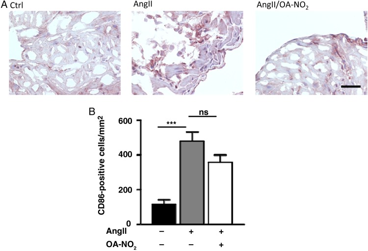
Aim: Atrial fibrosis, one of the most striking features in the pathology of atrial fibrillation (AF), is promoted by local and systemic inflammation. Electrophilic fatty acid nitroalkenes, endogenously generated by both metabolic and inflammatory reactions, are anti-inflammatory mediators that in synthetic form may be useful as drug candidates. Herein we investigate whether an exemplary nitro-fatty acid can limit atrial fibrosis and AF.

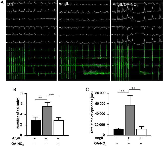
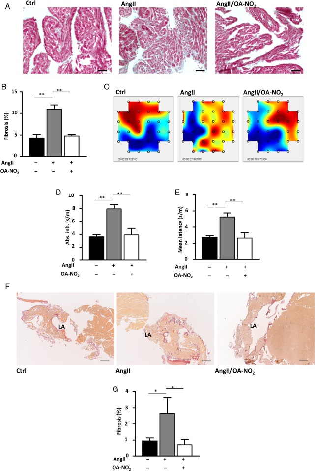
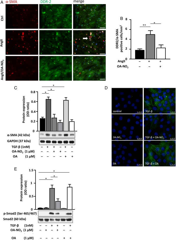
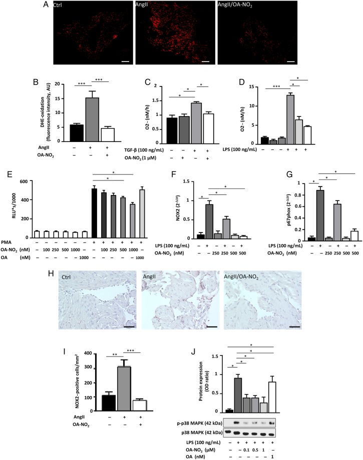
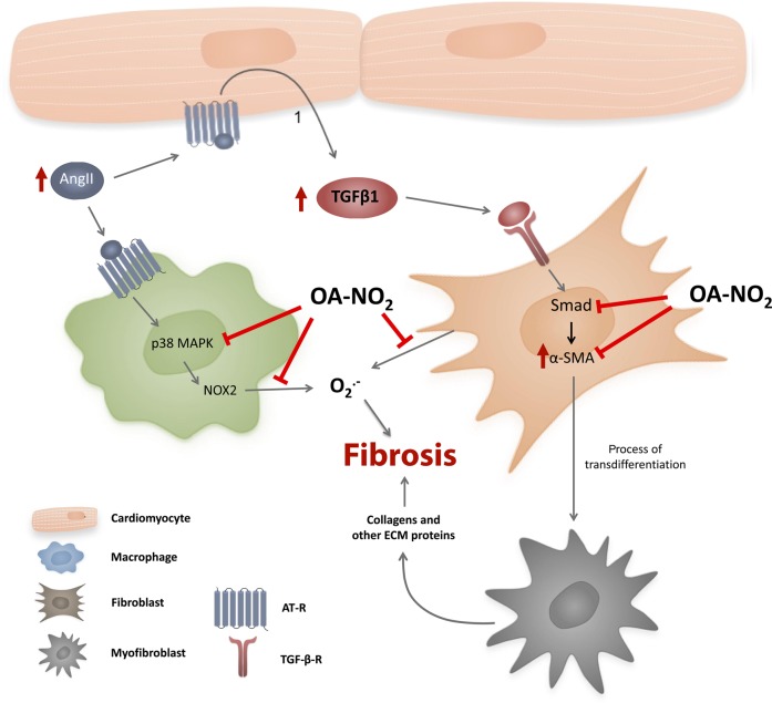
Methods and results: Wild-type C57BL6/J mice were treated for 2 weeks with angiotensin II (AngII) and vehicle or nitro-oleic acid (10-nitro-octadec-9-enoic acid, OA-NO2, 6 mg/kg body weight) via subcutaneous osmotic minipumps. OA-NO2 significantly inhibited atrial fibrosis and depressed vulnerability for AF during right atrial electrophysiological stimulation to levels observed for AngII-naive animals. Left atrial epicardial mapping studies demonstrated preservation of conduction homogeneity by OA-NO2. The protection from fibrotic remodelling was mediated by suppression of Smad2-dependent myofibroblast transdifferentiation and inhibition of Nox2-dependent atrial superoxide formation.

Conclusion: OA-NO2 potently inhibits atrial fibrosis and subsequent AF. Nitro-fatty acids and possibly other lipid electrophiles thus emerge as potential therapeutic agents for AF, either by increasing endogenous levels through dietary modulation or by administration as synthetic drugs.

Keywords: Atrial fibrillation; Fibrosis; Nitro-fatty acids; Reactive oxygen species.


Figures

Figure 1
Figure 1
OA-NO2 treatment attenuates elevated vulnerability for atrial fibrillation and restores electrical homogeneity in AngII-treated mice. AngII and OA-NO2 were administered over a period of 2 weeks via osmotic minipumps (Alzet), and afterwards, the mice underwent an electrophysiological study with right atrial stimulation. (A) Representative figures of ECGs with and without fibrillation episodes. (B) AngII treatment leads to a distinct elevation of the number of AF episodes in AngII(+)/OA-NO2(−) mice (n = 14), whereas notably in AngII(+)/OA-NO2(+) mice (n = 7), the number of AF episodes is comparable to the control group [AngII(−)/OA-NO2(−) mice] (n = 19). (C) The total time of AF episodes was calculated and the prolonged time of AF episodes was significantly reduced in AngII(+)/OA-NO2(+) (n = 7) compared with AngII(+)/OA-NO2(−) mice (n = 14). Data are expressed as mean ± SEM. **P < 0.01 and ***P < 0.001.
Figure 2
Figure 2
Electrical conductance and atrial fibrosis. OA-NO2 reduces extents of atrial fibrosis in AngII-treated mice. Paraffin-embedded atrial sections were subjected to Masson’s trichrome staining. (A) Representative images of trichrome-stained atrial sections. Scale bar indicates 750 µm. (B) Quantification of fibrotic areas. AngII(+)/OA-NO2(−) (n = 5) animals show an higher extent of atrial fibrosis, while concomitant OA-NO2 treatment leads to a significant reduction of atrial fibrotic areas AngII(+)/OA-NO2(+)-treated animals (n = 6). (C) Representative images of epicardial mappings performed with the same hearts subjected to the trichrome staining. (D and E) Measurement of absolute inhomogeneity and mean latency by epicardial mapping closely reflected changes observed in atrial fibrosis (n = 5, 6, 5), with a significant correlation between absolute inhomogeneity and the amount of atrial fibrosis. (F) Representative images of Picrosirius red-stained atrial sections. Scale bar indicates 1500 µm. (G) Quantification of the Picrosirius red staining corroborates the results of the trichrome staining (n = 9, 8, 7). Data are expressed as mean ± SEM. *P < 0.05 and **P < 0.01.
Figure 3
Figure 3
OA-NO2 inhibits fibroblast differentiation by inhibition of Smad-2 activation. (A and B) DDR2/α-SMA co-staining of murine atrial sections shows a significant higher amount of myofibroblasts in atrial tissue of AngII(+)/OA-NO2(−) mice (n = 5) compared with the control group [AngII(−)/OA-NO2(−)] (n = 5), while OA-NO2 treatment (n = 4) reduced the number of myofibroblasts to the level of control animals. Representative arrow indicates co-localization of DDR2 and α-SMA. Scale bar indicates 40 µm. (C and D) Treatment of 3T3 fibroblasts with TGF-β (5 ng/mL) and/or OA-NO2 (1 µM) for 8 h reveals an inhibition of TGF-β-induced fibroblast differentiation by OA-NO2 evidenced by western blot analysis and immunofluorescent staining. Pooled data from four independent experiments are shown. Scale bar indicates 25 µm. (E) OA-NO2 reduces TGF-β-induced phosphorylation of Smad-2 (Ser 465/467) in 3T3 fibroblasts, as detected by western blot. Pooled data from three independent experiments are shown. Data are expressed as mean ± SEM. *P < 0.05 and **P < 0.01.
Figure 4
Figure 4
OA-NO2 inhibits atrial generation of reactive inflammatory mediators. (A and B) DHE stainings of atrial sections reveal a significantly reduced ROS formation in AngII(+)/OA-NO2(+) mice (n = 8) compared with AngII(+)/OA-NO2(−) animals (n = 7), with ROS levels in AngII(+)/OA-NO2(+) equalling those of control animals (n = 8). Scale bar indicates 750 µm. (C) TGF-β-induced superoxide production measured by cytochrome c reduction assay in 3T3 fibroblasts is significantly blunted upon OA-NO2 treatment (n = 5). (D) LPS-induced production of superoxide in murine peritoneal RAW 264.7 macrophages measured by cytochrome c reduction assay is inhibited by OA-NO2 in a dose-dependent manner (n = 6). (E) Oxidative burst in murine peritoneal RAW 264.7 macrophages induced by phorbol-12-myristate-13-acetate (PMA) is also reduced by application of OA-NO2 (n = 4). (F and G) The LPS-induced expression of NOX2 as well as of its cytosolic regulatory protein p67phox is significantly attenuated by OA-NO2 treatment in a dose-dependent manner (n = 5). (H and I) NOX2 stainings of atrial sections reveal a significantly reduced NOX2 expression in AngII(+)/OA-NO2(+) mice (n = 8) compared with AngII(+)/OA-NO2(−) animals (n = 7), with ROS levels in AngII(+)/OA-NO2(+) equalling those of control animals (n = 8). Scale bar indicates 50 µm. (J) OA-NO2 decreases phosphorylation of p38 MAPK. Pooled data from four independent experiments are shown. Data are expressed as mean ± SEM. *P < 0.05, **P < 0.01, and ***P < 0.001.
Figure 5
Figure 5
Number of CD86-positive cells in atrial tissue. (A) Representative figures of an immunohistological staining of atrial sections by using an anti-CD86-specific antibody. (B) Quantification of CD86-positive cells in atrial tissue reveal a significant higher amount of these cells in AngII (+)/OA-NO2 (−) animals (n = 5/4/4). Furthermore, this was not significantly influenced by a parallel treatment with OA-NO2. For the staining of paraffin-embedded murine atrial sections with CD86 (1 : 500, rabbit IgG, NB110–55488, Novus Biologicals, USA), a standard protocol was used. For quantification, CD86-positive cells were counted in atrial sections. Scale bar = 50 µm. ***P < 0.001.
Figure 6
Figure 6
Proposed mechanism by which OA-NO2 reduces fibrosis in the atrium of AngII-treated mice. OA-NO2 decreases fibroblast transdifferentiation through inhibition of Smad-2 activation, as indicated by a reduction of α-SMA expression. Additionally, OA-NO2 reduces atrial oxidative stress due to attenuation of superoxide production from macrophages and fibroblasts by inhibition of NOX2 and p67 phox expression via p38 MAPK deactivation.

References

    1. Camm AJ, Kirchhof P, Lip GY, Schotten U, Savelieva I, Ernst S, Van Gelder IC, Al-Attar N, Hindricks G, Prendergast B, Heidbuchel H, Alfieri O, Angelini A, Atar D, Colonna P, De Caterina R, De Sutter J, Goette A, Gorenek B, Heldal M, Hohloser SH, Kolh P, Le Heuzey JY, Ponikowski P, Rutten FH. Guidelines for the management of atrial fibrillation: the Task Force for the Management of Atrial Fibrillation of the European Society of Cardiology (ESC). 2010;31:2369–2429. - PubMed
    1. Burstein B, Nattel S. Atrial fibrosis: mechanisms and clinical relevance in atrial fibrillation. 2008;51:802–809. - PubMed
    1. Kim YM, Guzik TJ, Zhang YH, Zhang MH, Kattach H, Ratnatunga C, Pillai R, Channon KM, Casadei B. A myocardial Nox2 containing NAD(P)H oxidase contributes to oxidative stress in human atrial fibrillation. 2005;97:629–636. - PubMed
    1. Rubbo H, Radi R, Trujillo M, Telleri R, Kalyanaraman B, Barnes S, Kirk M, Freeman BA. Nitric oxide regulation of superoxide and peroxynitrite-dependent lipid peroxidation. Formation of novel nitrogen-containing oxidized lipid derivatives. 1994;269:26066–26075. - PubMed
    1. Rudolph V, Schopfer FJ, Khoo NK, Rudolph TK, Cole MP, Woodcock SR, Bonacci G, Groeger AL, Golin-Bisello F, Chen CS, Baker PR, Freeman BA. Nitro-fatty acid metabolome: saturation, desaturation, beta-oxidation, and protein adduction. 2009;284:1461–1473. - PMC - PubMed

Publication types