. 2016 Jan 8;291(2):826-36.
doi: 10.1074/jbc.M115.674234. Epub 2015 Nov 23.

Runx1 Phosphorylation by Src Increases Trans-activation via Augmented Stability, Reduced Histone Deacetylase (HDAC) Binding, and Increased DNA Affinity, and Activated Runx1 Favors Granulopoiesis

Affiliations

Runx1 Phosphorylation by Src Increases Trans-activation via Augmented Stability, Reduced Histone Deacetylase (HDAC) Binding, and Increased DNA Affinity, and Activated Runx1 Favors Granulopoiesis

Wan Yee Leong et al. J Biol Chem. .

Abstract

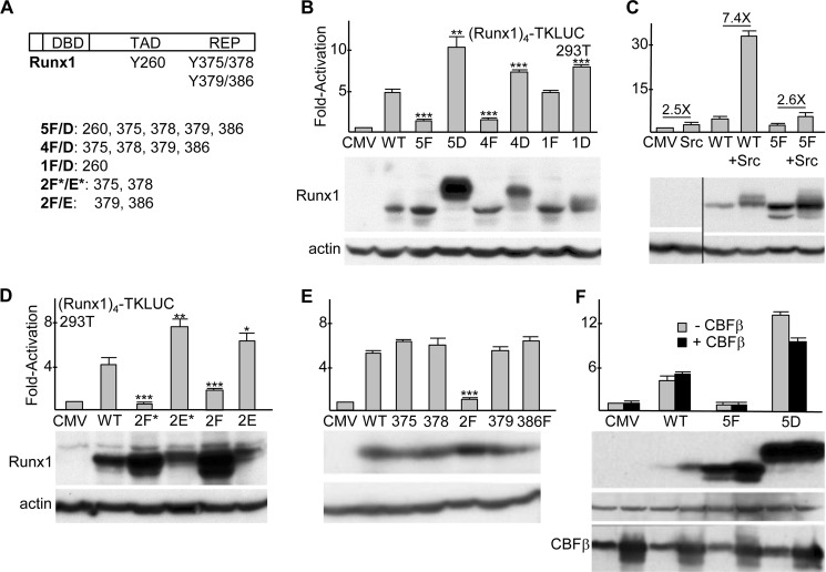
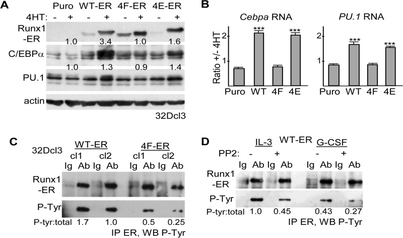
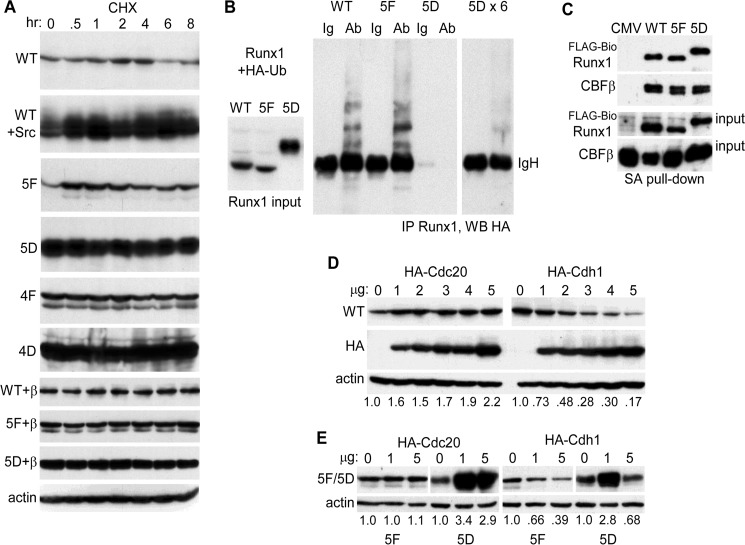
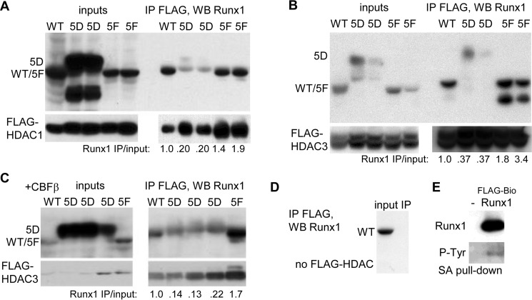
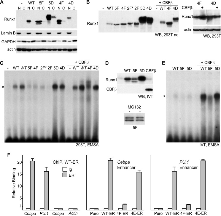
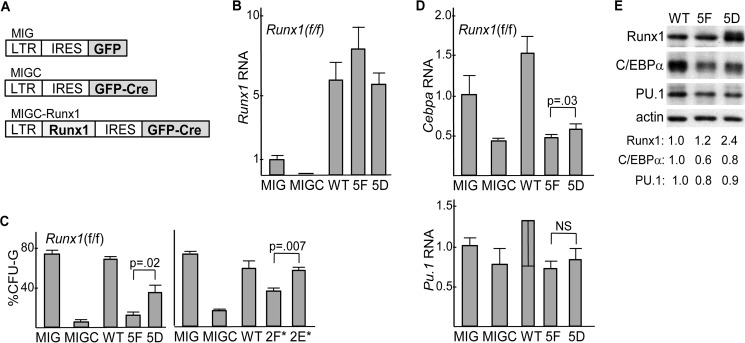
Src phosphorylates Runx1 on one central and four C-terminal tyrosines. We find that activated Src synergizes with Runx1 to activate a Runx1 luciferase reporter. Mutation of the four Runx1 C-terminal tyrosines to aspartate or glutamate to mimic phosphorylation increases trans-activation of the reporter in 293T cells and allows induction of Cebpa or Pu.1 mRNAs in 32Dcl3 myeloid cells, whereas mutation of these residues to phenylalanine to prevent phosphorylation obviates these effects. Three mechanisms contribute to increased Runx1 activity upon tyrosine modification as follows: increased stability, reduced histone deacetylase (HDAC) interaction, and increased DNA binding. Mutation of the five modified Runx1 tyrosines to aspartate markedly reduced co-immunoprecipitation with HDAC1 and HDAC3, markedly increased stability in cycloheximide or in the presence of co-expressed Cdh1, an E3 ubiquitin ligase coactivator, with reduced ubiquitination, and allowed DNA-binding in gel shift assay similar to wild-type Runx1. In contrast, mutation of these residues to phenylalanine modestly increased HDAC interaction, modestly reduced stability, and markedly reduced DNA binding in gel shift assays and as assessed by chromatin immunoprecipitation with the -14-kb Pu.1 or +37-kb Cebpa enhancers after stable expression in 32Dcl3 cells. Affinity for CBFβ, the Runx1 DNA-binding partner, was not affected by these tyrosine modifications, and in vitro translated CBFβ markedly increased DNA affinity of both the translated phenylalanine and aspartate Runx1 variants. Finally, further supporting a positive role for Runx1 tyrosine phosphorylation during granulopoiesis, mutation of the five Src-modified residues to aspartate but not phenylalanine allows Runx1 to increase Cebpa and granulocyte colony formation by Runx1-deleted murine marrow.

Keywords: Runx1; Src; hematopoiesis; histone deacetylase (HDAC); myeloid cell; transcription factor; tyrosine kinase.


Figures

FIGURE 1.
FIGURE 1.
A, diagram of murine Runx1b, with locations of the DNA-binding domain (DBD), trans-activation domain (TAD), C-terminal repression domain (REP), and five tyrosine residues shown previously to be modified by Src kinase (28) indicated (top). Tyrosine residues altered in Runx1 variants are also listed (bottom). B, 150 ng of (Runx1)4TKLUC was co-transfected into sub-confluent 293T cells in a 24-well plate with 0.8 ng of CMV-βGal and 5 ng of either pcDNA3 (CMV) or pcDNA3 vectors expressing wild-type Runx1 (WT) or its indicated tyrosine variants. The ratio of luciferase/β-galactosidase activity was assessed at 48 h, and the values relative to CMV, which was set on average to 1.0, are shown (mean and S.E. from three determinations). *, p < 0.05; **, p < 0.01; ***, p < 0.001 (top). 293T cells in 6-well plates were transfected with 1 μg of CMV or CMV-Runx1 expression vectors, and total cellular proteins were assessed at 48 h for Runx1 and β-actin expression by Western blotting (bottom). C, 293T cells were transfected with (Runx1)4TKLUC, CMV-βGal, and CMV, CMV-Runx1, or CMV-5F, alone or with 40 ng of CEFL-Src. Activation relative to CMV alone is shown (mean and S.E. from three determinations). The fold effect of Src expression on the average activity of CMV, Runx1, or 5F is also indicated (top). 293T cells in 6-well dishes were transfected with 250 ng of CMV, CMV-Runx1, or CMV-5F, alone or with 1 μg of CEFL-Src, and total cellular proteins were assessed at 48 h for Runx1 and β-actin expression (bottom). D, activation of the Runx1 reporter and protein expression was assessed as in A for WT Runx1 and its indicated variants (mean and S.E. from three determinations). E, activation by FLAG-BioRunx1 or its indicated variants and their expression in 293T cells is shown (mean and S.E. from three determinations). F, 293T cells were transfected with (Runx1)4TKLUC, CMV-βGal, and CMV, CMV-Runx1, or CMV-5F, alone or with 5 ng of CMV-CBFβ. Activation relative to CMV alone is shown (mean and S.E. from three determinations). 293T cells in 6-well dishes were transfected with 1 μg of CMV, CMV-Runx1, or CMV-5F, alone or with 1 μg of CMV-CBFβ, and total cellular proteins were assessed at 48 h for Runx1, β-actin, and CBFβ expression by Western blotting (bottom).
FIGURE 2.
FIGURE 2.
A, total cellular proteins from equal numbers (2E5) of 32Dcl3 cells stably transduced with pBABE Puro (Puro), Runx1-ER(T) (WT-ER), or its 4F-ER or 4E-ER variants and cultured ±4HT for 24 h were subjected to Western blotting using ERα, C/EBPα, PU. 1, or β-actin antibodies. Fold-increases in C/EBPα or PU.1 induced by 4HT, normalized to β-actin, are shown. B, total cellular RNAs from these same cultures were subjected to quantitative RT-PCR analysis for Cebpa and PU.1 and for actin as internal control. The expression of Cebpa or PU.1, normalized to actin, is shown as a ratio of expression ±4HT (mean and S.E. from three determinations). C, total cellular proteins from two WT-ER or 4F-ER 32Dcl3 subclones were subjected to ERα immunoprecipitation followed by Western blotting (WB) for phosphotyrosine. The ratios of phosphorylated/total WT-ER or 4F-ER are shown. D, pool of WT-ER-expressing 32Dcl3 cells was exposed to 20 μm PP2 or DMSO control for 8 h, either in IL-3 or 24 h after transfer to G-CSF. Total cellular proteins were then subjected to ERα immunoprecipitation followed by Western blotting for phosphotyrosine. The ratios of phosphorylated/total WT-ER are shown. Ab, antibody.
FIGURE 3.
FIGURE 3.
A, 293T cells in 6-well dishes were transfected with 1 μg of CMV-Runx1 (WT), 0.25 μg of WT + 1 μg of CMV-Src; 1 μg of CMV-5F, CMV-5D, CMV-4F, or CMV-4D; 1 μg of CMV-WT + 1 μg of CMV-CBFβ, 5F + CBFβ, or 5D + CBFβ. CHX was added for 0.5–8 h, and total cellular proteins from equal numbers of cells were then evaluated for Runx1 isoform expression by Western blotting. Actin expression was assessed on similarly cultured 293T cells exposed to CHX. B, 293T cells in 10-cm dishes were transfected with 2 μg of CMV-WT, 1 μg of CMV-5F, or 0.4 μg of CMV-5D with 3 μg of CMV-HA-ubiquitin (HA-Ub). Extracts prepared 2 days later were subjected to Runx1 Western blotting, with 1/12th volume of the 5D extract compared with the WT or 5F extracts loaded to equalize expression (left). In addition 0.5 mg of total cell lysates were subjected to rabbit anti-Runx1 immunoprecipitation followed by anti-HA (16B12) Western blotting, with 1/12th (center) or 1/2 (right) of the 5D immunoprecipitate loaded relative to WT or 5F. The position of immunoglobulin heavy chain (IgH) is shown. Ab, antibody. C, total cellular proteins from 293T cells in 10-cm dishes transfected with 1.5 μg of CMV-CBFβ, 1.5 μg of EF1α-birA, and 3 μg of EF1α-FLAG-BioWT, 3 μg of EF1α-FLAG-Bio5F, or 10 ng of EF1α-FLAG-Bio5D were subjected to streptavidin-agarose pulldown followed by Western blotting of 2.5% input or pulldown samples using Runx1 and CBFβ antisera. D, 293T cells in 6-well dishes were transfected with 2 μg of Runx1 and 0–5 μg of HA-Cdc20 or HA-Cdh1 expression vectors. Total cellular proteins were then analyzed by Western blotting using Runx1, HA (Y-11), and β-actin antibodies. The relative intensities of the Runx1 bands, normalized to β-actin expression, are shown below each lane. E, 293T cells were transfected with 1 μg of 5F or 0.5 μg of 5D along with 0, 1, or 5 μg of HA-Cdc20 or HA-Cdh1 expression vectors, followed by Western blot analysis using Runx1 or β-actin antibodies.
FIGURE 4.
FIGURE 4.
A, 293T cells in 10-cm dishes were transfected with 5 μg of CMV-WT, 0.4 or 0.2 μg of CMV-5D, or 1 or 0.5 μg of CMV-5F, together with 3 μg of CMV-FLAG-HDAC1, and total cell extracts were prepared 2 days later. After FLAG IP, pulldown, or 10% of input, samples were subjected to Western blotting (WB) with Runx1 or FLAG antibodies. The ratio of IP/input Runx1 band intensity for each reaction is shown. B, similar experiment was conducted substituting CMV-FLAG-HDAC3. C, 293T cells were transfected with 1 μg of CMV-WT, 0.5, 0.25, or 0.1 μg of CMV-5D, or 0.5 μg of CMV-5F together with 2 μg of CMV-FLAG-HDAC3 and 2 μg of CMV-CBFβ, followed by FLAG IP and Runx1 or FLAG Western blot analysis. D, 293T cells were transfected with 5 μg of CMV-WT, followed by FLAG IP and Runx1 Western blot analysis. E, 293T cells were transfected with 3 μg of EF1α-birA, alone or with 3 μg of EF1α-FLAG-BioRunx1, followed by streptavidin pulldown and Western blotting for Runx1 and phosphotyrosine.
FIGURE 5.
FIGURE 5.
A, 293T cells in 10-cm dishes were transfected with 6 μg of CMV (−) or CMV vectors expressing WT Runx1 or its 5F, 5D, 4F, or 4D variants. Nuclear (N) and cytoplasmic (C) extracts from equal cell numbers, prepared 48 h later, were subjected to Western blotting for Runx1, lamin B, GAPDH, or β-actin. B, 293T cells in 10-cm dishes were transfected with 6 μg of CMV (−) or CMV vectors expressing WT Runx1 or its 5F, 4F, 2F*, 2F, 5D, or 4D variants alone, or 3 μg of CMV or WT, 4F, or 4D with 3 μg of CMV-CBFβ. 20 μg of nuclear extracts prepared 48 h later were subjected to Western blotting (WB) using Runx1 antiserum (left). 293T cells in 6-well dishes were transfected with 1 μg of 4F or 4D, alone or with 1 μg of CBFβ, and total cellular proteins prepared 48 h later were subjected to Western blotting using Runx1 and β-actin antibodies (right). C, 6 μg of each nuclear extract were subjected to electrophoretic mobility shift analysis (EMSA) using a radiolabeled 32-bp double-stranded oligonucleotide containing a central consensus Runx1-binding site derived from the myeloperoxidase promoter. In addition, lanes with 12 μg of the WT or WT+CBFβ extracts (WT) were included. *indicates location of the specific gel shift band. D, Runx1 WT, 5F, or 5D, as well as CBFβ, were generated by in vitro transcription-translation (IVT) using 1 μg of linearized pcDNA3 expression vectors in a 50-μl reaction. Equal volumes, together with control reticulocyte lysate (−), were subjected to Western blotting for Runx1 and CBFβ (top). In addition, expression of 5F generated with or without 25 μm MG132 was analyzed similarly (bottom). E, after equalizing WT, 5F, and 5D protein based on densitometry, these samples as well as control lysate were analyzed for DNA binding by EMSA, alone or with addition of 7 μl of CBFβ lysate. F, 1E6 32Dcl3 Puro, WT-ER, 4F-ER, or 4E-ER cells cultured for 24 h in 4HT were subjected to ChIP assay using 2 μg of rabbit IgG or rabbit-anti-ERα (ER) antiserum followed by quantitative PCR using genomic primers specific for the +37-kb Cebpa enhancer, the −14 kb-PU.1 enhancer, the −2.5-kb Cebpa promoter region, or the β-actin promoter. Relative binding of WT-ER to each of these elements (left) or of WT-ER, 4F-ER, and 4E-ER to the Cebpa enhancer (center) or the PU.1 enhancer (right) is shown, with signal obtained with IgG in Puro cells set to 1.0 for each primer pair (mean and S.E. from three determinations).
FIGURE 6.
FIGURE 6.
A, diagram of MIG, MIGC, and MIGC-Runx1 retroviral vectors. LTR, long-terminal repeat; IRES, internal ribosome entry site; GFP, green fluorescent protein; GFP-CRE, fusion protein linking GFP and CRE. B, expression of Runx1 RNA, relative to mS16 ribosomal subunit RNA, in lineage-depleted (Lin), GFP+ cells isolated 4 days after transduction of Runx1(f/f) marrow cells with MIG, MIGC, MIGC-WT, MIGC-5F, or MIGC-5D. Average Runx1 expression after MIG transduction was set to 1.0 (mean and S.E. of three determinations). C, percentage of CFU-G relative to CFU-G + CFU-M obtained from equal numbers of lineage-depleted GFP+ cells plated in methylcellulose culture at 1E3/ml with HI-FBS and IL-3, IL-6, and stem cell factor after similar transduction which is shown (left, mean and S.E. from three determinations). A related set of experiments was conducted using MIG, MIGC, MIGC-WT, MIGC-2F*, and MIGC-2E* (right, mean and S.E. from three determinations). D, expression of Cebpa or Pu.1 RNAs, relative to mS16 ribosomal subunit RNA, in LinGFP+ cells isolated 4 days after transduction of Runx1(f/f) marrow cells with MIG, MIGC, MIGC-WT, MIGC-5F, or MIGC-5D. Average Cebpa or Pu.1 expression after MIG transduction was set to 1.0 (mean and S.E. of three determinations). E, total cellular proteins from equal numbers of LinGFP+ cells isolated 4 days after transduction of Runx1(f/f) marrow cells with MIGC-WT, MIGC-5F, or MIGC-5D were subjected to Western blotting for Runx1, C/EBPα, PU.1, or β-actin. Band intensities, normalized to that of β-actin and set to 1.0 after WT transduction, are also shown.

References

    1. Swiers G., de Bruijn M., and Speck N. A. (2010) Hematopoietic stem cell emergence in the conceptus and the role of Runx1. Int. J. Dev. Biol. 54, 1151–1163 - PMC - PubMed
    1. Ichikawa M., Asai T., Saito T., Seo S., Yamazaki I., Yamagata T., Mitani K., Chiba S., Ogawa S., Kurokawa M., and Hirai H. (2004) AML-1 is required for megakaryocytic maturation and lymphocytic differentiation, but not for maintenance of hematopoietic stem cells in adult hematopoiesis. Nat. Med. 10, 299–304 - PubMed
    1. Growney J. D., Shigematsu H., Li Z., Lee B. H., Adelsperger J., Rowan R., Curley D. P., Kutok J. L., Akashi K., Williams I. R., Speck N. A., and Gilliland D. G. (2005) Loss of Runx1 perturbs adult hematopoiesis and is associated with a myeloproliferative phenotype. Blood 106, 494–504 - PMC - PubMed
    1. Guo H., Ma O., Speck N. A., and Friedman A. D. (2012) Runx1 deletion or dominant inhibition reduces Cebpa transcription via conserved promoter and distal enhancer sites to favor monopoiesis over granulopoiesis. Blood 119, 4408–4418 - PMC - PubMed
    1. Huang G., Zhang P., Hirai H., Elf S., Yan X., Chen Z., Koschmieder S., Okuno Y., Dayaram T., Growney J. D., Shivdasani R. A., Gilliland D. G., Speck N. A., Nimer S. D., and Tenen D. G. (2008) PU.1 is a major downstream target of AML1 (RUNX1) in adult mouse hematopoiesis. Nat. Genet. 40, 51–60 - PubMed

Publication types

LinkOut - more resources