. 2016 Feb;15(2):506-22.
doi: 10.1074/mcp.M115.049965. Epub 2015 Nov 23.

Proteomic Analysis of Dynein-Interacting Proteins in Amyotrophic Lateral Sclerosis Synaptosomes Reveals Alterations in the RNA-Binding Protein Staufen1

Affiliations

Proteomic Analysis of Dynein-Interacting Proteins in Amyotrophic Lateral Sclerosis Synaptosomes Reveals Alterations in the RNA-Binding Protein Staufen1

Noga Gershoni-Emek et al. Mol Cell Proteomics. 2016 Feb.

Abstract

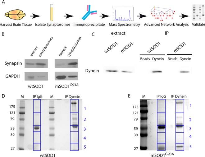
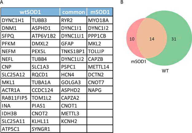
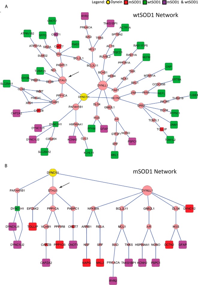
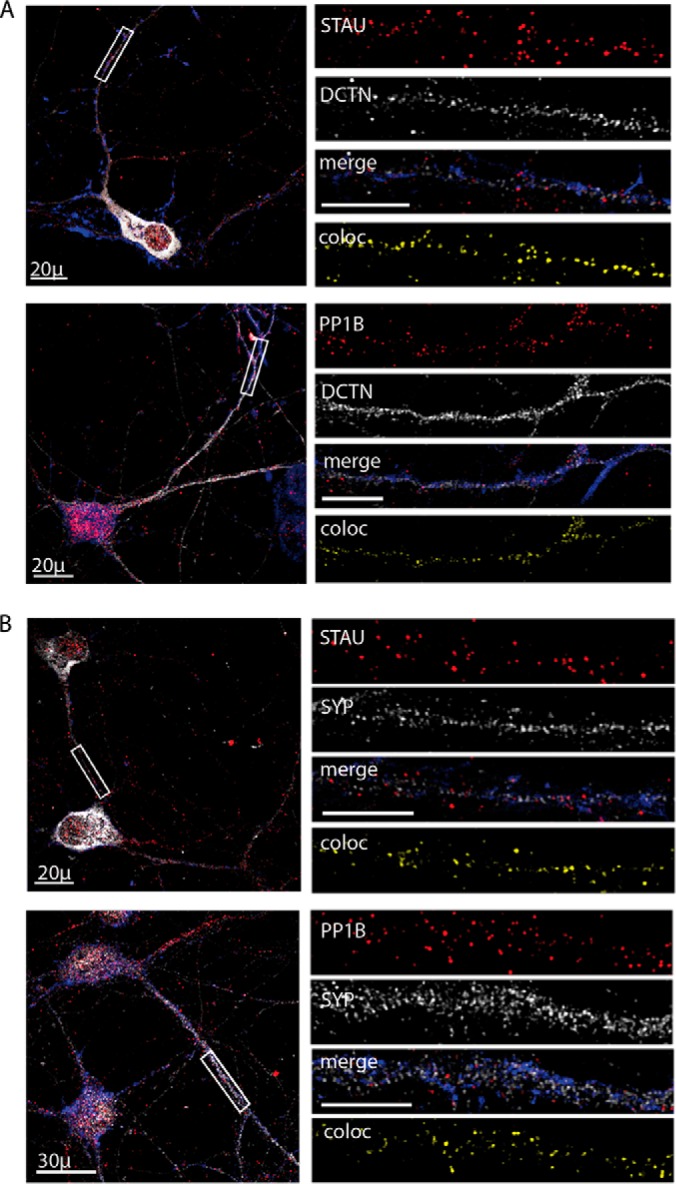
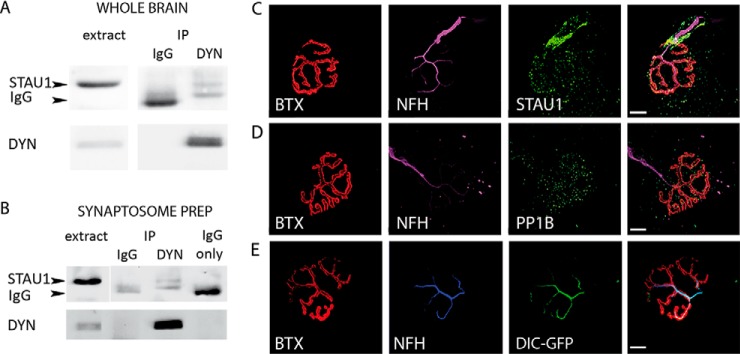
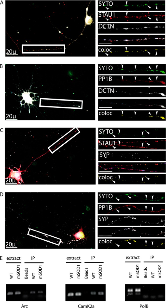
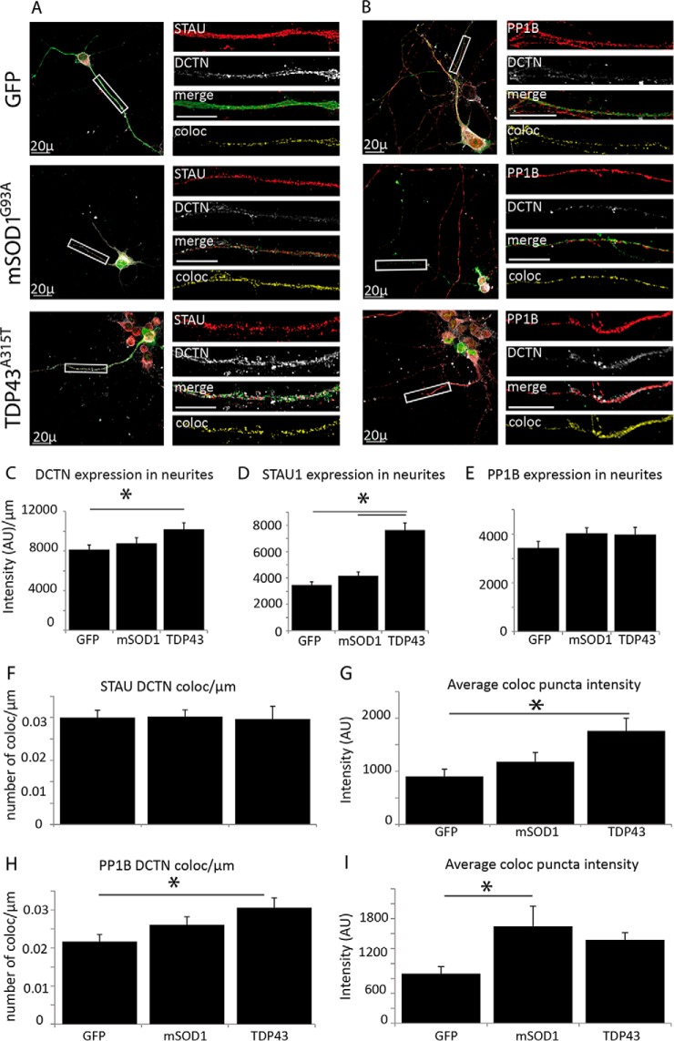
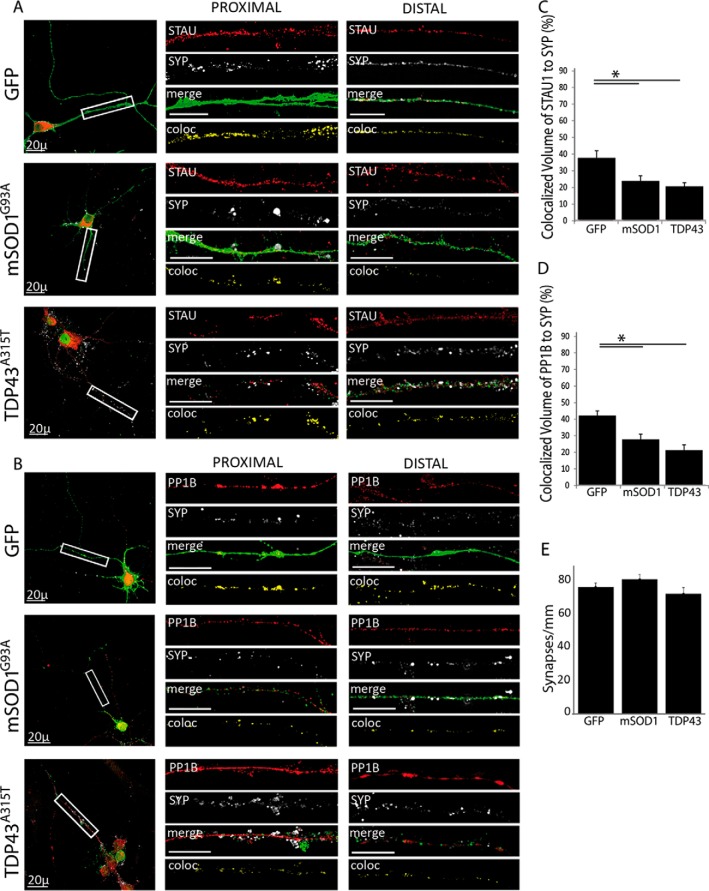
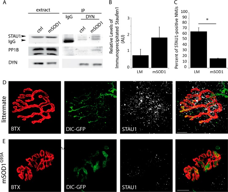
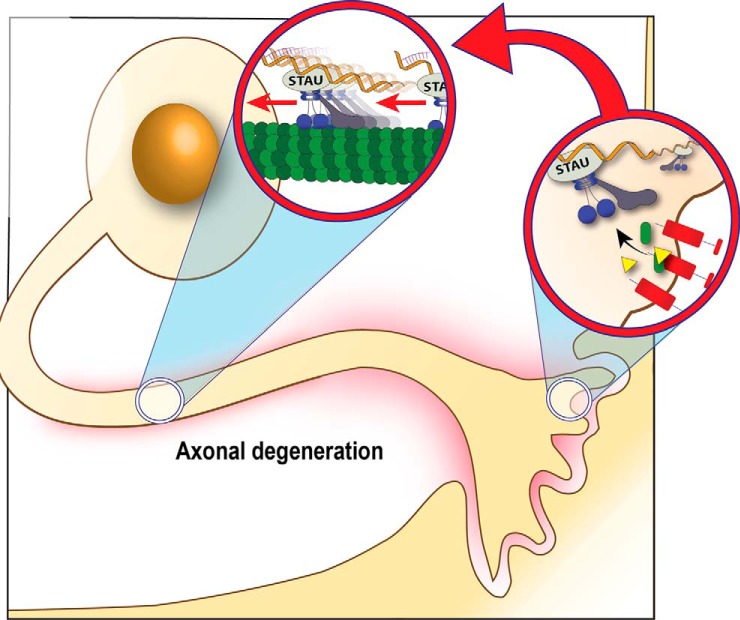
Synapse disruption takes place in many neurodegenerative diseases, including amyotrophic lateral sclerosis (ALS). However, the mechanistic understanding of this process is still limited. We set out to study a possible role for dynein in synapse integrity. Cytoplasmic dynein is a multisubunit intracellular molecule responsible for diverse cellular functions, including long-distance transport of vesicles, organelles, and signaling factors toward the cell center. A less well-characterized role dynein may play is the spatial clustering and anchoring of various factors including mRNAs in distinct cellular domains such as the neuronal synapse. Here, in order to gain insight into dynein functions in synapse integrity and disruption, we performed a screen for novel dynein interactors at the synapse. Dynein immunoprecipitation from synaptic fractions of the ALS model mSOD1(G93A) and wild-type controls, followed by mass spectrometry analysis on synaptic fractions of the ALS model mSOD1(G93A) and wild-type controls, was performed. Using advanced network analysis, we identified Staufen1, an RNA-binding protein required for the transport and localization of neuronal RNAs, as a major mediator of dynein interactions via its interaction with protein phosphatase 1-beta (PP1B). Both in vitro and in vivo validation assays demonstrate the interactions of Staufen1 and PP1B with dynein, and their colocalization with synaptic markers was altered as a result of two separate ALS-linked mutations: mSOD1(G93A) and TDP43(A315T). Taken together, we suggest a model in which dynein's interaction with Staufen1 regulates mRNA localization along the axon and the synapses, and alterations in this process may correlate with synapse disruption and ALS toxicity.


Figures

Fig. 1.
Fig. 1.
Preparation of samples for mass spectrometry. (A) Diagram depicting the workflow as described in this paper: Brains were harvested from mSOD1 and wtSOD1 mice, and synaptosomes were then isolated. Next, dynein was immunoprecipitated and the immunoprecipitates run on gels, followed by MS analysis. Identified proteins were subjected to filtration and used for reconstruction of an interaction subnetwork. Predicted interactions of interest were validated by biochemical and imaging techniques. (B) The synaptic preparation, along with complete brain extract, was subjected to Western blot analysis. Synaptosome preparations show enrichment for synapsin; glyceraldehyde 3-phosphate dehydrogenase serves as a loading control. (C) Synaptosome preparation was immunoprecipitated with either dynein antibodies or nonspecific IgG, followed by Western blot analysis for dynein. (D, E) Dynein precipitates from the synaptosomes were run on gel and stained with Coomassie Blue. The gel was then segmented into five slices at approximate molecular weight 121, 65, 45, and 32 KDa for mass spectrometry analysis.
Fig. 2.
Fig. 2.
Analysis of alterations in dynein-bound synapse fractions in mSOD1 mice and wild-type controls. The mass spectrometry data of dynein interactors in the synapse were filtered for reproducibility and confidence. Proteins that appeared in all separate biological repeats and not in the control with >99% confidentiality were chosen. (A) The list of identified proteins after the filtration process, segregated into correlating groups. (B) Venn diagram depicting distribution of the identified proteins, divided in to the same groups as in A.
Fig. 3.
Fig. 3.
Predicted networks of dynein interactors in the synapse. Subnetworks connecting the dynein intermediate chain-DYNC1I1 anchor to the genes transcribing the immunoprecipitated proteins identified by MS after filtration (terminals) in wtSOD1 (A) and mSOD1 (B). These subnetworks were reconstructed using the ANAT tool over the HIPPIE high-confidence PPI network and rendered using Cytoscape. Square nodes represent terminals (red—mSOD1 only; green—wtSOD1 only; purple—both). Due to the conversion of protein names to gene symbols, PP1B appears as PP1CB. Predicted nodes are in pink, with size increasing in accordance with centrality in the subnetworks.
Fig. 4.
Fig. 4.
Colocalization of Staufen1 and PP1B with dynactin and synaptic markers in spinal cord cultures. Primary neurons were seeded on cover slides and grown for 10 days and then fixed and immunostained for STAU1, PP1B, DCTN, and SYP. Phalloidin was used to visualize neurites. Representative images of the neurons were taken using a confocal microscope with a 40x objective, with high-magnification close-ups of the axon taken with a 100x objective. High-magnification images were used to analyze volume colocalization along the neurites using the Imaris software, represented in the Coloc channel. (A) Immunostaining for DCTN with STAU and PP1B. (B) Immunostaining for SYP with STAU and PP1B. Unlabeled scale bars represent 10 μm, n = 3, 5–8 images per experimental repetition.
Fig. 5.
Fig. 5.
In vivo analysis of dynein–Staufen1 interaction. (A) Protein extracts from p120 adult brains were subjected to immunoprecipitation with dynein antibody. As a control, we used nonspecific IgG. The immunoprecipitate was then subjected to Western blot analysis using antibodies against Stau1 (arrow) and dynein. (B) Synaptosomes were isolated from p120 adult brains, and then IP-ed for dynein and blotted for Staufen1 and dynein. The lower band (∼55kDa), visible in the IP and not in the extract, is the heavy chain of the anti-dynein IgG. (C–E) Gastrocnemius muscle was excised from p120 adult mice, fixed, and treated with bungarotoxin to stain the NMJ and then immunostained. Images were collected with a confocal microscope with a 100x objective. Representative images of NMJs showing enrichment of STAU1 (C), PP1B (D), and dynein intermediate chain (DIC)-GFP (E) at the NMJ, and colocalization with neurofilament (neurofilament heavy chain) are shown. Scale bars represent 10 μm.
Fig. 6.
Fig. 6.
Colocalization of Staufen1 and PP1B with mRNA in neurites. Primary spinal cord neurons were seeded on cover slides and grown for 10 days. They were treated with SYTO-14 for total RNA detection, fixed, and immunostained for STAU1, PP1B, DCTN, and SYP. Images were collected with a confocal microscope. Representative images of the neurons were taken with a 40x objective and high-magnification close-ups of the axon taken with a 100x objective. High-magnification images were used to analyze volume colocalization along the neurites using the Imaris software, represented in the Coloc channel. (A) Colocalization of RNA with STAU1 and DCTN in neurites. (B) Colocalization of RNA with PP1B and DCTN. (C) Colocalization of RNA with STAU1 and SYP. (D) Colocalization of RNA with PP1B and SYP. Unlabeled scale bars represent 10 μm. (E) RNA was harvested from dynein immunoprecipitated from mSOD1 mouse and littermate control brains. Following cDNA preparation, standard RT-PCR was carried out for CamK2a, Arc, and polymerase B (PolB).
Fig. 7.
Fig. 7.
ALS-linked mutations alter the association of STAU1 and PP1B with DCTN. (A and B) Primary spinal cord neurons were seeded on cover slides and infected with lentiviruses expressing GFP, GFP-tagged mSOD1G93A and TDP43A315T. After 10 days in vitro (DIV), cells were fixed and immunostained. Images were collected with a confocal microscope. Representative images of the neurons were taken with a 40x objective, with high-magnification close-ups of the axon taken with a 100x objective. DCTN, STAU, and PP1B localization along the axon are not altered as a result of ALS-linked mutations. (C and D) DCTN and STAU1 levels remain constant along the neurite for the GFP control and mSOD1 but are both increased in TDP43-infected neurons. (E) PP1B levels remain constant for all three groups. (F and G) The number of colocalized puncta (viewed in the Coloc channel) of DCTN with STAU1 is constant (F), with an increased intensity in ALS models (G). (H and I) Colocalization of DCTN with PP1B (shown in the Coloc channel) is similarly increased in ALS models. Statistical analysis was done with ANOVA, with Dunnet's post-hoc test, *p < .05. Unlabeled scale bars represent 10 μm, n = 3.
Fig. 8.
Fig. 8.
ALS-linked mutations decrease the association of STAU1 and PP1B with synaptic markers. Primary spinal cord neurons were seeded on cover slides and infected with lentiviruses expressing GFP, GFP-tagged mSOD1G93A, and TDP43A315T. After 10 DIV, cells were fixed and immunostained. Images were collected with a confocal microscope. Representative images of the neurons were taken with a 40x objective, with high-magnification close-ups of the proximal and distal axon taken with a 100x objective. High-magnification images were used to analyze volume colocalization along the neurites using the Imaris software, represented in the Coloc channel. The percentage of colocalized volume is shown. (A) Colocalization of SYP with STAU in infected neurons. (B) Colocalization of SYP with PP1B in infected neurons. (C) Quantification of SYP and STAU colocalization a ∼15% decrease in colocalized volume in ALS-infected neurons. (D) Quantification of SYP and PP1B colocalization showing a decrease of ∼15% in colocalized volume as a result of ALS-linked mutations. (E) The total number of synapses was quantified using the Image J software and normalized to neurite length. Statistical analysis was done with ANOVA, with Dunnet's post-hoc test, *p < .05. Unlabeled scale bars represent 10 μm, n = 3.
Fig. 9.
Fig. 9.
Expression of Staufen1 at the NMJ is depleted in adult mSOD1 mice. (A) Whole brain extracts from mSOD1 mice and healthy littermates were subjected to dynein immunoprecipitation, with nonspecific IgG antibody as a control. The immunoprecipitate was then subjected to Western blot analysis and blotted for Staufen1 (arrow) and dynein. The lower band (∼55 kDa), visible in the IP lanes, but not in the extract, is the heavy chain of either the anti-dynein or the IgG. (B) Densitometry analysis using Image J software shows a consistent but variable increase in the amount of immunoprecipitated Staufen1 in mSOD1 extracts, n = 3. (C-E) Gastrocnemius muscles from DIC-GFP p120 healthy littermates (D) and mSOD1 mice (E) were excised, fixed, and treated with bungarotoxin to stain the postsynaptic NMJ and then immunostained for STAU1 and neurofilament heavy chain. Images were acquired with a confocal microscope with a 100x objective. Analysis of >150 NMJs on three biological repeats show STAU1 localization at the synapse in 19% of NMJs in mSOD1, compared with 64% in the littermate. Statistical analysis was done with Student's t test, *p < .005, n = 3. Scale bar represents 10 μm.
Fig. 10.
Fig. 10.
Suggested model: Dynein plays a dual role in the synaptic localization of Staufen1. We propose a model in which dynein performs a dual role: in retrograde transport and in anchoring. In healthy neurons, the majority of Staufen1-containing mRNPs bound to dynein are anchored at sites of local protein synthesis, i.e. synapses or axonal branching points. As a result of neurodegeneration, dynein may switch its role to retrograde transport, clearing Staufen1 mRNPs from the distal synapse toward the cell body. Clearance of mRNPs from the distal axon contributes to reduced axonal protein synthesis and may serve to advance neurodegenerative processes.

References

    1. Moloney E. B., de Winter F., and Verhaagen J. (2014) ALS as a distal axonopathy: Molecular mechanisms affecting neuromuscular junction stability in the presymptomatic stages of the disease. Frontiers Neurosci 8, 252 - PMC - PubMed
    1. De Vos K. J., Grierson A. J., Ackerley S., and Miller C. C. (2008) Role of axonal transport in neurodegenerative diseases. Annu. Rev. Neurosci. 31, 151–173 - PubMed
    1. Perlson E., Maday S., Fu M. M., Moughamian A. J., and Holzbaur E. L. (2010) Retrograde axonal transport: pathways to cell death? Trends Neurosci. 33, 335–344 - PMC - PubMed
    1. Kardon J. R., and Vale R. D. (2009) Regulators of the cytoplasmic dynein motor. Nat. Rev. Mol. Cell Biol. 10, 854–865 - PMC - PubMed
    1. Fu M. M., and Holzbaur E. L. (2014) Integrated regulation of motor-driven organelle transport by scaffolding proteins. Trends Cell Biol. 24, 564–574 - PMC - PubMed

Publication types

MeSH terms