. 2015 Dec 8;112(49):15196-201.
doi: 10.1073/pnas.1513803112. Epub 2015 Nov 23.

High-resolution crystal structure of a hepatitis B virus replication inhibitor bound to the viral core protein

Affiliations

High-resolution crystal structure of a hepatitis B virus replication inhibitor bound to the viral core protein

Klaus Klumpp et al. Proc Natl Acad Sci U S A. .

Abstract

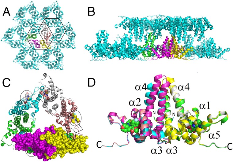
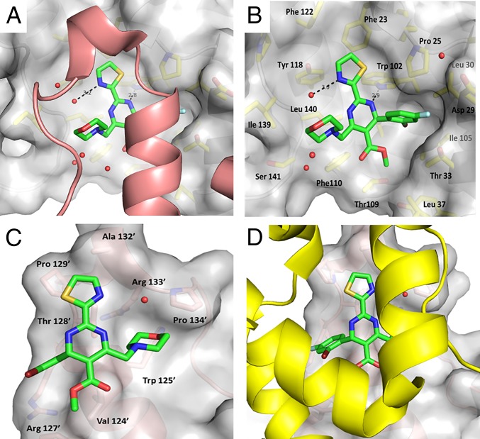
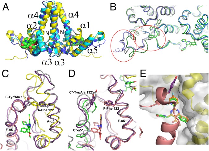
The hepatitis B virus (HBV) core protein is essential for HBV replication and an important target for antiviral drug discovery. We report the first, to our knowledge, high-resolution crystal structure of an antiviral compound bound to the HBV core protein. The compound NVR-010-001-E2 can induce assembly of the HBV core wild-type and Y132A mutant proteins and thermostabilize the proteins with a Tm increase of more than 10 °C. NVR-010-001-E2 binds at the dimer-dimer interface of the core proteins, forms a new interaction surface promoting protein-protein interaction, induces protein assembly, and increases stability. The impact of naturally occurring core protein mutations on antiviral activity correlates with NVR-010-001-E2 binding interactions determined by crystallography. The crystal structure provides understanding of a drug efficacy mechanism related to the induction and stabilization of protein-protein interactions and enables structure-guided design to improve antiviral potency and drug-like properties.

Keywords: HBV inhibitor; HBV treatment; capsid; core; protein–protein interaction.


Conflict of interest statement

Conflict of interest statement: The authors are employees of Novira Therapeutics (K.K., A.M.L., R.V., S.R., C.E., G.H., and O.A.F.), Beryllium (C.L., R.B., K.A., and J.A.), and Vista Informatics Corporation (G.L.).

Figures

Fig. 1.
Fig. 1.
Structure and activity of HBV core inhibitors. (A) Chemical structures. (B) Antiviral activity in HepG2.2.15 cells, NVR 010–001-E2 (squares), GLS4 (circles), BAY 41–4109 (triangles), and BAY 41–4109-IE (diamonds). (C) Thermal shift assay using wild-type CoreND protein in the absence of compound (DMSO, black line), in the presence of BAY 41–4109-IE (orange, wide dashed line), BAY 41–4109 (red, dotted line), GLS4 (NVR-010–002-E2, blue, dot-dashed line), and NVR-010–001-E2 (black, dashed line). (D) Antiviral activity in HepG2.2.15 cells. Data shown are mean values (± SD).
Fig. 2.
Fig. 2.
Crystal structure of CoreND-Y132A protein bound to NVR-010–001-E2. (A) The hexamer in the asymmetric unit is shown in color. (B) Packing of sheets derived from hexamers showing the head-to-head arrangement of the proteins. (C) Hexamer with the six bound NVR-010–001-E2 molecules highlighted by circles. Individual monomers are colored yellow (monomer A), magenta (monomer B), green (monomer C), cyan (monomer D), gray (monomer E), and salmon (monomer F). Monomers A and B are shown in space-filling mode. The arrows towards monomer A (yellow) and F (salmon) highlight the two different compound binding sites corresponding to intrahexamer (monomers A and F) and interhexamer (monomer F) interactions. (D) Overlay of the three dimers in the asymmetric unit. The structures vary only in the stalk region. For monomers C and D, the ends of the stalk helices are disordered, and helix 4 has a kink at amino acid 80. Two representative binding sites from monomers A and B are indicated by space-filling models of bound NVR-010–001-E2.
Fig. 3.
Fig. 3.
Binding site of NVR-010–001-E2. (A) The ligand sits in the groove of the contact domain (gray surface) on one dimer and is covered by a lid formed by helix 5 from the adjacent dimer, colored in salmon. (B) Binding surface to the contact domain. The lid is removed for clarity. (C and D) Binding surface to the lid. The contact domain is removed for clarity in C and shown in yellow in D.
Fig. 4.
Fig. 4.
Compound binding site comparisons. (A) Overlay of the dimer A-B (yellow, NVR-010–001-E2 shown in space-filling mode) with the A-B dimer of 3KXS (cyan, [18]) and 4BMG (blue, [19]). (B) Overlay of the NVR-010–001-E2 bound structure with wild-type capsid (2G33, blue, [15]) and wild-type capsid with compound HAP-1 putatively bound (3G34, gray, [15]). Helix 5 and the C-terminal tail are circled in red for the two corresponding dimers, with Tyr132 shown for the wild-type structures. (C and D) Overlay of the NVR 010–001-E2 bound structure with wild-type capsid (2G33, gray, [15]) and wild-type capsid with compound HAP-1 putatively bound (3G34, purple, [15]). (C) In the A-F interface, helix 5 of monomer A shifts so that Phe122 can fill the space normally occupied by Tyr132’ of monomer F (monomer A, yellow; monomer F, salmon). (D) The interface in the capsid between hexamers with monomers F (salmon) and C* from the next hexamer (green). (E) Overlay of the model of BAY 41–4109 (orange) on the structure of NVR-010–001-E2 (green).

References

    1. Cowie BC, Carville KS, MacLachlan JH. Mortality due to viral hepatitis in the Global Burden of Disease Study 2010: New evidence of an urgent global public health priority demanding action. Antivir Ther. 2013;18(8):953–954. - PubMed
    1. Klumpp K, Crépin T. Capsid proteins of enveloped viruses as antiviral drug targets. Curr Opin Virol. 2014;5:63–71. - PubMed
    1. Seeger C, Zoulim F, Mason WS. Hepadnaviruses. In: Knipe DM, Howley PM, editors. Fields Virology. Vol II. Lippincott Williams & Wilkins; Philadelphia: 2013. pp. 2185–2221.
    1. Dryden KA, et al. Native hepatitis B virions and capsids visualized by electron cryomicroscopy. Mol Cell. 2006;22(6):843–850. - PubMed
    1. Wynne SA, Crowther RA, Leslie AG. The crystal structure of the human hepatitis B virus capsid. Mol Cell. 1999;3(6):771–780. - PubMed

Publication types

MeSH terms

Associated data