Review
. 2016 Jan 5;371(1685):20150044.
doi: 10.1098/rstb.2015.0044.

A flexible genetic toolkit for arthropod neurogenesis

Affiliations
Review

A flexible genetic toolkit for arthropod neurogenesis

Angelika Stollewerk. Philos Trans R Soc Lond B Biol Sci. .

Abstract

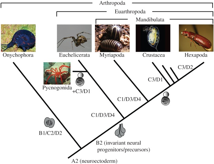
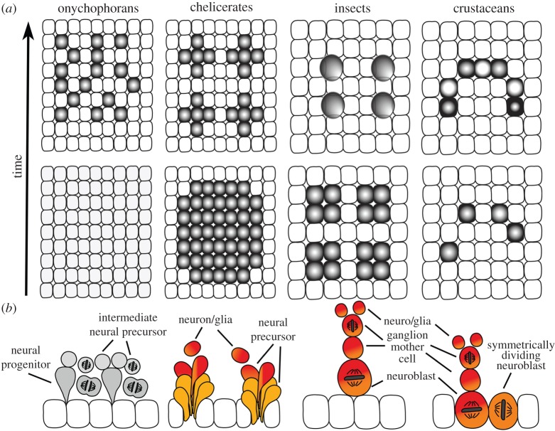
Arthropods show considerable variations in early neurogenesis. This includes the pattern of specification, division and movement of neural precursors and progenitors. In all metazoans with nervous systems, including arthropods, conserved genes regulate neurogenesis, which raises the question of how the various morphological mechanisms have emerged and how the same genetic toolkit might generate different morphological outcomes. Here I address this question by comparing neurogenesis across arthropods and show how variations in the regulation and function of the neural genes might explain this phenomenon and how they might have facilitated the evolution of the diverse morphological mechanisms of neurogenesis.

Keywords: Notch signalling; asymmetric division; neural precursors; neural progenitors; neurogenic potential; patterning genes.


Figures

Figure 1.
Figure 1.
Arthropod phylogeny and the evolution of the neurogenesis module. The arthropod relationships shown in the phylogenetic tree are the framework for the discussions in this review. A2–D4 represent parts of the module variants recently described by [1]. Module A: patterning cells with neurogenic potential. In contrast with animals with a generalized ectoderm (module A1, not shown), all arthropods have a restricted area with neurogenic potential (neuroectoderm) that gives rise to the central nervous system (module A2). Module B: patterning of neural progenitors. Neural progenitors/precursor groups show a similar, invariant pattern in all euarthropods (module B2), while the neural progenitors of onychophorans appear in random positions (module B1). The ancestral pattern of euarthropod neurogenesis is the selection of neural precursor groups. Modules C and D: proliferation and movement of neural progenitors, respectively. The neural precursors of chelicerates and myriapods (mainly) do not divide (module C1) and directly differentiate into neurons and glia. The neural precursor groups are internalized by ingression (module D3) or invagination (module D4). Both mechanisms are seen in chelicerates (including pycnogonids) and myriapods. In addition, pycnogonids have asymmetrically dividing progenitors (module C3) that remain in the epithelium (module D1). By contrast, the neural progenitors of onychophorans divide symmetrically (module C2) after their delamination (module D2). Insect and crustacean neuroblasts divide asymmetrically (module C3), however insect neuroblasts delaminate (module D2), while crustacean neuroblasts remain in the epithelium (module D1). The scheme and description refer to the processes in the ventral neuroectoderm.
Figure 2.
Figure 2.
Patterning of the neurogenic region in euarthropods. The dashed lines indicate the ventral midline of the neuroectoderm. (a) Flat preparation of a Glomeris marginata embryo (millipede) showing the RNA expression pattern of the SoxB1 gene in all neuroectodermal cells. (b,c) Comparison of the msh RNA expression pattern in flat preparations of three ventral neuromeres of G. marginata and Tribolium castaneum (beetle). Note the difference in the width of the neuroectoderm in these species. The boxes frame the neuromere of the second leg segment in G. marginata and the second thoracic neuromere of T. castaneum, respectively. (d,e) Schematic of the arrangement of the neural precursor groups in hemi-neuromeres of G. marginata and the spider Cupiennius salei. The precursor groups expressing msh are shown in brown. (f,g) Schematic drawings comparing the expression of the dorsoventral patterning genes vnd (blue), ind (red) and msh (brown) in the neuroblasts of Drosophila melanogaster and T. castaneum. The neuroblasts 6-2 and 7-2 express both vnd and ind in D. melanogaster, while neuroblasts 4-2 and 5-3 express ind and msh in T. castaneum. l1–l3, leg segment 1 to 3; pc, procephalic neuroectoderm; t1–t3, thoracic segments 1 to 3; vne, ventral neuroectoderm.
Figure 3.
Figure 3.
Pattern of neural precursor/progenitor specification and division. Small sections of the neuroectoderm (white squares: neuroectodermal cells) are shown for each group. (a) Onychophorans: ASH shows a low homogeneous expression in the neuroectoderm (light grey) and is upregulated in the neural progenitors (dark grey) after their delamination. Chelicerates: ASH is upregulated in large domains and becomes restricted to groups of cells. A similar pattern is seen in myriapods (not shown). Insects: ASH is expressed in small proneural clusters. Expression becomes restricted to single neuroblasts. Crustaceans (branchiopods): ASH is upregulated after neuroblast specification. Over time more neuroblasts express ASH and the gene is expressed at different levels indicated by lighter and darker grey. (b) Division pattern and movement of neural precursors/progenitors. Onychophorans: single neural progenitors delaminate and divide symmetrically to produce intermediate neural precursors, which divide again. The expression of snail and prospero has not been studied in onychophorans. Chelicerates: most neural precursors are postmitotic. Neural precursor groups express snail (orange). Neural precursors co-express prospero (red) before detaching from the group. Insects: neuroblasts express snail and prospero and divide asymmetrically to produce GMCs, which divide again to produce neurons and glia. Crustaceans show the same division pattern but snail is expressed long before prospero. Snail-positive neuroblasts can divide symmetrically.

References

    1. Hartenstein V, Stollewerk A. 2015. The evolution of early neurogensis. Dev. Cell 32, 390–407. (10.1016/j.devcel.2015.02.004) - DOI - PMC - PubMed
    1. Storch V, Welsch U. 2004. Systematische zoologie. Berlin, Germany: Spektrum akademischer Verlag.
    1. Regier JC, Shultz JW, Zwick A, Hussey A, Ball B, Wetzer R, Martin JW, Cunningham CW. 2010. Arthropod relationships revealed by phylogenomic analysis of nuclear protein-coding sequence. Nature 463, 1079–1084. (10.1038/nature08742) - DOI - PubMed
    1. Rehm P, Meusemann K, Borner J, Misof B, Burmester T. 2014. Phylogenetic position of Myriapoda revealed by 454 transcriptome sequencing. Mol. Phylogenet. Evol. 77, 25–33. (10.1016/j.ympev.2014.04.007) - DOI - PubMed
    1. Rota-Stabelli O, Campbell L, Brinkmann H, Edgecombe GD, Longhorn SJ, Peterson KJ, Pisani D, Philippe H, Telford MJ. 2011. A congruent solution to arthropod phylogeny: phylogenomics, microRNAs and morphology support monophyletic Mandibulata. Proc. R. Soc. B 278, 298–306. (10.1098/rspb.2010.0590) - DOI - PMC - PubMed

MeSH terms

LinkOut - more resources