. 2015 Nov 24;10(11):e0141506.
doi: 10.1371/journal.pone.0141506. eCollection 2015.

Multi-Parametric MRI and Texture Analysis to Visualize Spatial Histologic Heterogeneity and Tumor Extent in Glioblastoma

Affiliations

Multi-Parametric MRI and Texture Analysis to Visualize Spatial Histologic Heterogeneity and Tumor Extent in Glioblastoma

Leland S Hu et al. PLoS One. .

Abstract

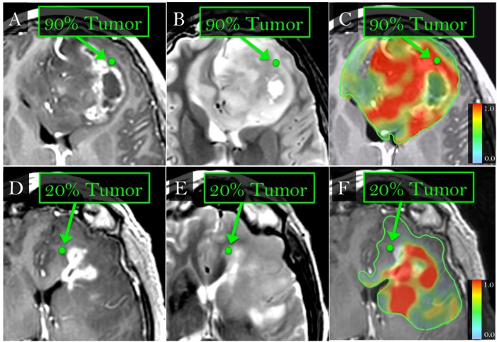
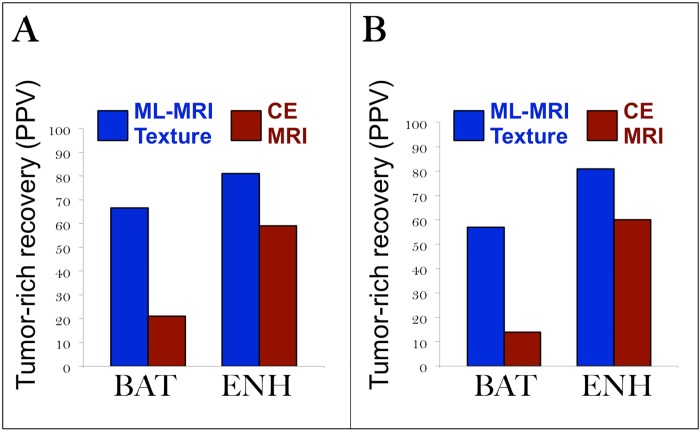
Background: Genetic profiling represents the future of neuro-oncology but suffers from inadequate biopsies in heterogeneous tumors like Glioblastoma (GBM). Contrast-enhanced MRI (CE-MRI) targets enhancing core (ENH) but yields adequate tumor in only ~60% of cases. Further, CE-MRI poorly localizes infiltrative tumor within surrounding non-enhancing parenchyma, or brain-around-tumor (BAT), despite the importance of characterizing this tumor segment, which universally recurs. In this study, we use multiple texture analysis and machine learning (ML) algorithms to analyze multi-parametric MRI, and produce new images indicating tumor-rich targets in GBM.

Methods: We recruited primary GBM patients undergoing image-guided biopsies and acquired pre-operative MRI: CE-MRI, Dynamic-Susceptibility-weighted-Contrast-enhanced-MRI, and Diffusion Tensor Imaging. Following image coregistration and region of interest placement at biopsy locations, we compared MRI metrics and regional texture with histologic diagnoses of high- vs low-tumor content (≥80% vs <80% tumor nuclei) for corresponding samples. In a training set, we used three texture analysis algorithms and three ML methods to identify MRI-texture features that optimized model accuracy to distinguish tumor content. We confirmed model accuracy in a separate validation set.

Results: We collected 82 biopsies from 18 GBMs throughout ENH and BAT. The MRI-based model achieved 85% cross-validated accuracy to diagnose high- vs low-tumor in the training set (60 biopsies, 11 patients). The model achieved 81.8% accuracy in the validation set (22 biopsies, 7 patients).

Conclusion: Multi-parametric MRI and texture analysis can help characterize and visualize GBM's spatial histologic heterogeneity to identify regional tumor-rich biopsy targets.


Conflict of interest statement

Competing Interests: The authors have read the journal's policy and the authors of this manuscript have the following competing interests: J. Ross Mitchell is scientific co-founder of Calgary Scientific (CSI). This does not alter the authors' adherence to all the PLOS ONE policies on sharing data and materials.

Figures

Fig 1
Fig 1. ML-based MRI invasion maps show tumor-rich (>80% tumor nuclei) extent throughout ENH and BAT.
(A,B,C,E) Biopsy locations within the non-enhancing BAT zone (green dots, arrows) on T1+C (A,D) and T2W (B,E) images correspond with high-tumor (>80% tumor nuclei) and low-tumor (<80% tumor nuclei) tissue samples on histologic analysis. (C,F) Color overlay maps with manual tracings (green) around BAT show the probability (range 0–1) of tumor-rich (red) vs tumor-poor (green/blue) content, based on ML analysis and multi-parametric MRI in 60 training biopsies and 22 validation biopsies. The maps show correspondence between tumor-rich (B, red) and tumor-poor (D, blue/gray) biopsy samples.
Fig 2
Fig 2. ML-based model improves tumor-rich biopsy delineation compared with CE-MRI.
(A) ML-based MRI texture model in the full dataset (n = 82, Table 1) shows higher positive predictive values (PPV) (66.7% in BAT, 81.3% in ENH) for recovering tumor-rich samples compared with CE-MRI (21.2% in BAT, 59.2% in ENH). These PPVs suggest that the ML-based model would help recover tumor-rich BAT samples with over three times greater efficiency compared with CE-MRI guidance. (B) ML-based MRI texture model in the subanalysis (n = 76, S2 Appendix) provides higher positive predictive values (PPV) (57.1% in BAT, 80.6% in ENH) for recovering tumor-rich samples (>80% tumor nuclei) compared with CE-MRI (13.8% in BAT, 59.6% in ENH). Based on these PPVs, the ML-based model would enable four times more efficient tumor-rich recovery from BAT compared with CE-MRI guidance.

References

    1. Bonavia R, Inda MM, Cavenee WK, Furnari FB. Heterogeneity maintenance in glioblastoma: a social network. Cancer Res. 2011. June 15;71(12):4055–60. 10.1158/0008-5472.CAN-11-0153 - DOI - PMC - PubMed
    1. Marusyk A, Almendro V, Polyak K. Intra-tumour heterogeneity: a looking glass for cancer? Nat Rev Cancer. 2012. April 19;12(5):323–34. 10.1038/nrc3261 - DOI - PubMed
    1. Agarwal S, Sane R, Oberoi R, Ohlfest JR, Elmquist WF. Delivery of molecularly targeted therapy to malignant glioma, a disease of the whole brain. Expert Rev Mol Med. 2011. May 13;13:e17 Review. 10.1017/S1462399411001888 - DOI - PMC - PubMed
    1. Cancer Genome Atlas Research Network. Comprehensive genomic characterization defines human glioblastoma genes and core pathways. Nature. 2008. October 23;455(7216):1061–8. 10.1038/nature07385 - DOI - PMC - PubMed
    1. Price SJ, Jena R, Burnet NG, Hutchinson PJ, Dean AF, Peña A, et al. Improved delineation of glioma margins and regions of infiltration with the use of diffusion tensor imaging: an image-guided biopsy study. AJNR Am J Neuroradiol. 2006. October;27(9):1969–74. - PMC - PubMed

Publication types