. 2015 Nov 24;10(11):e0143679.
doi: 10.1371/journal.pone.0143679. eCollection 2015.

Development of an Aquaporin-4 Orthogonal Array of Particle-Based ELISA for Neuromyelitis Optica Autoantibodies Detection

Affiliations

Development of an Aquaporin-4 Orthogonal Array of Particle-Based ELISA for Neuromyelitis Optica Autoantibodies Detection

Francesco Pisani et al. PLoS One. .

Abstract

Serological markers of Nuromyelitis Optica (NMO), an autoimmune disorder of the central nervous system, are autoantibodies targeting the astrocytic water channel aquaporin-4 (AQP4). We have previously demonstrated that the main epitopes for these autoantibodies (AQP4-IgG) are generated by the supramolecular arrangement of AQP4 tetramers into an Orthogonal Array of Particles (OAPs). Many tests have been developed to detect AQP4-IgG in patient sera but several procedural issues affect OAP assembly and consequently test sensitivity. To date, the protein based ELISA test shows the lowest sensitivity while representing a valid alternative to the more sensitive cell based assay (CBA), which, however, shows economic, technical and interpretation problems. Here we have developed a high perfomance ELISA in which native OAPs are used as the molecular target. To this aim a native size exclusion chromatography method has been developed to isolate integral, highly pure and AQP4-IgG-recognized OAPs from rat brain. These OAPs were immobilized and oriented on a plastic plate by a sandwich approach and 139 human sera were tested, including 67 sera from NMO patients. The OAP-ELISA showed a 99% specificity and a higher sensitivity (91%) compared to the CBA test. A comparative analysis revealed an end-point titer three orders of magnitude higher than the commercial ELISA and six times higher than our in-house CBA test. We show that CNS-extracted OAPs are crucial elements in order to perform an efficient AQP4-IgG test and the OAP-ELISA developed represents a valid alternative to the CBA currently used.


Conflict of interest statement

Competing Interests: The authors have declared that no competing interests exist.

Figures

Fig 1
Fig 1. OAP-ELISA development workflow.
Plasma membrane proteins were extracted under native conditions from rat brain plasma membrane vescicles and subjected to nSEC to isolate AQP4-OAPs. Native AQP4-OAPs were then immobilized on a plastic plate using a commercial AQP4 antibody with the sandwich approach. Indirect anti-human-biotin/streptavidin-HRP based AQP4-IgG detection was performed. Note that the commercial antibody recognizes the intracellular region of AQP4, while AQP4-IgG autoantibodies recognize the extracellular regions. Thus, the C-terminal anti-AQP4 antibody allows the correct orientation of AQP4-OAPs for AQP4-IgG binding.
Fig 2
Fig 2. Extraction evaluation for maintaining OAP integrity and AQP4-IgG binding.
(A) AQP4 immunoblotting after BN-PAGE of rat brain membrane vescicles extracted in native conditions using five different 3% DDM containing buffers. Buffers containing 0%, 2% or 10% of glycerol in combination with 12 or 150 mM NaCl were tested. Note that all tested extraction conditions were able to preserve OAP structure. (B) Left, AQP4 immunoblotting after immunoprecipitation with commercial AQP4 antibody, three NMO and one MS sera, using BN-Bufferand nSEC compatible buffer (arrowhead in A). High AQP4-IgG titer (NMO1 and 2), and low titer (NMO3) were used. Right, densitometric analysis showing the commercial anti-AQP4 normalized AQP4 immunoprecipitated by AQP4-IgG (n = 3, *p<0.05, nSEC VS BN-buffer).
Fig 3
Fig 3. OAPpurification by nSEC.
(A) Chromatograms obtained by S-300 (left) and S-500 (right)-based nSEC (columns 16/60). AQP4 levels in each fraction were evaluated by dot blot and data are reported on the graph displayed at the bottom of the chromatogram. Note that using S-300, AQP4 was only eluted in the first fractions (38-40ml), while using S-500, AQP4 was detected between 50 and 90ml. (B) BN-PAGE analysis of AQP4-OAP distribution in nSEC fractions. (C) SDS-PAGE analysis of the M23/M1 ratio in S-500 fractions. Note that the M23/M1 ratio reported in the histogram correlates with the OAP dimension of each fraction shown in B.
Fig 4
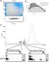
Fig 4. Analysis of OAP complexity and purification grade using 16/60 Sephacryl S-500-based nSEC.
(A) Analysis of nSEC fractions obtained using 16/60 Sephacryl S-500. The E17-E30 fraction analysis was performed by SDS-PAGE followed by Coomassie blue staining and AQP4 immunoblotting. Note that 10 times more proteins were used for Coomassie blue staining than for AQP4 immunoblotting. (B) Quantification of AQP4 enrichment (ng of AQP4/ul) and purification grade (AQP4/total protein (%)) in fractions E17-E30. The red arrow indicates the fraction with the highest AQP4 purification and enrichment grade. (C) Chromatogram obtained by S-500 based nSEC and analysis. Fractions 17 and 21 were analyzed by 2DE and immunoblot (bottom). The red arrow indicates the position, in the SEC elution profile, of the OAP-containing fraction with maximal enrichment and purification grade used for 2DE.
Fig 5
Fig 5. AQP4-IgG specific OAP-ELISA.
(A) Analysis of AQP4 IgG binding on E21 (M23 enriched fraction), E27 (M1 enriched fraction) and total extract using a pool of High (H) and of Low (L) titer sera each from five different NMO patients. The results have been normalized on a control pool serum from five different MS patients. *P<0.01 versus E27 and total extract. (B) Representative OAP-ELISA result. Commercial Anti-AQP4 antibody-based ELISA (AQP4), two sera from AQP4 IgG positive NMO patients (NMO1 and NMO 2) and one MS control serum were analyzed using native (columns A,C) and denatured (B,D) AQP4 positive (A,B,C,D) and AQP4 negative (E) nSEC fractions (AQP4 negative fraction was E35). Empty wells were in column F. Note that AQP4-IgG binding (blue reaction in NMO 1 and NMO 2) was only obtained using AQP4 IgG positive NMO sera on native AQP4 positive fractions, while all other conditions were negative. (C) Cartoon of conformational epitope disassembly after denaturation using in-house OAP-ELISA.
Fig 6
Fig 6. OAPs-ELISA performance.
(A) Scatter plot of sera tested by OAP-ELISA. Among the 67 NMO sera analyzed, 61 were found to be positive by OAP-ELISA, while in the control group 71/72 were negative by OAPs-ELISA. The cut-off lane represents the limit value for positivity/negativity. In house CBA negative were reported in red. (B) intra- and iter-assay reproducibility of OAPs-ELISA using four NMO and four control MS sera. (C) Sensitivity as a function of specificity ROC analysis curve.
Fig 7
Fig 7. Detection limits of commercial ELISA, OAP-ELISA and CBA.
(A) Positive controls (CI, CII), High- (H), Low- (L) titer NMO sera and MS sera were tested using limiting dilution analysis by commercial ELISA. (B) MS and H and L NMO sera serial dilutions tested by OAP-ELISA. Arrows highlight end-point titer. (C) Table summarizing the end-point titer obtained with commercial ELISA, OAPs-ELISA and CBA.

References

    1. Lennon VA, Kryzer TJ, Pittock SJ, Verkman AS, Hinson SR. IgG marker of optic-spinal multiple sclerosis binds to the aquaporin-4 water channel. J Exp Med. 2005;202(4):473–7. - PMC - PubMed
    1. Pisani F, Sparaneo A, Tortorella C, Ruggieri M, Trojano M, Mola MG, et al. Aquaporin-4 autoantibodies in Neuromyelitis Optica: AQP4 isoform-dependent sensitivity and specificity. PloS one. 2013;8(11):e79185 10.1371/journal.pone.0079185 - DOI - PMC - PubMed
    1. Bergamaschi R, Ghezzi A. Devic's neuromyelitis optica: clinical features and prognostic factors. Neurol Sci. 2004;25 Suppl 4:S364–7. - PubMed
    1. Hinson SR, Pittock SJ, Lucchinetti CF, Roemer SF, Fryer JP, Kryzer TJ, et al. Pathogenic potential of IgG binding to water channel extracellular domain in neuromyelitis optica. Neurology. 2007;69(24):2221–31. - PubMed
    1. Lennon VA, Wingerchuk DM, Kryzer TJ, Pittock SJ, Lucchinetti CF, Fujihara K, et al. A serum autoantibody marker of neuromyelitis optica: distinction from multiple sclerosis. Lancet. 2004;364(9451):2106–12. - PubMed

Publication types

LinkOut - more resources