. 2015 Nov 24;10(11):e0143420.
doi: 10.1371/journal.pone.0143420. eCollection 2015.

Mutant SOD1 Increases APP Expression and Phosphorylation in Cellular and Animal Models of ALS

Affiliations

Mutant SOD1 Increases APP Expression and Phosphorylation in Cellular and Animal Models of ALS

Polina Rabinovich-Toidman et al. PLoS One. .

Abstract

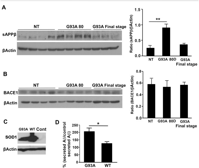
Amyotrophic lateral sclerosis (ALS) is a fatal neurodegenerative disease and it is the most common adult onset neurodegenerative disorder affecting motor neurons. There is currently no effective treatment for ALS and our understanding of the pathological mechanism is still far away from prevention and/or treatment of this devastating disease. Amyloid precursor protein (APP) is a transmembrane protein that undergoes processing either by β-secretase or α-secretase, followed by γ-secretase. In the present study, we show that APP levels, and aberrant phosphorylation, which is associated with enhanced β-secretase cleavage, are increased in SOD1G93A ALS mouse model. Fluorescence resonance energy transfer (FRET) analysis suggests a close interaction between SOD1 and APP at hippocampal synapses. Notably, SOD1G93A mutation induces APP-SOD1 conformational changes, indicating a crosstalk between these two signaling proteins. Inhibition of APP processing via monoclonal antibody called BBS that blocks APP β-secretase cleavage site, resulted in reduction of mutant SOD1G93A levels in animal and cellular models of ALS, significantly prolonged life span of SOD1G93A mice and diminished inflammation. Beyond its effect on toxic mutant SOD1G93A, BBS treatment resulted in a reduction in the levels of APP, its processing product soluble APPβ and pro-apoptotic p53. This study demonstrates that APP and its processing products contribute to ALS pathology through several different pathways; thus BBS antibody could be a promising neuroprotective strategy for treatment of this disease.


Conflict of interest statement

Competing Interests: The therapeutic approach described here is under a patent application (Methods and compositions with immune therapy for treatment of Dementia. PCT/US2014/26377). None of the authors hold any equity in NasVax Ltd, Israel. There are no further patents, products in development or marketed products to declare. This does not alter the authors' adherence to all the PLOS ONE policies on sharing data and materials, as detailed online in the guide for authors.

Figures

Fig 1
Fig 1. APP expression and phosphorylation in spinal cords of pre-symptomatic and symptomatic mice.
Spinal cords from 30 -and 80-day-old and end stage SOD1G93A mice were homogenized and their membrane fractions were subjected to immunoblot analysis. Age matched NT littermates served as control. For APP detection 22C11 antibody was used. For detection of phosphorylated APP (pAPP) a specific polyclonal anti pAPP T688 was used. β-Actin was used as an internal loading control. Immunoblot blot and densitometric analysis of the relative expression and phosphorylation levels of APP in the spinal cord of (A) 30-day-old mice, (B) 80-day-old and end stage SOD1G93A mice. All values are expressed as the mean ± SEM. Statistical comparisons were performed using one-way ANOVA followed by Tukey–Kramer post hoc test. N(NT 30D) = 3, N(G93A 30D) = 3, N(NT = 4), N(G93A 80D) = 4, N(G93A end stage = 3).*p < 0.05; **p < 0.01 (v.s. NT littermates).
Fig 2
Fig 2. Mutant SOD1 leads to enhanced APP processing in vivo and in vitro.
Soluble and membrane fractions from 80-day-old and end stage SOD1G93A mice were subjected to immunoblot analysis. Age matched NT littermates served as control. A. sAPPβ was detected using polyclonal antibody specific for the neoepitope generated after BACE cleavage in the spinal cords soluble fractions. The levels of sAPPβ were normalized to β-Actin levels. B. Detection of BACE1 in the spinal cord homogenates membrane fraction was performed using a specific anti C terminal MAb. Spinal cord BACE1 levels were normalized to β-Actin levels. All values are expressed as the mean ± SEM. Statistical comparisons were performed using one-way ANOVA followed by Tukey–Kramer post hoc test. N(NT = 4), N(G93A 80D) = 4, N(G93A Final stage = 3).*p < 0.05; **p < 0.01 (v.s. NT littermates). CHO cells overexpressing APP 751 were transiently transfected with WT SOD1 or with mutant SOD1G93A. C. Representative Western blot of mutant and WT SOD1 expression in CHO cells as a result of transient transfection. Human HA -tagged SOD1 was detected using anti HA antibody. Non-tranfected cells served as negative control D. Aβ42 peptides levels were measured in the medium using sandwich ELISA and the ratio between secreted Aβ42 in each treatment group and the untreated group was calculated and presented in percentage. The experiment was repeated three times. Statistical comparisons were performed using Student's t-test. *p value <0.05.
Fig 3
Fig 3. BBS treatment results in reduction of APP and mutant SOD1G93A levels in vitro and in vivo.
Primary astroglial cells were isolated from brains of mutant SOD1G93A pups. After 7 days in culture cells were treated with 13nM BBS for 24h. Induction of mutant SOD1G93A fused to GFP expression in NSC34 stable line was performed by incubating the cells with Doxycycline for 48h. 24h after the induction the cells were treated with 26nM BBS for 24h. The levels of mutant SOD1, APP and actin were measured using Western blot analysis. SOD1 was detected by polyclonal anti human SOD1 antibody and APP was detected using 22C11 antibody. SOD1 levels were normalized to β-Actin levels. A. Levels of APP and SOD1 in primary astroglial cells. B. Levels of APP and SOD1 fused to GFP in NSC34 stable line. All values are expressed as the mean ± SEM. Statistical comparisons were performed using Student's t-test. *p < 0.05. For in vivo analysis of the effect of BBS, spinal cords of i.c.v. and i.p. treated SODG93A mice were homogenized and subjected to immunoblot analysis. Analysis of the effect of BBS, in spinal cords of i.c.v. treated SODG93A mice C. sAPPβ was detected in spinal cord soluble fractions and normalized to β-Actin levels. D. APP and SOD1 in spinal cords of i.c.v. treated mice were detected in membrane fractions and normalized to β-Actin. All values are expressed as the mean ± SEM. Statistical comparisons were performed using Student's t-test. N(BBS) = 4, N(Non relevant) = 5. *p < 0.05; **p < 0.01 (v.s. Non relevant). Analysis of the effect of BBS, in spinal cords of i.p. treated SODG93A mice. E. The levels of mutant SOD1, APP and tubulin in the membrane fraction were measured using Western blot analysis. Statistical comparisons were performed using Student's t-test. N(BBS) = 12, N(Control) = 11.*p < 0.05.
Fig 4
Fig 4. APP molecules closely associate with SOD1 at single hippocampal synapses.
A. Representative confocal images of boutons of a hippocampal neuron that was co-transfected with APPYFP and SOD1CFP. Scale bar: 2 μm. B. Pseudocolor-coded fluorescence images of a bouton expressing APPYFP and SOD1CFP before and after YFP photobleaching, Scale bar: 2 μm. C. Summary of Em data for APPYFP / GB1aCFP (n = 26), APPYFP / SOD1CFP (n = 39), APPYFP / SOD1CFP (n = 42). Error bars represent SEM.
Fig 5
Fig 5. BBS treatment leads to decrease in p53 levels and astrogliosis marker GFAP.
A. The levels of p53were quantified using immunoblot analysis in the soluble fraction of spinal cord homogenates of SODG93A mice treated with BBS or non relevant MAb for 42 days. p53 levels were detected using PAb 240 antibody and normalized to β-Actin. All values are expressed as the mean ± SEM. Statistical comparisons were performed using Student's t-test. N(BBS) = 4, N(Non relevant) = 5. *p < 0.05; **p < 0.01 (v.s. Non relevant). B. Lumber spinal cords were subjected to immunohistochemical analysis. Spinal cord sections were stained with anti-GFAP antibody, and the intensity of the staining was analyzed using Image-J Software. Scale bars in panel B correspond to 1 mm, respectively. Statistical comparisons were performed using Student's t-test. N(BBS) = 5, N(Non relevant) = 5. **p < 0.01 (v.s. Non relevant).
Fig 6
Fig 6. BBS treatment leads increase in survival of SOD1G93A transgenic mice.
55-day-old female and male SODG93A mice received a weekly dose of 3mg/kg MAb BBS via i.p. injection. Controls were treated with PBS. A. Weight of each animal was recorded weekly. B. Motor functions of the i.p. treated mice were assessed by performing Rotarod test. Mice were trained to run on the 2-cm-diameter rod, which rotated at a fixed speed of 13 turns per minute. Mice were allowed to run for up to 1 min in each trial, or until they fell off. C. Plots of disease onset (Median BBS = 95; Median PBS = 91.5; *p <0.05; Average BBS = 97.2; Average = 91.9; *p <0.05), survival(Median BBS = 95; Median PBS = 91.5; *p <0.05; Average BBS = 97.2; Average = 91.9; *p <0.05) and the average survival in days. N (PBS) = 24, N (BBS) = 24.

References

    1. Leigh PN. Chapter 13 Amyotrophic lateral sclerosis. Handb Clin Neurol / Ed by PJ Vinken GW Bruyn. 2007;82: 249–78. 10.1016/S0072-9752(07)80016-9 - DOI - PubMed
    1. Pasinelli P, Brown RH. Molecular biology of amyotrophic lateral sclerosis: insights from genetics. Nat Rev Neurosci. 2006;7: 710–23. 10.1038/nrn1971 - DOI - PubMed
    1. Gurney ME, Pu H, Chiu AY, Dal Canto MC, Polchow CY, Alexander DD, et al. Motor neuron degeneration in mice that express a human Cu,Zn superoxide dismutase mutation. Science. 1994;264: 1772–5. Available: http://www.ncbi.nlm.nih.gov/pubmed/8209258. - PubMed
    1. Slunt HH, Thinakaran G, Von Koch C, Lo AC, Tanzi RE, Sisodia SS. Expression of a ubiquitous, cross-reactive homologue of the mouse beta-amyloid precursor protein (APP). J Biol Chem. 1994;269: 2637–44. Available: http://www.ncbi.nlm.nih.gov/pubmed/8300594. - PubMed
    1. Zheng H, Koo EH. The amyloid precursor protein: beyond amyloid. Mol Neurodegener. 2006;1: 5 10.1186/1750-1326-1-5 - DOI - PMC - PubMed

Publication types

MeSH terms