Is in vivo amyloid distribution asymmetric in primary progressive aphasia?
- PMID: 26600088
- PMCID: PMC4789129
- DOI: 10.1002/ana.24566
Is in vivo amyloid distribution asymmetric in primary progressive aphasia?
Abstract
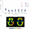
We aimed to determine whether (18) F-florbetapir amyloid positron emission tomography imaging shows a clinically concordant, left-hemisphere-dominant pattern of deposition in primary progressive aphasia (PPA). Elevated cortical amyloid (Aβ(+) ) was found in 19 of 32 PPA patients. Hemispheric laterality of amyloid burden was compared between Aβ(+) PPA and an Aβ(+) amnestic dementia groups (n = 22). The parietal region showed significantly greater left lateralized amyloid uptake in the PPA group than the amnestic group (p < 0.007), consistent with the left lateralized pattern of neurodegeneration in PPA. These results suggest that the cortical distribution of amyloid may have a greater clinical concordance than previously reported.
© 2016 American Neurological Association.
Figures


References
-
- Mesulam MM. Primary progressive aphasia--a language-based dementia. The New England journal of medicine. 2003 Oct 16;349(16):1535–42. - PubMed
Publication types
MeSH terms
Substances
Grants and funding
LinkOut - more resources
Full Text Sources
Other Literature Sources

