. 2015 Nov 23;10(11):e0143423.
doi: 10.1371/journal.pone.0143423. eCollection 2015.

Mutually Exclusive Roles of SHARPIN in Integrin Inactivation and NF-κB Signaling

Affiliations

Mutually Exclusive Roles of SHARPIN in Integrin Inactivation and NF-κB Signaling

Nicola De Franceschi et al. PLoS One. .

Abstract

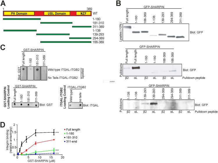
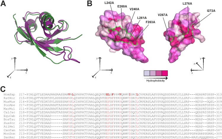
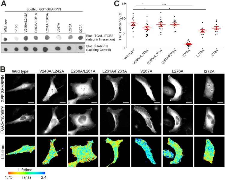
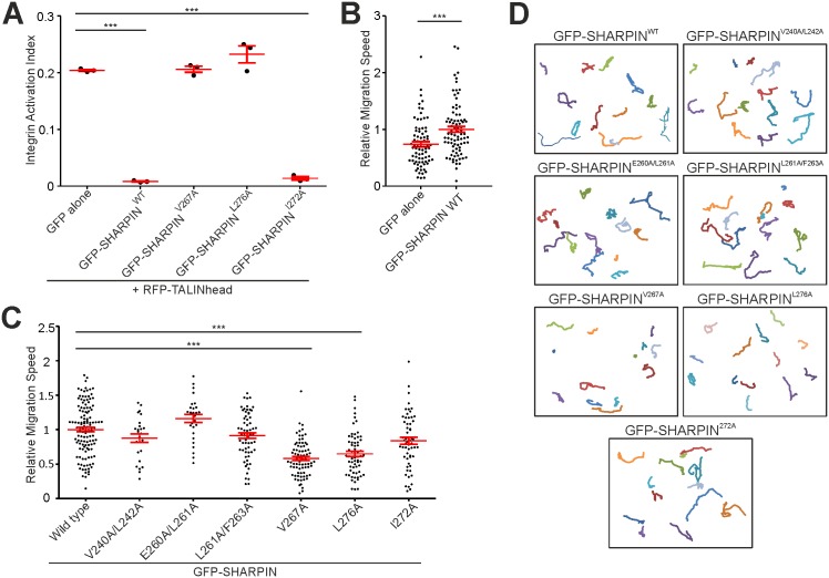
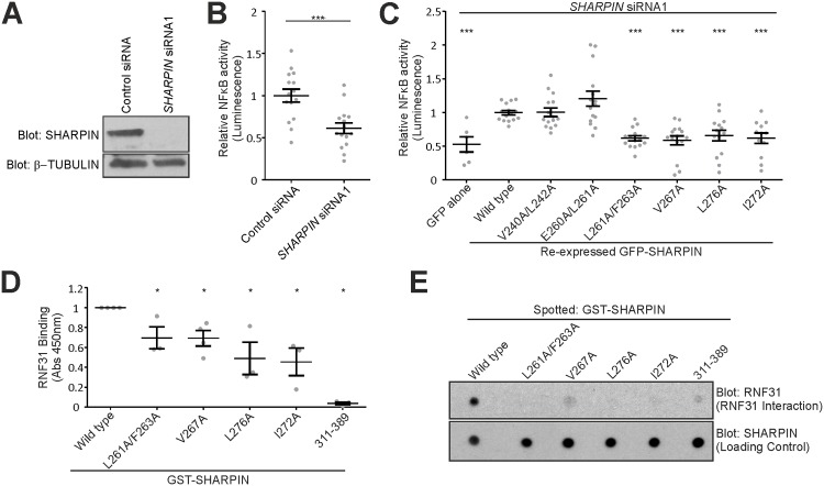
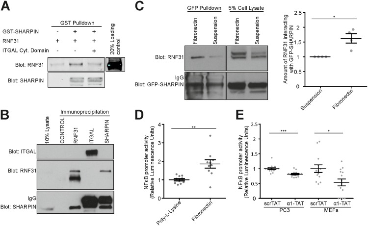
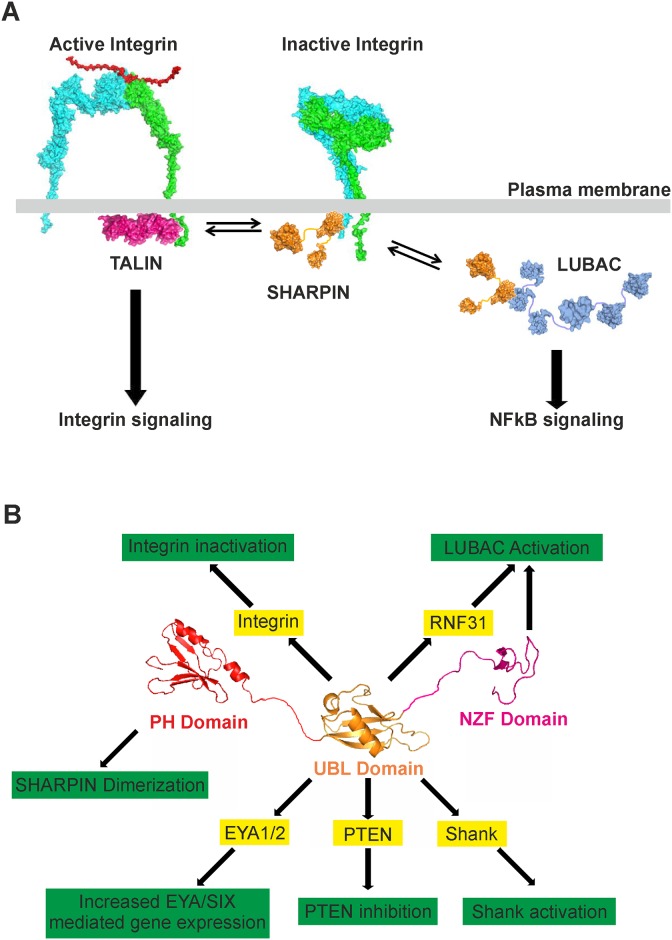
SHANK-associated RH domain interactor (SHARPIN) inhibits integrins through interaction with the integrin α-subunit. In addition, SHARPIN enhances nuclear factor-kappaB (NF-κB) activity as a component of the linear ubiquitin chain assembly complex (LUBAC). However, it is currently unclear how regulation of these seemingly different roles is coordinated. Here, we show that SHARPIN binds integrin and LUBAC in a mutually exclusive manner. We map the integrin binding site on SHARPIN to the ubiquitin-like (UBL) domain, the same domain implicated in SHARPIN interaction with LUBAC component RNF31 (ring finger protein 31), and identify two SHARPIN residues (V267, L276) required for both integrin and RNF31 regulation. Accordingly, the integrin α-tail is capable of competing with RNF31 for SHARPIN binding in vitro. Importantly, the full SHARPIN RNF31-binding site contains residues (F263A/I272A) that are dispensable for SHARPIN-integrin interaction. Importantly, disrupting SHARPIN interaction with integrin or RNF31 abolishes SHARPIN-mediated regulation of integrin or NF-κB activity, respectively. Altogether these data suggest that the roles of SHARPIN in inhibiting integrin activity and supporting linear ubiquitination are (molecularly) distinct.


Conflict of interest statement

Competing Interests: The authors have declared that no competing interests exist.

Figures

Fig 1
Fig 1. The UBL domain of SHARPIN mediates binding to integrin.
(A) Schematic representation of SHARPIN with its functional domains and the SHARPIN fragments used in this study. (B) Pull-down experiments to determine the interaction between GFP-SHARPIN (full-length or fragments) and peptides corresponding to the cytoplasmic domain of ITGAL and ITGB2. (C) Far-Western analysis of GST-SHARPIN (full-length or fragments) binding to full-length ITGAL-ITGB2 or ITGAL-ITGB2 lacking both cytoplasmic tails. Loading controls for GST-SHARPIN (full-length or fragments) and both ITGAL-ITGB2s are shown. (D) Fluorescence polarization-based titration of GST-SHARPIN (full-length or fragments) binding to an integrin peptide corresponding to the conserved domain within the cytoplasmic tail of ITGA2. Average normalized binding curves are shown (mean ± s.e.m. ***: p<0.001).
Fig 2
Fig 2. Designing SHARPIN mutants using a SHARPIN UBL domain model.
(A) Superposition of the SHARPIN UBL domain model backbone (purple) with the UBL domain of HOIL1L (Green, PDB accession code: 4DBG). (B) Surface representation of the SHARPIN UBL model. The surface is color-coded according to residue hydrophobicity from light to deep purple. Footprints of residues mutated in this study are indicated as green outlines. (C) Alignment of part of the SHARPIN UBL domain across different species. Conserved residues mutated in this study are indicated in red.
Fig 3
Fig 3. Fine mapping of the integrin binding site in SHARPIN.
(A) Interaction of different GST-SHARPIN (WT and all point mutants) with full-length ITGAL-ITGB2 was determined using Far-Western assays. GST-SHARPIN1-180 was used as negative control. (B) HEK293 cancer cells, overexpressing WT or mutant GFP-SHARPIN in combination with ITGA5–mCherry, subjected to FRET analysis by FLIM. Fluorescence lifetimes, mapping spatial FRET in cells, are depicted using a pseudo-color scale (blue, normal lifetime; red, FRET (reduced lifetime)). Scale bars: 10 μm. (C) Quantification of FRET efficiency for all mutants (n ≥ 12 cells). All numerical data are mean ± s.e.m. ***: p<0.001, *: p<0,05.
Fig 4
Fig 4. Residues V267 and L276 are essential for SHARPIN-mediated integrin inhibition.
(A) FACS analysis of CHO cells overexpressing GFP alone, or WT or mutant GFP-SHARPIN, together with RFP-TALIN head. The Integrin Activation Index was calculated by dividing active cell-surface integrin levels (FN7-10 binding minus FN7-10 binding in the presence of EDTA) by total cell-surface integrin levels (Mb1.2 staining minus secondary antibody alone) (n = 3). (B) Quantification of migration speed of cpdm MEFs overexpressing GFP alone or GFP-SHARPINWT on 50 μg/ml collagen (n = 78 and 83 cells, respectively). (C,D) Quantification of migration speed (n = 27–125 cells) (C), and representative cell tracks (D) of cpdm MEFs overexpressing WT or mutant GFP-SHARPIN on 50 μg/ml collagen. All numerical data are mean ± s.e.m. ***: p<0.001, *: p<0.05.
Fig 5
Fig 5. Fine mapping of the RNF31 binding site in SHARPIN.
(A) Western blot analysis of SHARPIN and β-tubulin levels in control- or SHARPIN-silenced PC3 cells. (B) TNF-induced NF-κB promoter activity of SHARPIN- or control-silenced PC3 cells was measured using a luciferase reporter assay. (n = 3 with 5 replicates each). (C) TNF-induced NF-κB promoter activity of SHARPIN-silenced PC3 cells, expressing GFP alone, WT or mutant GFP-SHARPIN (n = 6–15 measurements from 2–3 experiments). (D,E) Interaction between RNF31 and WT or mutant GST-SHARPIN was determined using an ELISA-based binding assay (n = 3) (D) or Far-Western analysis (E). All numerical data are mean ± s.e.m. ***: p<0.001, **: p<0.01, *: p<0.05.
Fig 6
Fig 6. Integrin and RNF31 binding to SHARPIN are mutually exclusive.
(A) Pull-down assay showing that the presence of a peptide corresponding to the cytoplasmic domain of ITGAL prevents interaction between GST-SHARPIN and RNF31. (B) Co-immunoprecipitation of endogenous SHARPIN, RNF31 and ITGAL from Jurkat cells (a GFP antibody was used as negative control). (C) Pull-down of GFP-SHARPIN from HEK293 cells demonstrated that cells in suspension show decreased RNF31-SHARPIN interaction compared to adherent cells (n = 4). RNF31 binding was normalized to total RNF31 levels and the amount of pulled-down GFP-SHARPIN. (D) TNF-induced NF-κB promoter activity of PC3 cells, adherent to either 5 μg/ml fibronectin or poly-L-lysin (a substratum for integrin-independent cell adhesion), was measured using a luciferase reporter assay (n = 2 with 5 replicates each). (E) TNF-induced NF-κB promoter activity of PC3 cells or WT MEFs, incubated with a membrane-permeable ITGA1-tail peptide (α1-TAT) or a scrambled peptide (ScrTAT), was measured using a luciferase reporter assay (n = 2 with 6–9 replicates, and n = 3 with 3–5 replicates, respectively). All numerical data are mean ± s.e.m. ***: p<0.001, **: p<0.01, *: p<0.05.
Fig 7
Fig 7. Current model of SHARPIN function.
(A) SHARPIN inhibits integrin activation by binding to the α-integrin cytoplasmic domain and preventing binding of the integrin activator TALIN. In addition, SHARPIN is part of LUBAC, which is required for activation of the canonical NF-κB pathway. We now demonstrate that integrin and RNF31 binding are mutually exclusive as they are mediated by partially overlapping binding sites within the SHARPIN UBL domain. (B) An overview of SHARPIN interactions and how these interactions affect diverse signaling pathways. SHARPIN is depicted schematically with its functional domains (an N-terminal PH domain, a central UBL domain and a C-terminal NZF domain). The UBL domain is a multi-faceted protein interaction hub that has been shown to interact with a number of proteins, highlighted in yellow bars. These interactions each have specific functional consequences, highlighted in green bars. In addition, the N-terminal PH domain of SHARPIN mediates SHARPIN homodimerization and the C-terminal NZF domain is required for LUBAC function.

References

    1. Bouvard D, Pouwels J, De Franceschi N, Ivaska J. Integrin inactivators: balancing cellular functions in vitro and in vivo. Nat Rev Mol Cell Biol. 2013;14: 430–442. 10.1038/nrm3599 - DOI - PubMed
    1. Winograd-Katz SE, Fassler R, Geiger B, Legate KR. The integrin adhesome: from genes and proteins to human disease. Nat Rev Mol Cell Biol. 2014;15: 273–288. 10.1038/nrm3769 - DOI - PubMed
    1. Cabodi S, del Pilar Camacho-Leal M, Di Stefano P, Defilippi P. Integrin signalling adaptors: not only figurants in the cancer story. Nat Rev Cancer. 2010;10: 858–870. 10.1038/nrc2967 - DOI - PubMed
    1. Rantala JK, Pouwels J, Pellinen T, Veltel S, Laasola P, Mattila E, et al. SHARPIN is an endogenous inhibitor of beta1-integrin activation. Nat Cell Biol. 2011;13: 1315–1324. 10.1038/ncb2340 - DOI - PMC - PubMed
    1. Pouwels J, De Franceschi N, Rantakari P, Auvinen K, Karikoski M, Mattila E, et al. SHARPIN Regulates Uropod Detachment in Migrating Lymphocytes. Cell Rep. 2013;5: 619–628. 10.1016/j.celrep.2013.10.011 - DOI - PMC - PubMed

Publication types

MeSH terms