. 2016 Jun;54(6):831-42.
doi: 10.1165/rcmb.2015-0040OC.

Fibrogenic Lung Injury Induces Non-Cell-Autonomous Fibroblast Invasion

Affiliations

Fibrogenic Lung Injury Induces Non-Cell-Autonomous Fibroblast Invasion

Neil Ahluwalia et al. Am J Respir Cell Mol Biol. 2016 Jun.

Abstract

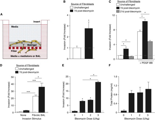
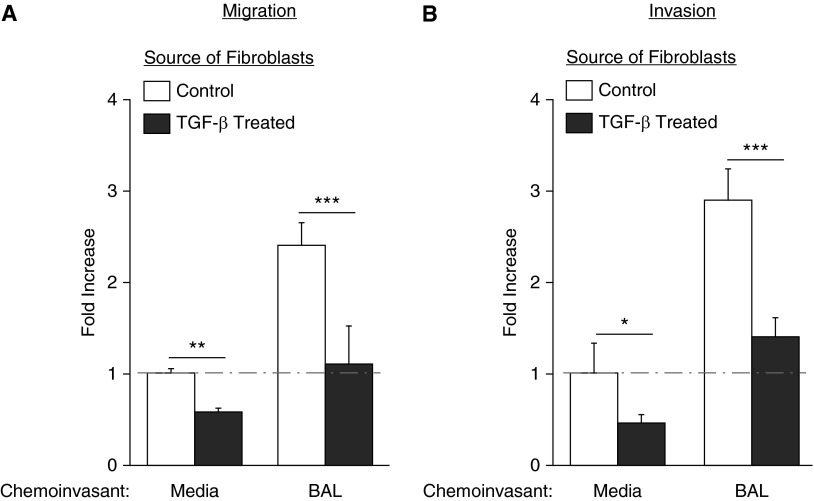
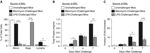
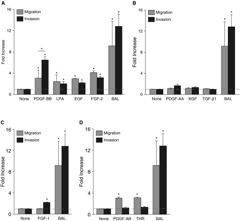
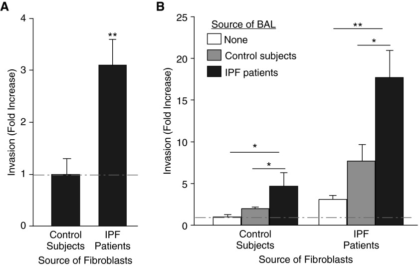
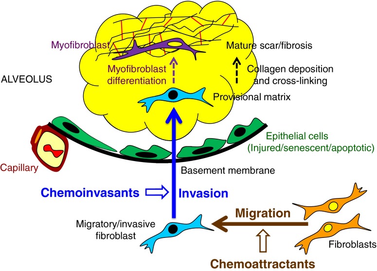
Pathologic accumulation of fibroblasts in pulmonary fibrosis appears to depend on their invasion through basement membranes and extracellular matrices. Fibroblasts from the fibrotic lungs of patients with idiopathic pulmonary fibrosis (IPF) have been demonstrated to acquire a phenotype characterized by increased cell-autonomous invasion. Here, we investigated whether fibroblast invasion is further stimulated by soluble mediators induced by lung injury. We found that bronchoalveolar lavage fluids from bleomycin-challenged mice or patients with IPF contain mediators that dramatically increase the matrix invasion of primary lung fibroblasts. Further characterization of this non-cell-autonomous fibroblast invasion suggested that the mediators driving this process are produced locally after lung injury and are preferentially produced by fibrogenic (e.g., bleomycin-induced) rather than nonfibrogenic (e.g., LPS-induced) lung injury. Comparison of invasion and migration induced by a series of fibroblast-active mediators indicated that these two forms of fibroblast movement are directed by distinct sets of stimuli. Finally, knockdown of multiple different membrane receptors, including platelet-derived growth factor receptor-β, lysophosphatidic acid 1, epidermal growth factor receptor, and fibroblast growth factor receptor 2, mitigated the non-cell-autonomous fibroblast invasion induced by bronchoalveolar lavage from bleomycin-injured mice, suggesting that multiple different mediators drive fibroblast invasion in pulmonary fibrosis. The magnitude of this mediator-driven fibroblast invasion suggests that its inhibition could be a novel therapeutic strategy for pulmonary fibrosis. Further elaboration of the molecular mechanisms that drive non-cell-autonomous fibroblast invasion consequently may provide a rich set of novel drug targets for the treatment of IPF and other fibrotic lung diseases.

Keywords: fibroblast; idiopathic pulmonary fibrosis; invasion; migration; pathogenesis.


Figures

Figure 1.
Figure 1.
Fibrogenic lung injury increases lung fibroblast invasion. (A) Schematic drawing of the filter-based assay used to model fibroblast invasion through basement membrane. We assessed primary lung fibroblast invasion through Matrigel covering polyethylene filters with 8-μm pores, in response to media with or without defined mediators or bronchoalveolar lavage (BAL). (B) Lung fibroblasts isolated from mice at Day 7 after bleomycin challenge demonstrated increased cell-autonomous invasion compared with lung fibroblasts from unchallenged mice. Data are presented as mean (±SEM) fold increase in number of invading cells, normalized to invasion of lung fibroblasts from unchallenged mice (n = 5 experiments; *P < 0.05). (C) Lung fibroblasts isolated from mice at Day 21 after bleomycin challenge demonstrated decreased cell-autonomous and non–cell-autonomous invasion compared with lung fibroblasts from unchallenged mice. Non–cell-autonomous invasion was induced by platelet-derived growth factor-BB (PDGF-BB) (10−9 M). Data are presented as mean (±SEM) fold increase in invasion, normalized to lung fibroblasts from unchallenged mice (n = 3 mice per group; *P < 0.05). (D) BAL recovered from mice at Day 7 after 1.2 U/kg bleomycin challenge induced non–cell-autonomous invasion of lung fibroblasts isolated from either unchallenged or bleomycin-challenged mice, which greatly exceeded the cell-autonomous invasion of these cells. Data are presented as mean (±SEM) fold increase in invasion, normalized to the invasion of lung fibroblasts from unchallenged mice in the absence of an exogenous stimulus (n = 5 experiments; *P < 0.05, ***P < 0.001). (E) BAL collected from mice at Day 7 after challenge with 3 U/kg bleomycin induced greater non–cell-autonomous invasion of unchallenged mouse lung fibroblasts than did BAL collected from mice at Day 7 after challenge with 1 or 2 U/kg bleomycin. Data are presented as mean (±SEM) fold increase in invasion, normalized to the cell-autonomous invasion of lung fibroblasts from unchallenged mice (n = 5 mice per group as the source of lung fibroblasts, and n = 5 mice per group as the source of BAL; *P < 0.05). (F) Higher-dose bleomycin did not increase vascular leak at Day 7 after challenge, as assessed by BAL total protein concentration (n = 5 mice per group as the source of BAL).
Figure 2.
Figure 2.
Transforming growth factor (TGF)-β1 stimulation decreases migration and invasion of lung fibroblasts. (A) Primary lung fibroblasts pretreated with TGF-β1 for 48 hours had decreased migration (A) and invasion (B), both in the absence and presence of BAL from injured mice. Data are presented as mean (±SEM) fold increase in migration or invasion, normalized to the control fibroblast to media condition (n = 3 mice as source of lung fibroblasts and BAL; *P < 0.05, **P < 0.01, ***P < 0.001).
Figure 3.
Figure 3.
Comparison of fibroblast invasion induced by profibrotic bleomycin injury with proinflammatory LPS injury. (A) BAL recovered from mice at Day 1 after 4 mg/kg LPS challenge had greater percentages of neutrophils (Segs) than BAL recovered from mice at Day 1 after 1.2 U/kg bleomycin challenge, whereas BAL from bleomycin-challenged mice had greater percentages of macrophages (Mono) and lymphocytes (Lymphs). Data are presented as mean (±SEM) percentage (n = 3 mice per group; *P < 0.05, ***P < 0.001). (B) Determination of total protein concentrations in BAL from unchallenged mice and from mice at Days 1, 4, and 7 after 1.2 U/kg bleomycin or 4 mg/kg LPS challenge, indicated protein concentration was greater in LPS-challenged mice than in bleomycin-challenged mice at Day 1, similar between these groups at Day 4, and greater in bleomycin-challenged mice than in LPS-challenged mice at Day 7. Data are presented as mean (±SEM) total protein concentration (n = 3 mice per group; *P < 0.05, **P < 0.01). (C) BAL from mice at Days 4 and 7 after 1.2 U/kg bleomycin challenge induced greater non–cell-autonomous invasion of lung fibroblasts isolated from unchallenged mice than did BAL from mice at these days after 4 mg/kg LPS challenge. Data are presented as mean (±SEM) fold increase in invasion, normalized to the cell-autonomous invasion of lung fibroblasts from unchallenged mice (n = 3 mice per group as the source of lung fibroblasts, and n = 3 mice per group as the source of BAL; *P < 0.05, ***P < 0.001). ND, none detected.
Figure 4.
Figure 4.
Different effects of fibroblast-active mediators on migration versus invasion. Assessment of the migration and invasion of lung fibroblasts from unchallenged mice induced by multiple fibroblast-active mediators indicated that: (A) PDGF-BB, lysophosphatidic acid (LPA), epidermal growth factor (EGF), and fibroblast growth factor 2 (FGF-2) induced both migration and invasion; (B) PDGF-AA, keratinocyte growth factor (KGF), and TGF-β1 induced neither migration nor invasion; (C) FGF-1 induced invasion, but not migration; and (D) PDGF-AB and thrombin induced migration but not invasion. BAL from mice at Day 7 after 1.2 U/kg bleomycin challenge induced greater invasion and migration than any individual mediator tested (AD). The mediator concentrations used were 10−9 M PDGF-BB, 10−7 M LPA, 100 ng/ml EGF, 15 ng/ml FGF-2, 50 ng/ml PDGF-AA, 100 ng/ml KGF, 5 ng/ml TGF-β1, 20 ng/ml FGF-1, 10 ng/ml PDGF-AB, 10 ng/ml and thrombin, in serum-free Dulbecco’s modified Eagle's medium media, as determined by pilot dose–response studies (data not shown). Each of the panels shares the same BAL condition. Data are presented as mean (±SEM) fold increase in invasion, normalized to the cell-autonomous invasion of lung fibroblasts from unchallenged mice (n = 3 mice as the source of lung fibroblasts, and n = 3 as the source of BAL; *P < 0.05). THR, thrombin.
Figure 5.
Figure 5.
Effects of fibroblast receptor knockdown on fibroblast non–cell-autonomous invasion. (A) Quantitative PCR demonstrating the mRNA expression of the relevant genes after small interfering RNA (siRNA)-mediated receptor knockdown. (B) Transfection with siRNA targeting platelet-derived growth factor receptor-β (PDGFRβ) reduced the invasion of lung fibroblasts from unchallenged mice that was induced by PDGF-BB. (C) Transfection with siRNAs targeting PDGFRβ, LPA1, epidermal growth factor receptor (EGFR), and fibroblast growth factor receptor 2 (FGFR2), but not transforming growth factor-β receptor 1 (TGF-βR1), reduced the invasion of lung fibroblasts from unchallenged mice that was induced by BAL recovered from mice at D7 after challenge with 1.2 U/kg bleomycin. Data are presented as percentage of the invasion of fibroblasts transfected with nontargeting (NT) siRNA that was induced by the same BAL samples acting on fibroblasts transfected with targeting siRNAs (n = 3 mice as the source of lung fibroblasts, and n = 3 as the source of BAL; *P < 0.05, ***P < 0.001).
Figure 6.
Figure 6.
BAL from patients with idiopathic pulmonary fibrosis (IPF) induces non–cell-autonomous invasion of lung fibroblasts from patients with IPF. (A) Lung fibroblasts isolated from patients with IPF demonstrated increased cell-autonomous invasion compared with lung fibroblasts from control subjects. (B) BAL recovered from patients with IPF induced non–cell-autonomous invasion of lung fibroblasts isolated from either patients with IPF or control subjects, which greatly exceeded the cell-autonomous invasion of these cells. BAL from control subjects also induced invasion of both IPF and control lung fibroblasts but to a significantly lesser extent than IPF BAL. Data are presented as mean (±SEM) fold increase in invasion, normalized to the cell-autonomous invasion of lung fibroblasts from control subjects (n = 5 persons with IPF and n = 5 control subjects as the source of lung fibroblasts; n = 3 persons with IPF and n = 3 control subjects as the source of BAL; *P < 0.05, **P < 0.01).
Figure 7.
Figure 7.
Regulation of fibroblast/myofibroblast accumulation in pulmonary fibrosis. The data presented in this study support a model of fibroblast/myofibroblast accumulation at sites of lung injury depicted in this figure. In this figure, thick solid arrows represent cell movements, whereas thinner dashed arrows represent cell and matrix maturations. Early after lung injury, fibroblasts acquire a migratory/invasive phenotype characterized by the acquisition of cell-autonomous invasive capacity. Concurrent with the development of this intrinsic fibroblast phenotype, potent soluble mediators that markedly augment fibroblast migration (chemoattractants) and invasion (chemoinvasants) are induced in the lungs of patients with IPF and in the bleomycin mouse model of pulmonary fibrosis. After invasion into provisional matrices, fibroblasts lose their initial migratory/invasive phenotype and transition to the matrix-synthetic and contractile phenotype characteristic of myofibroblasts, directed by cytokines, such as TGF-β. Once they have differentiated into myofibroblasts, these cells remain in the provisional matrix, where they secrete increased amounts of collagen and other matrix proteins. Collagen cross-linking and wound contraction then convert the myofibroblast-rich provisional matrices into mature scar/fibrosis.

References

    1. Selman M, King TE, Pardo A American Thoracic Society; European Respiratory Society; American College of Chest Physicians. Idiopathic pulmonary fibrosis: prevailing and evolving hypotheses about its pathogenesis and implications for therapy. Ann Intern Med. 2001;134:136–151. - PubMed
    1. Ahluwalia N, Shea BS, Tager AM. New therapeutic targets in idiopathic pulmonary fibrosis: aiming to rein in runaway wound-healing responses. Am J Respir Crit Care Med. 2014;190:867–878. - PMC - PubMed
    1. Ridley AJ, Schwartz MA, Burridge K, Firtel RA, Ginsberg MH, Borisy G, Parsons JT, Horwitz AR. Cell migration: Integrating signals from front to back. Science. 2003;302:1704–1709. - PubMed
    1. Wolf K, Alexander S, Schacht V, Coussens LM, von Andrian UH, van Rheenen J, Deryugina E, Friedl P. Collagen-based cell migration models in vitro and in vivo. Semin Cell Dev Biol. 2009;20:931–941. - PMC - PubMed
    1. Louis SF, Zahradka P. Vascular smooth muscle cell motility: from migration to invasion. Exp Clin Cardiol. 2010;15:e75–e85. - PMC - PubMed

Publication types