

Unilateral Vogt-Koyanagi-Harada Disease: A Clinical Case Report
- PMID: 26600790
- PMCID: PMC4649709
- DOI: 10.1159/000441616
Unilateral Vogt-Koyanagi-Harada Disease: A Clinical Case Report
Abstract
Purpose: To report a case of a 20-year-old female with decreased visual acuity (VA) in the left eye (LE).
Methods: This is a retrospective and descriptive case report based on data from clinical records, patient observation and analysis of diagnostic tests.
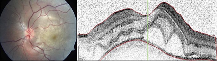
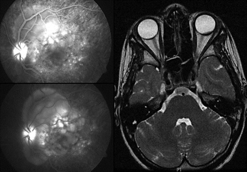
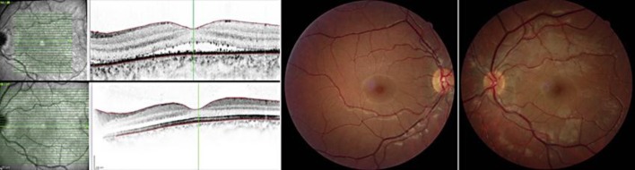
Results: A 20-year-old female presented with decreased VA in the LE for 3 days. Best-corrected visual acuity (BCVA) was 20/20 in the right eye (RE) and 20/40 in the LE. Pupillary function, intraocular pressure, results of external segment examinations and slit-lamp biomicroscopy were normal, bilaterally. RE fundoscopy was normal, and in the LE it revealed papillitis and posterior pole exudative retinal detachment. Optical coherence tomography (OCT) confirmed the macular serous retinal detachment and showed thickening of the posterior choroid also revealed by orbital ultrasound and magnetic resonance imaging (MRI). Fluorescein angiography showed angiographic features typical of Vogt-Koyanagi-Harada (VKH) disease: disseminated spotted choroidal hyperfluorescence and choroidal multifocal hypofluorescence, multifocal profuse leakage in the retina with pooling, serous retinal detachment and optic disc hyperfluorescence. Serological testing for the diagnosis of infectious pathologies was negative, and the review of systems was normal. The patient received systemic steroids and cyclosporine. LE BCVA improved up to 20/20 at 18 months after the diagnosis, with complete reabsorption of subretinal fluid and normal retinal and choroidal thickness by OCT.
Conclusion: Despite the unilateral involvement, the clinical and angiographic features were typical of VKH disease, and ophthalmologists should be aware to recognize this rare clinical variant of the disease.
Keywords: Corticosteroid therapy; Cyclosporine; Fundus fluorescein angiography; Unilateral Vogt-Koyanagi-Harada disease.
Figures
References
-
- Sakata VM, da Silva FT, Hirata CE, et al. Diagnosis and classification of Vogt-Koyanagi-Harada disease. Autoimmun Rev. 2014;13:550–555. - PubMed
-
- American Academy of Ophthalmology – The Eye MD Association (2014) Vogt-Koyanagi-Harada syndrome; in Basic and Clinical Science Course, Section 9: Intraocular Inflammation and Uveitis, chapter 6: Noninfectious (Autoimmune) Ocular Inflammatory Disease. San Francisco: AAO; 2014. pp. 183–190.
-
- Forster DJ, Green RL, Rao NA. Unilateral manifestation of Vogt-Koyanagi-Harada syndrome in a 7-year old child. Am J Ophthalmol. 1991;111:380–382. - PubMed
-
- Usui Y, Goto H, Sakai J, Takeuchi M, Usui M, Rao NA, et al. Presumed Vogt Koyanagi Harada disease with unilateral ocular involvement: report of three cases. Graefes Arch Clin Exp Opthalmol. 2009;247:1127–1132. - PubMed
Publication types
LinkOut - more resources
Full Text Sources
Other Literature Sources
