Case Reports
. 2015 Nov 1:80:490-5.
doi: 10.12659/PJR.894977. eCollection 2015.

Oncotic Cerebral Aneurysms in a Case of Left Atrial Myxoma, Role of Imaging in Diagnostics and Treatment

Affiliations
Case Reports

Oncotic Cerebral Aneurysms in a Case of Left Atrial Myxoma, Role of Imaging in Diagnostics and Treatment

Madan Mohan Gupta et al. Pol J Radiol. .

Abstract

Background: Myxomatous cerebral (oncotic) aneurysms following atrial myxoma is a rare neurological complication.

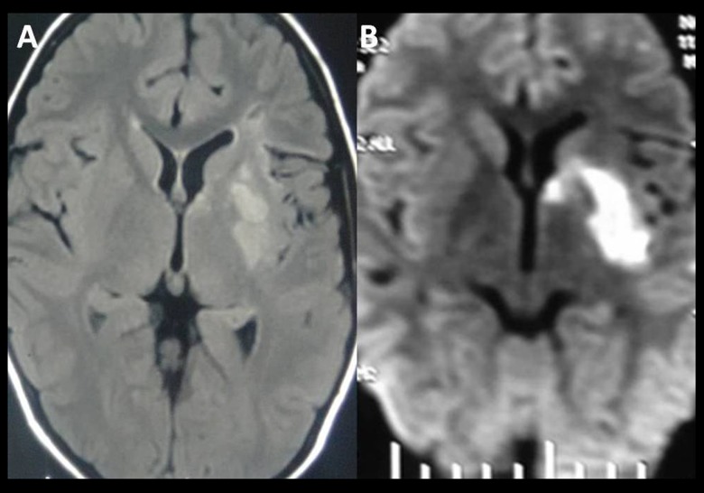
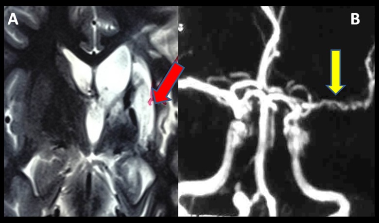
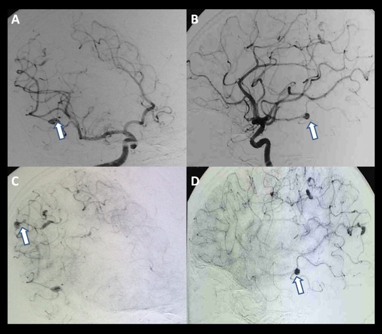
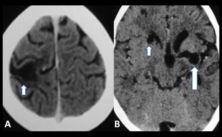
Case report: We report an 11-year- old boy with left atrial myxoma and multiple cerebral oncotic aneurysms. The characteristics of these aneurysms are indefinite and variable. The "Metastasize and Infiltrate" theory may be the key mechanism in the formation of these aneurysms.

Conclusions: Magnetic resonance imaging (MRI), computed tomography (CT) and angiography are useful in the diagnostics while digital subtraction angiography (DSA) is the best option. There are no definite guidelines for therapy of these aneurysms. Resection of cardiac myxomas, chemotherapy, radiotherapy, coil embolization and surgical treatment could be helpful.

Keywords: Cerebral Angiography; Heart Atria; Intracranial Aneurysm.


Figures

Figure 1
Figure 1
An 11-year-old boy left atrial myxoma with oncotic aneurysms; (A) FLAIR image of the brain shows acute infarct in the form of hyper-intensity in the left gangliocapsular region. (B) Diffusion weighted image shows restricted diffusion in the corresponding area.
Figure 2
Figure 2
An 11-year-old boy with left atrial myxoma and oncotic aneurysms; MR Angiogram (Time of flight sequence) shows complete cut-off of M1 segment of the left middle cerebral artery (MCA) (red arrow) and an aneurysm in the intradural segment of the right vertebral artery (yellow arrow).
Figure 3
Figure 3
An 11-year-old boy with left atrial myxoma and oncotic aneurysms; Follow-up MR, (A) shows gliotic area in the left gangliocapsular region (red arrow). (B) MR Angiogram (Time of flight sequence) shows partially recanalized, tortuous, narrowed left MCA (yellow arrow).
Figure 4
Figure 4
An 11-year-old boy with left atrial myxoma and oncotic aneurysms; Digital subtraction angiography (DSA), Left ICA angiogram shows – (A) irregular, narrowed and tortuous left MCA (small white arrow). (B) An aneurysm in the distal branch of left MCA (large white arrow).
Figure 5
Figure 5
An 11-year-old boy with left atrial myxoma and oncotic aneurysms; Digital subtraction angiography (DSA), Right ICA angiogram (A–D) – show multiple aneurysms in distal branches of the right middle cerebral artery (white arrows).
Figure 6
Figure 6
An 11-year-old boy with left atrial myxoma and oncotic aneurysms; Digital subtraction angiography (DSA), Right vertebral angiogram (A, B) – show an aneurysm in the intradural segment of the right vertebral artery before its junction with the basilar artery (arrow).
Figure 7
Figure 7
An 11-year-old boy with left atrial myxoma and oncotic aneurysms; One year after non-contrast computed tomography shows – (A) – infracts (not visible one year before) in the head of the right caudate nucleus (small white arrow) and (B) – right high parietal region (small white arrow). In addition (B) – also show an old infarct in the left gangliocapsular area (large white arrow).
Figure 8
Figure 8
An 11-year-old boy with left atrial myxoma and oncotic aneurysms; One year after CT angiogram shows – (A) – tortuous, dilated and fusiform left MCA (large white arrow). (B) show multiple aneurysms in the distal branches of bilateral middle cerebral and vertebral arteries (small white arrows).

References

    1. Xu Q, Zhang X, Wu P, et al. Multiple intracranial aneurysms followed left atrial myxoma: case report and literature review. J Thorac Dis. 2013;5(6):E227–31. - PMC - PubMed
    1. Pinede L, Duhaut P, Loire R. Clinical presentation of left atrial cardiac myxoma. A series of 112 consecutive cases. Medicine. 2001;80:159–72. - PubMed
    1. Lee VH, Connolly HM, Brown RD., Jr Central nervous system manifestations of cardiac myxoma. Arch Neurol. 2007;64:1115–20. - PubMed
    1. Sharma S, Tsyvine D, Maldjian PD, et al. An intriguing co-existence: atrial myxoma and cerebral cavernous malformations: case report and review of literature. J Am Soc Echocardiogr. 2011;24:110.e1–4. - PubMed
    1. Nucifora PG, Dillon WP. MR diagnosis of myxomatous aneurysms: report of two cases. Am J Neuroradiol. 2001;22:1349–52. - PMC - PubMed

Publication types

LinkOut - more resources