. 2015 Oct 19;6(11):4433-46.
doi: 10.1364/BOE.6.004433. eCollection 2015 Nov 1.

All-plastic, miniature, digital fluorescence microscope for three part white blood cell differential measurements at the point of care

Affiliations

All-plastic, miniature, digital fluorescence microscope for three part white blood cell differential measurements at the point of care

Alessandra Forcucci et al. Biomed Opt Express. .

Abstract

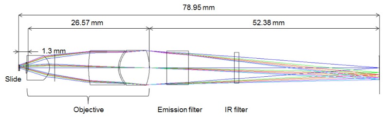
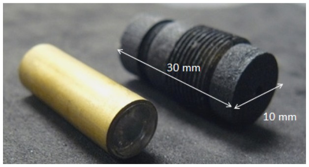
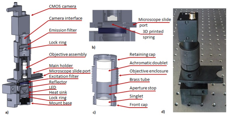
Three-part differential white blood cell counts are used for disease diagnosis and monitoring at the point-of-care. A low-cost, miniature achromatic microscope was fabricated for identification of lymphocytes, monocytes, and granulocytes in samples of whole blood stained with acridine orange. The microscope was manufactured using rapid prototyping techniques of diamond turning and 3D printing and is intended for use at the point-of-care in low-resource settings. The custom-designed microscope requires no manual adjustment between samples and was successfully able to classify three white blood cell types (lymphocytes, granulocytes, and monocytes) using samples of peripheral whole blood stained with acridine orange.

Keywords: (170.1470) Blood or tissue constituent monitoring; (170.2520) Fluorescence microscopy; (170.3880) Medical and biological imaging.


Figures

Fig. 1
Fig. 1
Optical schematic of the miniature achromatic objective.
Fig. 2
Fig. 2
Performance metrics of the miniature tunable fluorescent microscope for nominal working conditions: (a) modulation transfer function for expected performance at 525 nm, (b) modulation transfer function for expected performance at 650 nm, (c) spot diagram for expected performance at 525 nm (shown in blue) and 625 nm (shown in green). All plots are for image surface for design wavelength of 590nm, for following object points: 0.0mm, 0.25mm, 0.35mm, 0.45 mm, 0.50 mm, 0.55 mm, 0.6 mm. Airy disk radii for 525 nm and 650 nm: 4.179 and 5.126µm, respectively.
Fig. 3
Fig. 3
Assembled plastic objective inside brass tubing (left). 3D printed threaded objective holder (right).
Fig. 4
Fig. 4
Images of high resolution USAF target taken with the miniature 0.35 NA achromatic objective. (a) Image acquired at 525 nm, (b) Image acquired at 650 nm. Magnified inlays of the smallest resolvable elements are shown outlined in each image. Contrast has been enhanced for presentation purposes.
Fig. 5
Fig. 5
(a) Optomechanical schematic of the miniature WBC microscope, (b) cross-section through sample chamber with 3D printed microscope slide positioning springs, (c) details of the optomechanical assembly of the prototype of the WBC objective, (d) photograph of assembled 3D printed WBC microscope is shown on an optical bench. The spacing between the holes is 1 inch for reference.
Fig. 6
Fig. 6
(a) Schematic of measurement system used to evaluate sample insertion repeatability. (b) Slide insertion repeatability test: blue line – measured distance, black line – mean distance, red lines – ± 1σ standard deviation boarder lines.
Fig. 7
Fig. 7
(a) Image of acridine orange taken at 400 ms exposure stained white blood cells in whole blood. Image acquired with the miniature all-plastic objective. (b) Magnified image of granulocyte. (c) Magnified image of a lymphocyte. (d) Magnified image of a monocyte. Contrast has been enhanced for presentation purposes. Red-to-green ratios were calculated using raw data.
Fig. 8
Fig. 8
Cumulative percentage of samples falling within percent difference from gold standard measurement on AcT Diff2 hematology analyzer.

References

    1. Buttarello M., Plebani M., “Automated blood cell counts: state of the art,” Am. J. Clin. Pathol. 130(1), 104–116 (2008).10.1309/EK3C7CTDKNVPXVTN - DOI - PubMed
    1. Osei-Bimpong A., Jury C., McLean R., Lewis S. M., “Point-of-care method for total white cell count: an evaluation of the HemoCue WBC device,” Int. J. Lab. Hematol. 31(6), 657–664 (2009).10.1111/j.1751-553X.2008.01093.x - DOI - PMC - PubMed
    1. Shi W., Guo L., Kasdan H., Tai Y. C., “Four-part leukocyte differential count based on sheathless microflow cytometer and fluorescent dye assay,” Lab Chip 13(7), 1257–1265 (2013).10.1039/c3lc41059e - DOI - PubMed
    1. Charache S., Nelson L., Keyser E., Metzger P., “A Clinical Trial of Three-Part Electronic Differential White Blood Cell Counts,” Arch. Intern. Med. 145(10), 1852–1855 (1985).10.1001/archinte.1985.00360100114019 - DOI - PubMed
    1. Blumenreich M. S., Clinical Methods: The History, Physical, and Laboratory Examinations (Butterworths, 1990). - PubMed

LinkOut - more resources