. 2015 Aug 7;1(7):e1500391.
doi: 10.1126/sciadv.1500391. eCollection 2015 Aug.

Why do animal eyes have pupils of different shapes?

Affiliations

Why do animal eyes have pupils of different shapes?

Martin S Banks et al. Sci Adv. .

Abstract

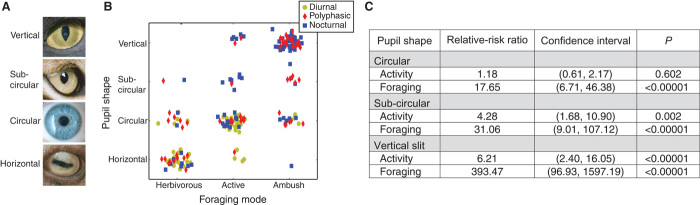
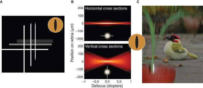
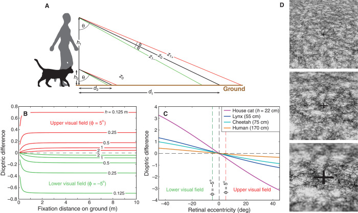
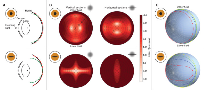
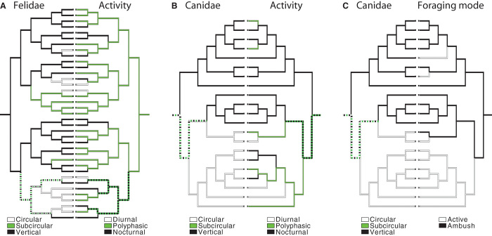
There is a striking correlation between terrestrial species' pupil shape and ecological niche (that is, foraging mode and time of day they are active). Species with vertically elongated pupils are very likely to be ambush predators and active day and night. Species with horizontally elongated pupils are very likely to be prey and to have laterally placed eyes. Vertically elongated pupils create astigmatic depth of field such that images of vertical contours nearer or farther than the distance to which the eye is focused are sharp, whereas images of horizontal contours at different distances are blurred. This is advantageous for ambush predators to use stereopsis to estimate distances of vertical contours and defocus blur to estimate distances of horizontal contours. Horizontally elongated pupils create sharp images of horizontal contours ahead and behind, creating a horizontally panoramic view that facilitates detection of predators from various directions and forward locomotion across uneven terrain.

Keywords: anatomy; aperture; blur; chromatic aberration; depth of field; evolution; eye; pupil; stereopsis.


Figures

Fig. 1
Fig. 1. Activity time, foraging mode, and pupil shape.
(A) Different pupil shapes. From top to bottom: vertical-slit pupil of the domestic cat, vertically elongated (subcircular) pupil of the lynx, circular pupil of man, and horizontal pupil of the domestic sheep. (B) Pupil shape as a function of foraging mode and diel activity. The axes are pupil shape [vertically elongated, subcircular (but elongated vertically), circular, or horizontally elongated] and foraging mode (herbivorous prey, active predator, or ambush predator). Each dot represents a species. Colors represent diel activity: yellow, red, and blue for diurnal, polyphasic, and nocturnal, respectively. The dots in each bin have been randomly offset to avoid overlap. (C) Results of statistical tests on the relationship between foraging, activity, and pupil shape. Multinomial logistic regression tests were conducted with foraging mode, activity time, and pupil shape as factors and genus as a covariate. Relative-risk ratios were computed for having a circular, subcircular, or vertical-slit pupil relative to having a horizontal pupil as a function of foraging mode or diel activity. Activity time proceeded from diurnal to polyphasic to nocturnal. Foraging mode proceeded from herbivorous prey to active predator to ambush predator. When the relative-risk ratio is greater than 1, the directional change in the independent variable (foraging or activity) was associated with a greater probability of having the specified pupil shape than a horizontal pupil.
Fig. 2
Fig. 2. Image quality for different amounts of defocus and pupil shapes.
(A) Astigmatic depth of field with vertical-slit pupil (12 × 1.5 mm). Three crosses are presented at different distances (0D, 0.4D, and 0.8D). The camera is focused on the nearest cross, so the other two are farther than the focal plane. The vertical limbs of all three crosses are relatively sharp, whereas the horizontal limbs of the two farther crosses are quite blurred. (B) Horizontal and vertical cross sections of point spread functions (PSFs) as a function of focal distance for an eye with a vertical-slit pupil (12 × 1.5 mm). The object was white. The PSFs incorporate diffraction and chromatic aberration. Log intensity in the PSF is represented by brightness (brighter corresponding to higher amplitude). Intensities lower than 10−3 of the peak amplitude have been clipped. The upper panel shows horizontal cross sections (relevant for imaging vertical contours). The icon in the lower middle of the panel represents the cross sections by a nominal PSF with a horizontal cut through it. The lower panel shows vertical cross sections (for imaging horizontals). The icon in the lower middle of the panel represents those cross sections. The dashed white lines are from Eqs. 3 and 4 and show that the equations are a good approximation to the PSF cross sections. (C) Photograph of a depth-varying scene taken with a camera with a vertical-slit aperture. The camera was focused on the toy bird, so objects nearer and farther are blurred, but more vertically than horizontally because of the aperture elongation. Movie S2 shows PSF cross sections and the scene as the aperture rotates from vertical to horizontal and back to vertical.
Fig. 3
Fig. 3. Height and defocus.
(A) Two viewers—human and domestic cat—with different eye heights, h1 and h2, fixate the ground. Fixation direction relative to earth vertical is θ. Fixation distances along the ground are d1 and d2, and distances along the lines of sight are z0. The eyes are focused at z0, so points above and below the fixation point are defocused. (B) Defocus (difference in dioptric distances: 1/z0 − 1/z1+ and 1/z0 − 1/z1−) as a function of fixation distance along the ground. Red and green curves correspond to the defocus 5° above and below fixation, respectively (ϕ = ±5°). Different curves represent different eye heights. How does pupil size vary with eye height? In vertebrates, AM0.196, where A is axial length and M is body mass (26). In quadrapeds, LM0.40, where L is limb length, an excellent proxy for eye height (27). Combining those equations, AL0.49, which means that axial length is proportional to the square root of eye height. Under the assumption that pupil size is proportional to eye size, the analysis shows that the defocus signal is indeed weaker in taller animals. (C) Defocus (difference in dioptric distances) for different vertical eccentricities. The viewer is fixating the ground. Different curves represent animals of different heights. The eccentricities corresponding to ϕ = ±5° are represented by dashed vertical lines. Because defocus in (B) is nearly independent of fixation distance, we represent the relationship between defocus and retinal eccentricity with one curve for each eye height. (D) Images of the ground for viewers of different heights. A virtual camera with a field of view of 30° and an aperture diameter of 4.5 mm was aimed toward a plane with θ = 56°. The camera was focused on the black cross at distance z0. From top to bottom, z0 was 0.6, 0.2, and 0.1 m (1.7D, 5D, and 10D, respectively).
Fig. 4
Fig. 4. Pupil shape and image quality in the model sheep eye.
(A) Schematic sheep eyes viewed from above. The upper plot is for a circular pupil and the lower plot for a horizontally elongated pupil with the same area. The black curves represent, from left to right, the anterior and posterior surfaces of the cornea (radius 11.66 and 13 mm, thickness 0.8 mm, refractive index 1.382), the anterior and posterior surfaces of the lens (radius 9.17 and −8.12 mm, thickness 9 mm, refractive index 1.516), and the retina (radius 12 mm). The red and green dashed curves respectively represent the focal surfaces for vertical and horizontal contours. (B) Widths of sections through the PSF for different pupils and retinal positions. The upper and lower plots were computed with circular (2.8 × 2.8 mm) and horizontally elongated (8 × 1 mm) pupils, respectively. The optic axis is in the center of each circular plot. Black concentric dashed circles represent different eccentricities. Colors correspond to the SD of the PSF (a measure of the spread of the PSF cross section) for vertical (left) and horizontal cross sections (right); lighter red corresponds to the smallest SD (that is, the sharpest image) and darker red corresponds to the largest SD (least sharp image). (C) Throughput for circular and horizontal pupils. The contour lines represent regions of constant throughput: red, blue, green, and yellow for 80, 60, 40, and 20%, respectively.
Fig. 5
Fig. 5. Ancestral reconstruction of pupil shape, activity, and foraging mode for Felidae and Canidae using parsimony.
Line colors indicate estimated state at each branch. Dashed lines indicate uncertain states; the two colors composing the dash indicate the two possible states. Pupil shape is indicated by the cladogram on the left in each panel. Activity or foraging mode is indicated by the cladogram on the right in each panel. (A) Changes in pupil shape compared to changes in activity time for Felidae. (B) Changes in pupil shape compared to changes in activity time for Canidae. (C) Changes in pupil shape compared to changes in foraging mode for Canidae. Comparison of changes in pupil shape to those in foraging mode for Felidae was omitted because of the lack of variation in foraging mode among the species.

References

    1. G. L. Walls, The Vertebrate Eye and Its Adaptive Radiation (Hafner, New York, 1942).
    1. Detwiler S. R., The eye and its structural adaptations. Proc. Am. Philos. Soc. 99, 224–238 (1955).
    1. Hammond P., Mouat G. S. V., The relationship between feline pupil size and luminance. Exp. Brain Res. 59, 485–490 (1985). - PubMed
    1. Wilcox J. G., Barlow H. B., The size and shape of the pupil in lightly anaesthetized cats as a function of luminance. Vision Res. 15, 1363–1365 (1975). - PubMed
    1. Roth L. S. V., Lundström L., Kelber A., Kröger R. H. H., Unsbo P., The pupils and optical systems of gecko eyes. J. Vis. 9, 27.1–27.11 (2009). - PubMed