. 2015 Sep 4;1(8):e1500503.
doi: 10.1126/sciadv.1500503. eCollection 2015 Sep.

Cis-regulatory mechanisms governing stem and progenitor cell transitions

Affiliations

Cis-regulatory mechanisms governing stem and progenitor cell transitions

Kirby D Johnson et al. Sci Adv. .

Abstract

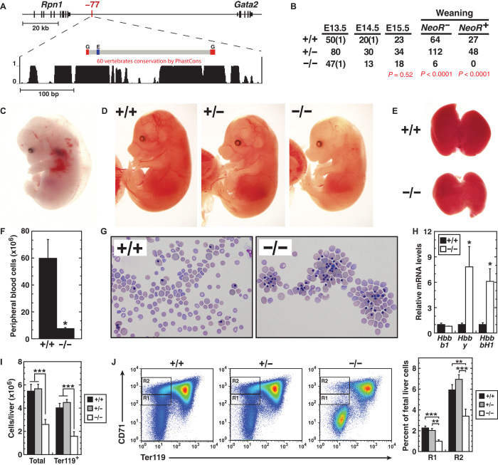
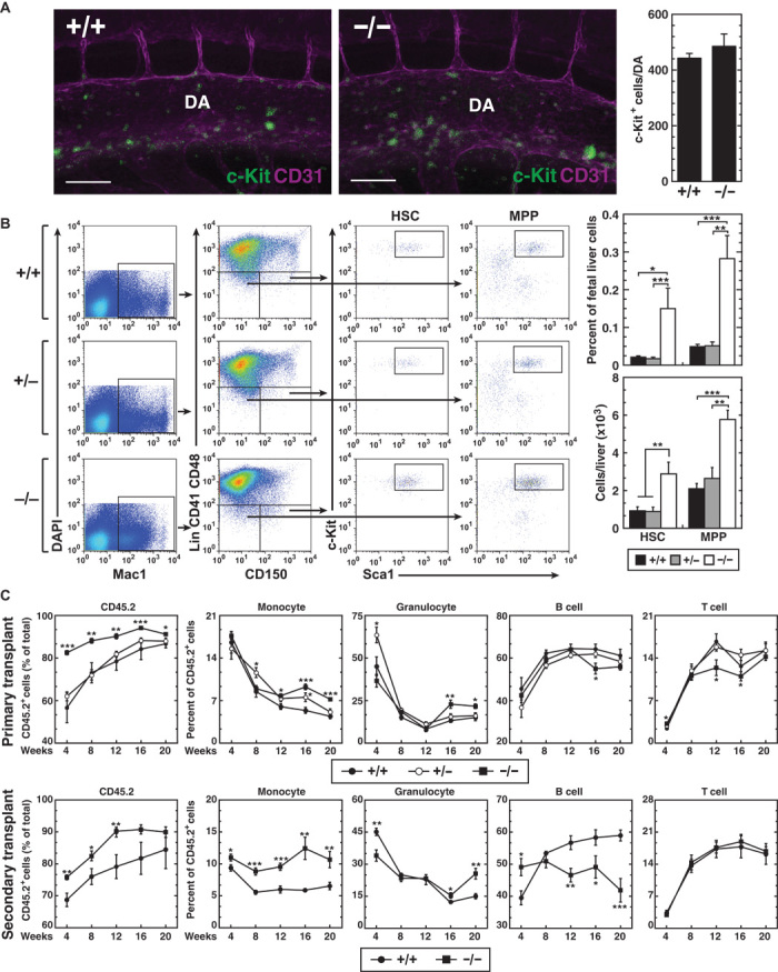
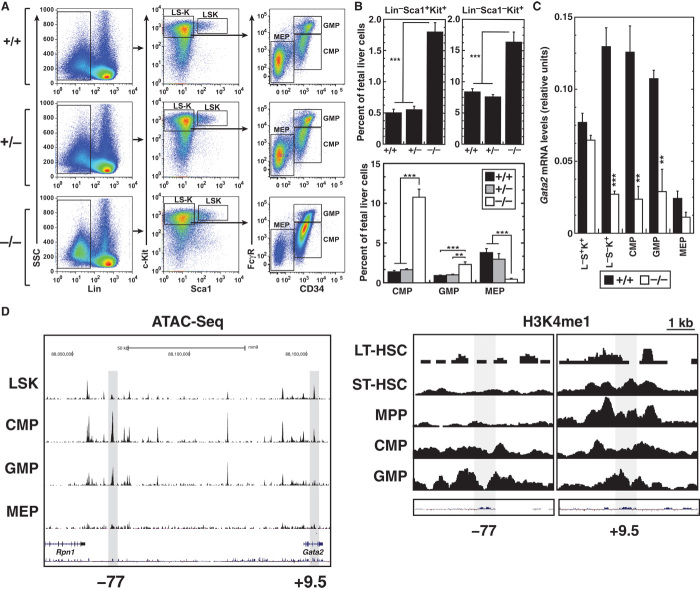
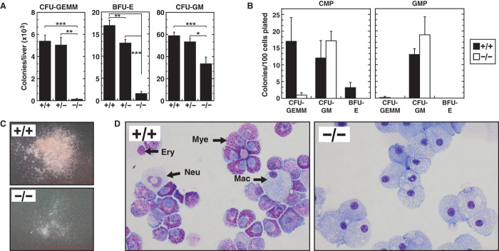
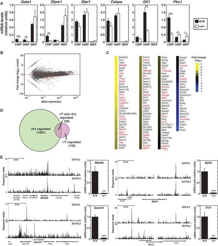
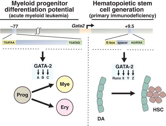
Cis-element encyclopedias provide information on phenotypic diversity and disease mechanisms. Although cis-element polymorphisms and mutations are instructive, deciphering function remains challenging. Mutation of an intronic GATA motif (+9.5) in GATA2, encoding a master regulator of hematopoiesis, underlies an immunodeficiency associated with myelodysplastic syndrome (MDS) and acute myeloid leukemia (AML). Whereas an inversion relocalizes another GATA2 cis-element (-77) to the proto-oncogene EVI1, inducing EVI1 expression and AML, whether this reflects ectopic or physiological activity is unknown. We describe a mouse strain that decouples -77 function from proto-oncogene deregulation. The -77(-/-) mice exhibited a novel phenotypic constellation including late embryonic lethality and anemia. The -77 established a vital sector of the myeloid progenitor transcriptome, conferring multipotentiality. Unlike the +9.5(-/-) embryos, hematopoietic stem cell genesis was unaffected in -77(-/-) embryos. These results illustrate a paradigm in which cis-elements in a locus differentially control stem and progenitor cell transitions, and therefore the individual cis-element alterations cause unique and overlapping disease phenotypes.

Keywords: GATA factor; GATA-2; cis-element; differentiation; hematopoiesis; myeloid; progenitor; stem cell; transcriptome.


Figures

Fig. 1
Fig. 1. −77 leukemogenic, long-range enhancer is essential for embryogenesis and hematopoiesis.
(A) Vertebrate conservation plot of −77 showing positions of GATA motifs (G; WGATAR) and E-box (E; CANNTG) within the deleted region. (B) Genotypes of −77+/+, −77+/−, and −77−/− NeoR- embryos at timed developmental stages and genotypes of NeoR and NeoR+ pups at time of weaning. (C) Representative E13.5 +9.5−/− embryo exhibiting anemia, hemorrhage, and edema. (D) Representative E15.5 −77+/+, −77+/−, and −77−/− littermates. (E) Representative E15.5 fetal livers from −77+/+ and −77−/− littermates. (F and G) E15.5 peripheral blood quantitation [−77+/+ (n = 3) and −77−/− (n = 3)] and representative Wright-Giemsa staining. (H) Relative expression of fetal and adult β-globin mRNA in peripheral blood at E15.5. (I) Total cells [−77+/+ (n = 10), −77+/− (n = 24), and −77−/− (n = 13)] and Ter119+ cells [−77+/+ (n = 7), −77+/− (n = 17), and −77−/− (n = 5)] in E13.5 fetal livers. (J) Representative flow cytometric analysis and quantitation of E13.5 fetal livers for CD71+Ter119 R1 and R2 erythroid progenitors [−77+/+ (n = 7), −77+/− (n = 17), and −77−/− (n = 5)]. Graphs show means ± SEM; *P < 0.05, **P < 0.01, ***P < 0.001.
Fig. 2
Fig. 2. Long-term repopulating HSC generation and function do not require −77.
(A) Whole-mount immunostaining of E10.5 −77+/+ and −77−/− embryos showing CD31+ cells (magenta) and c-Kit+ cells (green) within the dorsal aorta (DA). Scale bars, 100 μm. Quantitation of c-Kit+ cells within the whole dorsal aorta [−77+/+ (n = 3) and −77−/− (n = 3)] is shown on the right. (B) Representative flow cytometric analysis of E13.5 fetal livers for HSCs (LinMac1+CD41CD48CD150+Sca1+Kit+) and MPPs (LinMac1+CD41CD48CD150Sca1+Kit+). Quantitation of HSCs and MPPs is presented both as a percentage of total fetal liver cells (top) and as the number of cells per liver (bottom) [−77+/+ (n = 5), −77+/− (n = 6), and −77−/− (n = 4)]. (C) Contribution of −77+/− and −77−/− versus −77+/+ fetal liver cells in a competitive transplantation assay [−77+/+ (4 livers; 10 recipients), −77+/− (6 livers; 12 recipients), and −77−/− (3 livers; 11 recipients)]. After 20 weeks, secondary transplants were performed with bone marrow from fetal liver–transplanted −77−/− versus −77+/+ mice (2 bone marrow donors for each genotype; 8 recipient mice). The peripheral blood of recipient mice was analyzed for CD45.2 expression by flow cytometry at 4-week intervals after transplantation. Flow cytometric analysis of the proportions of CD45.2+ monocytes (Mac1+Gr), granulocytes (Mac1+Gr+), B cells (CD19+), and T cells (Thy1.2+) in the peripheral blood of recipient mice. Graphs show means ± SEM; *P < 0.05, **P < 0.01, ***P < 0.001.
Fig. 3
Fig. 3. Selective loss of Gata2 expression in −77−/− myeloid progenitors disrupts homeostasis.
(A) Representative flow cytometric analysis of E13.5 fetal livers for LinSca1+Kit+ and LinSca1Kit+ cells, CMPs (LinCD34+FcγRlowKit+Sca1), GMPs (LinCD34+FcγRhighKit+Sca1), and MEPs (LinCD34FcγRlowKit+Sca1). (B) Percentages of LinSca1+Kit+ and LinSca1Kit+ cells [top; −77+/+ (n = 7), −77+/− (n = 17), and −77−/− (n = 6)] and CMPs, GMPs, and MEPs [bottom; −77+/+ (n = 8), −77+/− (n = 6), and −77−/− (n = 9)]. (C) Quantitation of Gata2 mRNA levels in LinSca1+Kit+ and LinSca1Kit+ cells [−77+/+ (n = 5) and −77−/− (n = 7)] and CMPs, GMPs, and MEPs [−77+/+ (n = 4) and −77−/− (n = 4)]. (D) Profiles for chromatin accessibility via the assay for transposase-accessible chromatin (ATAC) and histone H3K4 monomethylation mined from existing data (53). Graphs show means ± SEM; **P < 0.01, ***P < 0.001.
Fig. 4
Fig. 4. Limited differentiation potential of −77−/− fetal liver progenitors.
(A) Colony-forming activity of hematopoietic progenitors from E14.5 fetal livers [−77+/+ (n = 12), −77+/− (n = 17), and −77−/− (n = 3)]. (B) Colony-forming activity of CMPs and GMPs sorted from E13.5 fetal livers [−77+/+ (n = 3) and −77−/− (n = 3)]. (C and D) Representative colonies and Wright-Giemsa–stained cells obtained from plating of CMPs. Scale bars, 2 mm. Mac, macrophage; Ery, erythroblast; Neu, neutrophil; Mye, myeloid. Graphs show means ± SEM; *P < 0.05, **P < 0.01, ***P < 0.001.
Fig. 5
Fig. 5. −77 establishes a functionally vital sector of the myeloid progenitor cell transcriptome.
(A) Quantitative gene expression analysis in CMPs, GMPs, and MEPs from −77+/+ and 77−/− E13.5 fetal livers [−77+/+ (n = 4) and −77−/− (n = 4)]. (B) MA plot of RNA-seq–based comparison of CMP transcriptomes from −77+/+ and −77−/− E13.5 fetal livers [−77+/+ (n = 3) and −77−/− (n = 3)]. Red points indicate down- or up-regulated genes [false discovery rate (FDR) <0.05]. (C) Heatmap depicting statistically significant genes down- or up-regulated by >1.5-fold. (D) Venn diagram depicting the extent of overlap between genes regulated by +9.5 in AGM (13) and −77 in CMPs. Genes shared between data sets are indicated in red in (C). (E) Evidence for direct GATA-2 regulation of −77 target genes. ChIP-seq profiles of GATA-2 and Scl/TAL1 in HPC-7 cells (58) and quantitative gene expression analysis in CMPs from −77+/+ and −77−/− E13.5 fetal livers [−77+/+ (n = 4) and −77−/− (n = 4)]. Graphs show means ± SEM; *P < 0.05, **P < 0.01, ***P < 0.001.
Fig. 6
Fig. 6
Selective control of stem and progenitor cell transitions by distinct cis-elements within a genetic locus. The model depicts differential +9.5 and −77 functions to control critical stages of hematopoiesis. Disruption of −77 and +9.5 in humans causes acute myeloid leukemia and primary immunodeficiency, respectively. Whereas both enhancers are essential for GATA-2 regulation, they operate in distinct cellular contexts, and genomic analyses indicate major differences in their target gene ensembles. Prog, myeloerythroid progenitor; Mye, myeloid progenitor; Ery, erythroid; DA, dorsal aorta.

References

    1. Yue F., Cheng Y., Breschi A., Vierstra J., Wu W., Ryba T., Sandstrom R., Ma Z., Davis C., Pope B. D., Shen Y., Pervouchine D. D., Djebali S., Thurman R. E., Kaul R., Rynes E., Kirilusha A., Marinov G. K., Williams B. A., Trout D., Amrhein H., Fisher-Aylor K., Antoshechkin I., DeSalvo G., See L.-H., Fastuca M., Drenkow J., Zaleski C., Dobin A., Prieto P., Lagarde J., Bussotti G., Tanzer A., Denas O., Li K., Bender M. A., Zhang M., Byron R., Groudine M. T., McCleary D., Pham L., Ye Z., Kuan S., Edsall L., Wu Y.-C., Rasmussen M. D., Bansal M. S., Kellis M., Keller C. A., Morrissey C. S., Mishra T., Jain D., Dogan N., Harris R. S., Cayting P., Kawli T., Boyle A. P., Euskirchen G., Kundaje A., Lin S., Lin Y., Jansen C., Malladi V. S., Cline M. S., Erickson D. T., Kirkup V. M., Learned K., Sloan C. A., Rosenbloom K. R., Lacerda de Sousa B., Beal K., Pignatelli M., Flicek P., Lian J., Kahveci T., Lee D., Kent W. J., Santos M. R., Herrero J., Notredame C., Johnson A., Vong S., Lee K., Bates D., Neri F., Diegel M., Canfield T., Sabo P. J., Wilken M. S., Reh T. A., Giste E., Shafer A., Kutyavin T., Haugen E., Dunn D., Reynolds A. P., Neph S., Humbert R., Scott Hansen R., De Bruijn M., Selleri L., Rudensky A., Josefowicz S., Samstein R., Eichler E. E., Orkin S. H., Levasseur D., Papayannopoulou T., Chang K.-H., Skoultchi A., Gosh S., Disteche C., Treuting P., Wang Y., Weiss M. J., Blobel G. A., Cao X., Zhong S., Wang T., Good P. J., Lowdon R. F., Adams L. B., Zhou X.-Q., Pazin M. J., Feingold E. A., Wold B., Taylor J., Mortazavi A., Weissman S. M., Stamatoyannopoulos J. A., Snyder M. P., Guigo R., Gingeras T. R., Gilbert D. M., Hardison R. C., Beer M. A., Ren, B. The Mouse ENCODE Consortium , A comparative encyclopedia of DNA elements in the mouse genome. Nature 515, 355–364 (2014). - PMC - PubMed
    1. Kellis M., Wold B., Snyder M. P., Bernstein B. E., Kundaje A., Marinov G. K., Ward L. D., Birney E., Crawford G. E., Dekker J., Dunham I., Elnitski L. L., Farnham P. J., Feingold E. A., Gerstein M., Giddings M. C., Gilbert D. M., Gingeras T. R., Green E. D., Guigo R., Hubbard T., Kent J., Lieb J. D., Myers R. M., Pazin M. J., Ren B., Stamatoyannopoulos J. A., Weng Z., White K. P., Hardison R. C., Defining functional DNA elements in the human genome. Proc. Natl. Acad. Sci. U.S.A. 111, 6131–6138 (2014). - PMC - PubMed
    1. Tsai F. Y., Keller G., Kuo F. C., Weiss M., Chen J., Rosenblatt M., Alt F. W., Orkin S. H., An early haematopoietic defect in mice lacking the transcription factor GATA-2. Nature 371, 221–226 (1994). - PubMed
    1. Tsai F.-Y., Orkin S. H., Transcription factor GATA-2 is required for proliferation/survival of early hematopoietic cells and mast cell formation, but not for erythroid and myeloid terminal differentiation. Blood 89, 3636–3643 (1997). - PubMed
    1. de Pater E., Kaimakis P., Vink C. S., Yokomizo T., Yamada-Inagawa T., van der Liden R., Kartalaei P. S., Camper S. A., Speck N., Dzierzak E., Gata2 is required for HSC generation and survival. J. Exp. Med. 210, 2843–2850 (2013). - PMC - PubMed