. 2015 Oct 23;1(9):e1500758.
doi: 10.1126/sciadv.1500758. eCollection 2015 Oct.

Three-dimensional printing of complex biological structures by freeform reversible embedding of suspended hydrogels

Affiliations

Three-dimensional printing of complex biological structures by freeform reversible embedding of suspended hydrogels

Thomas J Hinton et al. Sci Adv. .

Abstract

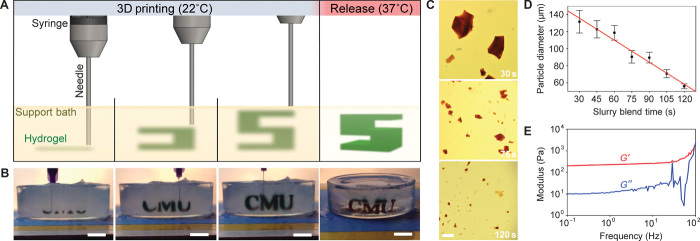
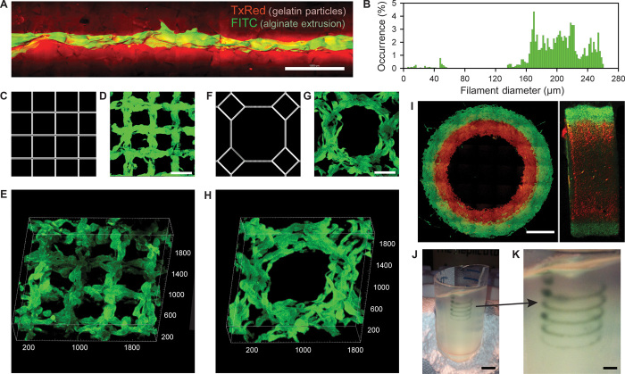
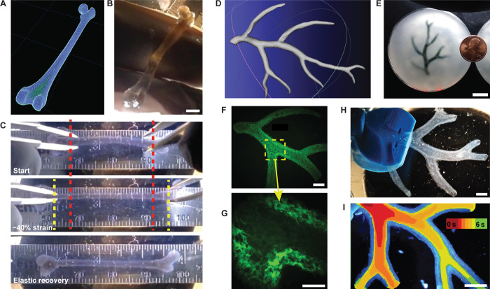
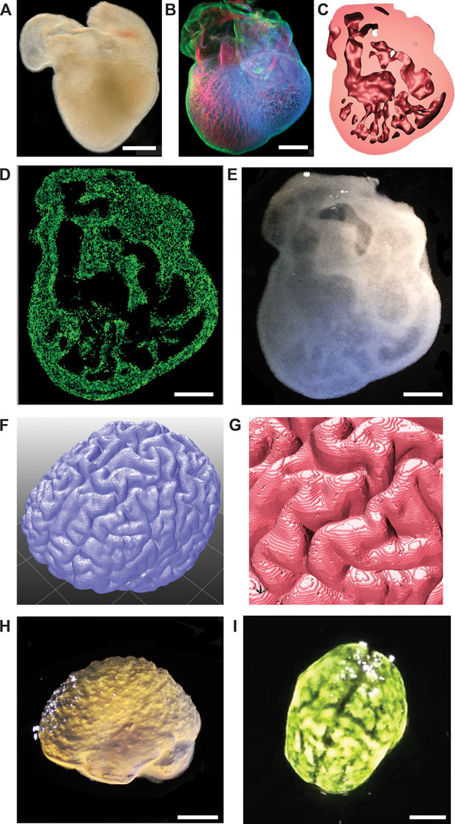
We demonstrate the additive manufacturing of complex three-dimensional (3D) biological structures using soft protein and polysaccharide hydrogels that are challenging or impossible to create using traditional fabrication approaches. These structures are built by embedding the printed hydrogel within a secondary hydrogel that serves as a temporary, thermoreversible, and biocompatible support. This process, termed freeform reversible embedding of suspended hydrogels, enables 3D printing of hydrated materials with an elastic modulus <500 kPa including alginate, collagen, and fibrin. Computer-aided design models of 3D optical, computed tomography, and magnetic resonance imaging data were 3D printed at a resolution of ~200 μm and at low cost by leveraging open-source hardware and software tools. Proof-of-concept structures based on femurs, branched coronary arteries, trabeculated embryonic hearts, and human brains were mechanically robust and recreated complex 3D internal and external anatomical architectures.

Keywords: 3D printing; alginate; biomimetic; collagen; fibrin; heart; hydrogels; perfusable vasculature; tissue engineering.


Figures

Fig. 1
Fig. 1. FRESH printing is performed by depositing a hydrogel precursor ink within the thermoreversible support bath consisting of gelatin microparticles and initiating gelling in situ through one of multiple cross-linking mechanisms.
(A) A schematic of the FRESH process showing the hydrogel (green) being extruded and cross-linked within the gelatin slurry support bath (yellow). The 3D object is built layer by layer and, when completed, is released by heating to 37°C and melting the gelatin. (B) Images of the letters “CMU” FRESH printed in alginate in Times New Roman font (black) and released by melting the gelatin support (gray material in the petri dish). When the gelatin support melts the change in optical properties, convective currents and diffusion of black dye out of the alginate make it appear that the letters are deforming, although they are not. (C) Representative images of gelatin particles produced by blending for 30, 75, or 120 s. (D) The mean Feret diameter of gelatin particles as a function of blending time from 30 to 120 s (n > 1000 per time point; the red line is a linear fit and error bars indicate SD). (E) Rheological analysis of storage (G′) and loss (G″) modulus for gelatin support bath showing Bingham plastic behavior. Scale bars, 1 cm (B) and 1 mm (C).
Fig. 2
Fig. 2. Analysis of the hydrogel filaments and structures fabricated using FRESH.
(A) A representative alginate filament (green) embedded within the gelatin slurry support bath (red). (B) Histogram of the diameter of isolated alginate filaments within the gelatin support bath showing a range from 160 to 260 μm. (C to E) A standard square lattice pattern commonly used for infill in 3D printing FRESH printed in fluorescent alginate (green) and viewed (D) top down and (E) in 3D. (F to H) An octagonal infill pattern FRESH printed in fluorescent alginate (green) and viewed (G) top down and (H) in 3D. (I) Example of a two-material print of coaxial cylinders in red and green fluorescently labeled alginate with a continuous interface shown in top down and lateral cross sections. (J) An example of a freeform, nonplanar FRESH print of a helix shown embedded in the gelatin support bath. (K) A zoomed-in view of the helix demonstrating that FRESH can print in true freeform and is not limited to standard layer-by-layer planar fabrication. Scale bars, 1 mm (A), 500 μm (D and G), 2 mm (I), 10 mm (J), and 2.5 mm (K).
Fig. 3
Fig. 3. FRESH printing of biological structures based on 3D imaging data and functional analysis of the printed parts.
(A) A model of a human femur from 3D CT imaging data is scaled down and processed into machine code for FRESH printing. (B) The femur is FRESH printed in alginate, and after removal from the support bath, it closely resembles the model and is easily handled. (C) Uniaxial tensile testing of the printed femur demonstrates the ability to be strained up to 40% and elastically recover. (D) A model of a section of a human right coronary arterial tree from 3D MRI is processed at full scale into machine code for FRESH printing. (E) An example of the arterial tree printed in alginate (black) and embedded in the gelatin slurry support bath. (F) A section of the arterial trees printed in fluorescent alginate (green) and imaged in 3D to show the hollow lumen and multiple bifurcations. (G) A zoomed-in view of the arterial tree shows the defined vessel wall that is <1 mm thick and the well-formed lumen. (H) A dark-field image of the arterial tree mounted in a perfusion fixture to position a syringe in the root of the tree. (I) A time-lapse image of black dye perfused through the arterial tree false-colored at time points of 0 to 6 s to show flow through the lumen and not through the vessel wall. Scale bars, 4 mm (B), 10 mm (E), 2.5 mm (F), 1 mm (G), and 2.5 mm (H and I).
Fig. 4
Fig. 4. FRESH printed scaffolds with complex internal and external architectures based on 3D imaging data from whole organs.
(A) A dark-field image of an explanted embryonic chick heart. (B) A 3D image of the 5-day-old embryonic chick heart stained for fibronectin (green), nuclei (blue), and F-actin (red) and imaged with a confocal microscope. (C) A cross section of the 3D CAD model of the embryonic heart with complex internal trabeculation based on the confocal imaging data. (D) A cross section of the 3D printed heart in fluorescent alginate (green) showing recreation of the internal trabecular structure from the CAD model. The heart has been scaled up by a factor of 10 to match the resolution of the printer. (E) A dark-field image of the 3D printed heart with internal structure visible through the translucent heart wall. (F) A 3D rendering of a human brain from MRI data processed for FRESH printing. (G) A zoomed-in view of the 3D brain model showing the complex, external architecture of the white matter folds. (H) A lateral view of the brain 3D printed in alginate showing major anatomical features including the cortex and cerebellum. The brain has been scaled down to ~3 mm in length to reduce printing time and test the resolution limits of the printer. (I) A top down view of the 3D printed brain with black dye dripped on top to help visualize the white matter folds printed in high fidelity. Scale bars, 1 mm (A and B) and 1 cm (D, E, H, and I).

References

    1. Shah A. M., Jung H., Skirboll S., Materials used in cranioplasty: A history and analysis. Neurosurg. Focus 36, E19 (2014). - PubMed
    1. FDA 510(k) summary statement for Osteofab Patient Specific Cranial Device. Report Number K121818, Center for Devices and Radiological Health (2013), http://www.accessdata.fda.gov/cdrh_docs/pdf12/K121818.pdf (viewed 10/11/15).
    1. Zopf D. A., Hollister S. J., Nelson M. E., Ohye R. G., Green G. E., Bioresorbable airway splint created with a three-dimensional printer. N. Engl. J. Med. 368, 2043–2045 (2013). - PubMed
    1. Bhatia S. K., Sharma S., 3D-printed prosthetics roll off the presses. Chem. Eng. Prog. 110, 28–33 (2014), www.aiche.org/sites/default/files/cep/20140528.pdf (viewed 10/12/15).
    1. Tumbleston J. R., Shirvanyants D., Ermoshkin N., Janusziewicz R., Johnson A. R., Kelly D., Chen K., Pinschmidt R., Rolland J. P., Ermoshkin A., Samulski E. T., DeSimone J. M., Additive manufacturing. Continuous liquid interface production of 3D objects. Science 347, 1349–1352 (2015). - PubMed