Review
. 2016 Jan;17(1):16-29.
doi: 10.1038/nrm.2015.5. Epub 2015 Nov 25.

The molecular basis for centromere identity and function

Affiliations
Review

The molecular basis for centromere identity and function

Kara L McKinley et al. Nat Rev Mol Cell Biol. 2016 Jan.

Abstract

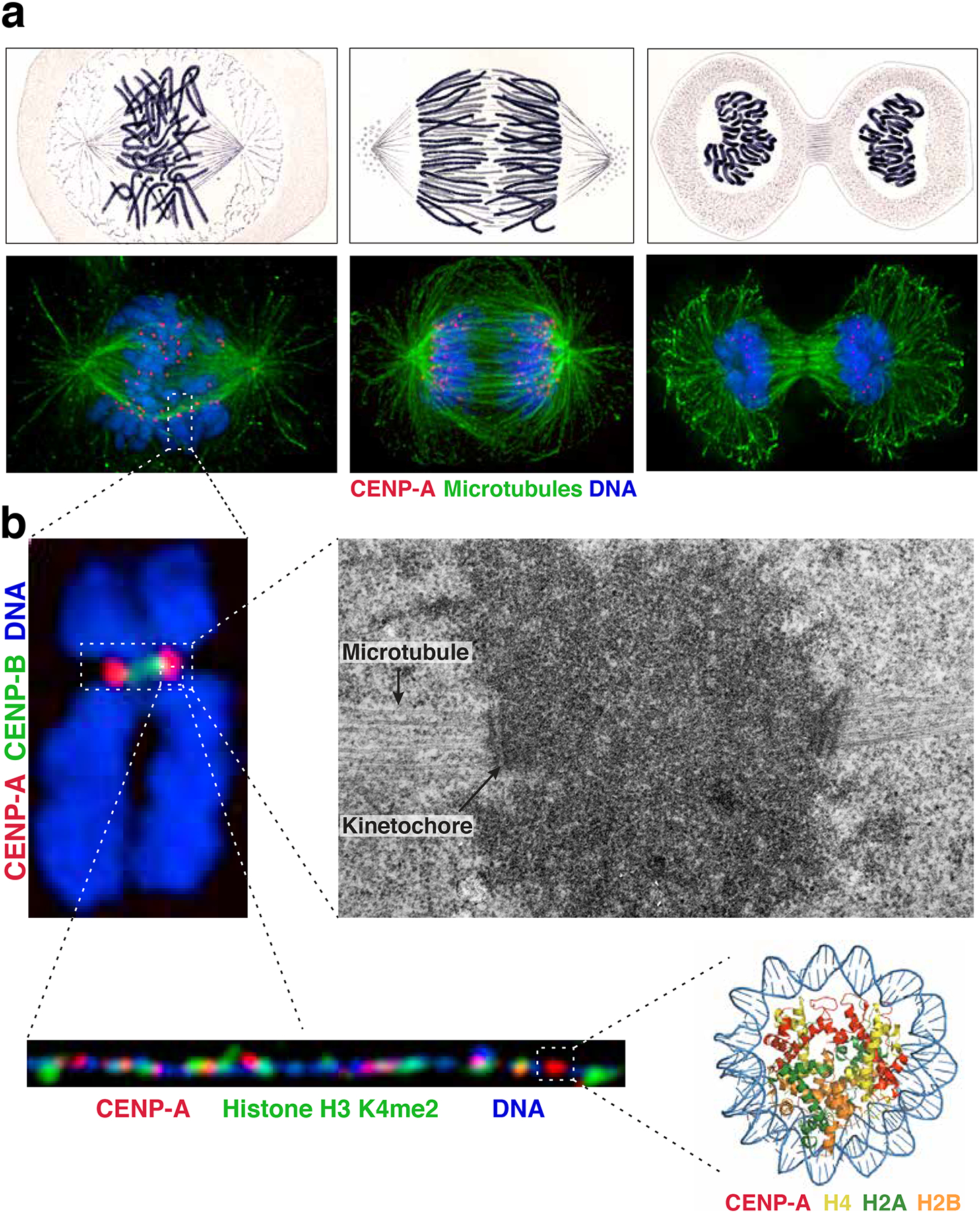
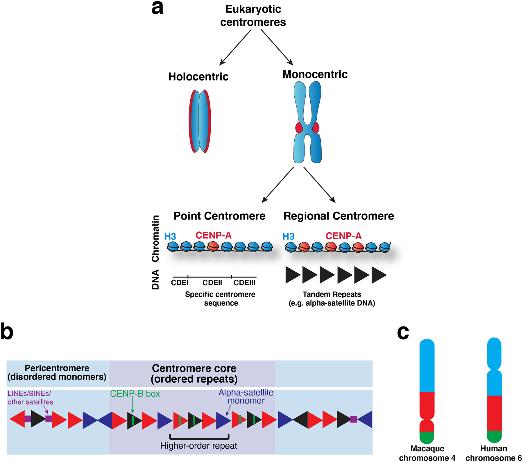
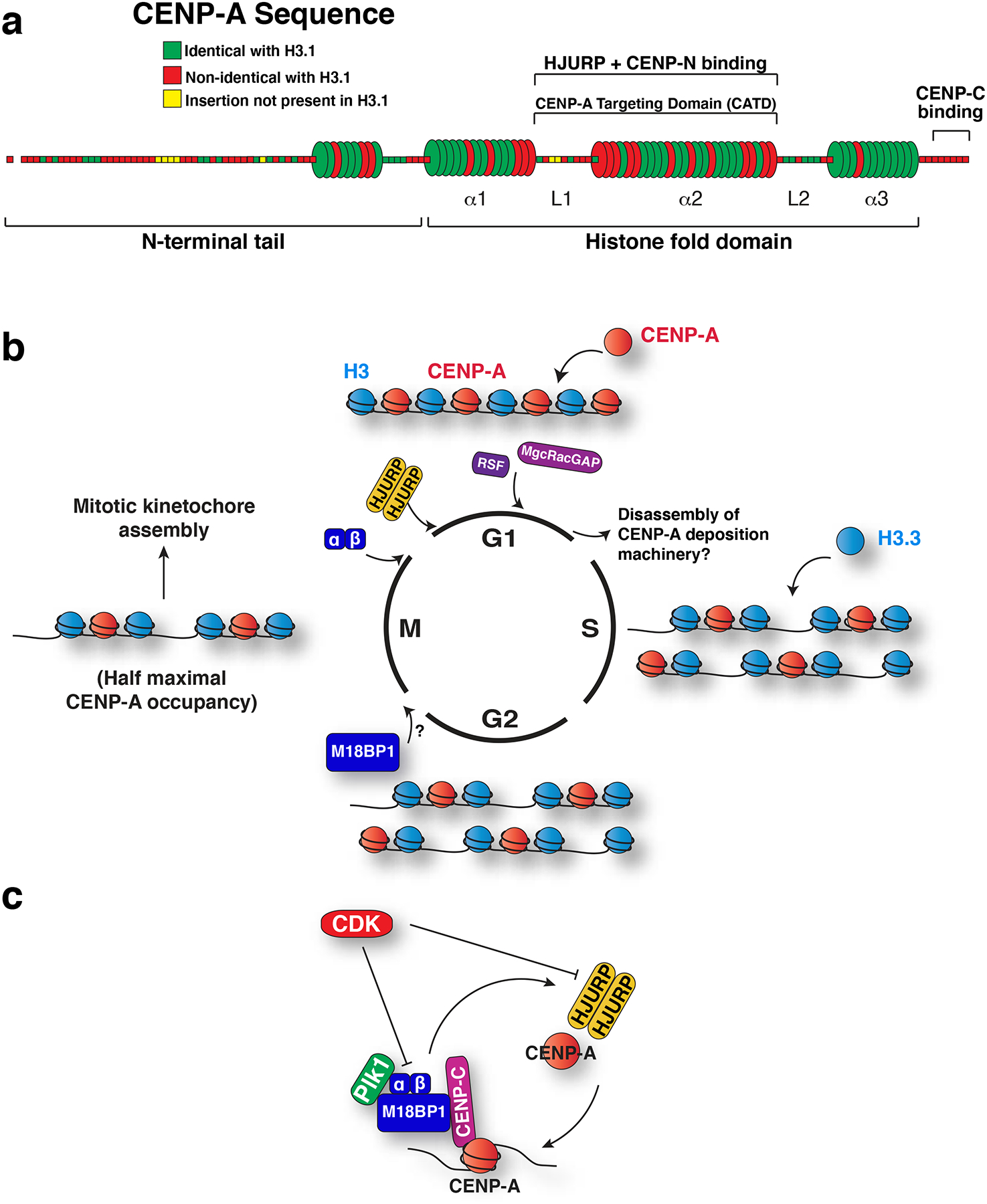
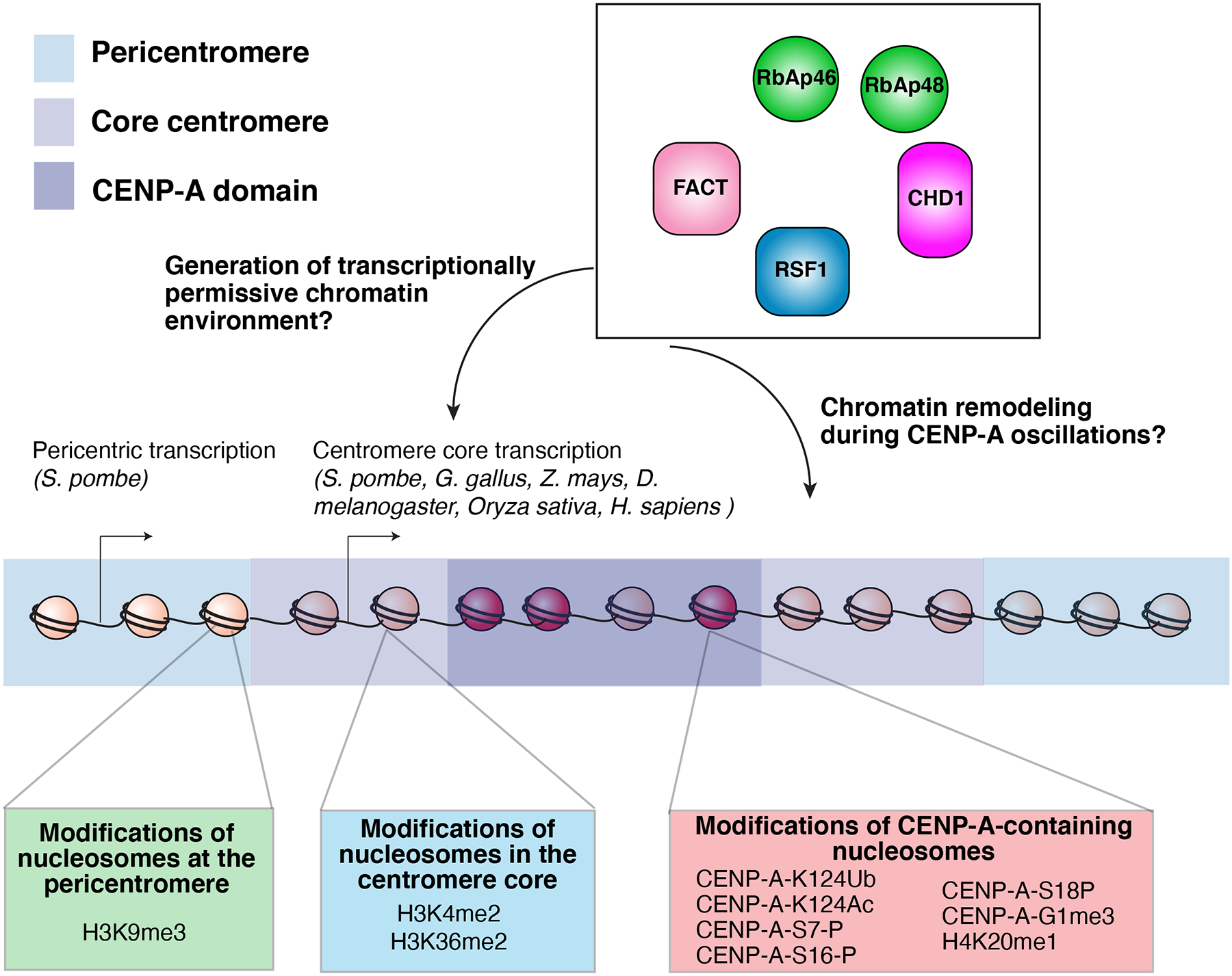
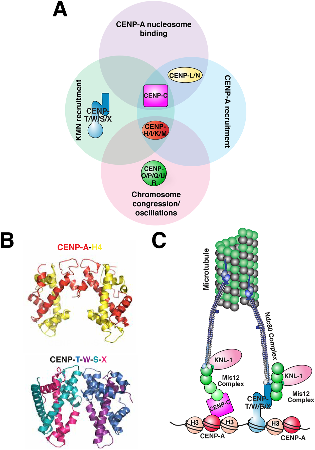
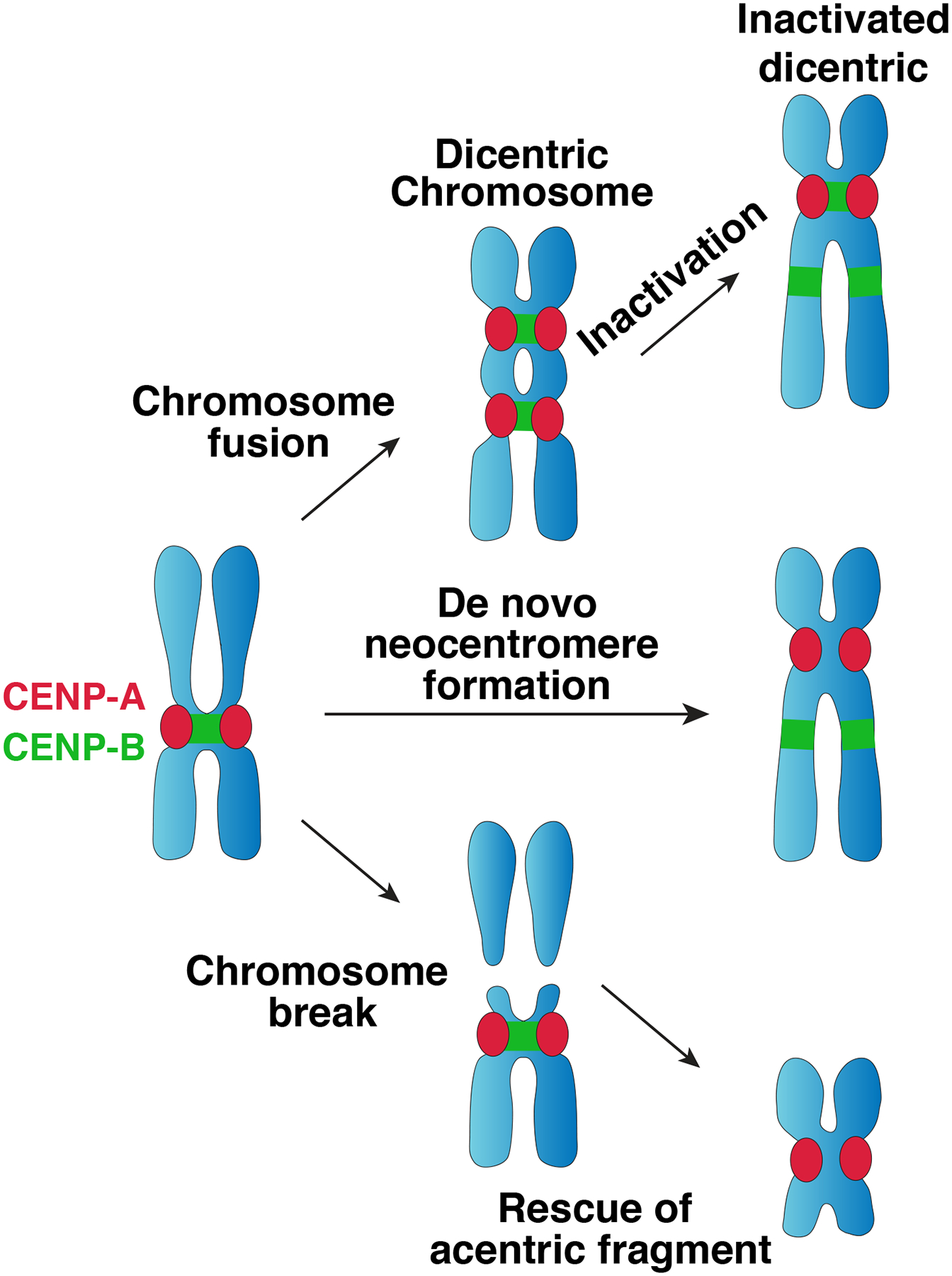
The centromere is the region of the chromosome that directs its segregation in mitosis and meiosis. Although the functional importance of the centromere has been appreciated for more than 130 years, elucidating the molecular features and properties that enable centromeres to orchestrate chromosome segregation is an ongoing challenge. Most eukaryotic centromeres are defined epigenetically and require the presence of nucleosomes containing the histone H3 variant centromere protein A (CENP-A; also known as CENH3). Ongoing work is providing important molecular insights into the central requirements for centromere identity and propagation, and the mechanisms by which centromeres recruit kinetochores to connect to spindle microtubules.


Figures

Figure 1.
Figure 1.. Visualization of the centromere.
a) Comparison of images of mitotic Salamander cells hand-drawn by Walther Flemming in 1882 (top) with immunofluorescence images of human cells (bottom) stained for microtubules (green), CENP-A (red) and DNA (blue). The images show cells at different phases of a mitotic cell cycle: late prometaphase-metaphase (left), anaphase (middle) and telophase (right). b) Images of the centromere at increasing resolution. Top left: immunofluorescence image of a mitotic chromosome stained for DNA (blue), CENP-A (red) and CENP-B (a marker for the alpha-satellite DNA repeats present at most human centromeres, green). Top right: electron micrograph of centromeric region of a mitotic chromosome showing centromeric chromatin (dark cloud), kinetochore, and microtubules (indicated by arrows). Image courtesy of Conly Rieder. Bottom left: Immunofluorescence image of stretched centromeric chromatin fibers showing patches of CENP-A (red) interspersed with H3, in this case specifically H3 dimethylated on lysine 4 (H3K4me2, green). Image courtesy of Elaine Dunleavy. Bottom right: Crystal structure of the CENP-A nucleosome. PDB ID: 3AN2
Figure 2.
Figure 2.. Centromere specification.
a) Diagram of the diverse types of centromeres found across eukaryotes. Holocentric chromosomes assemble a diffuse centromere across the whole chromosome. Monocentric chromosomes assemble a centromere at a single localized site on the chromosome, which is visible as a constriction between the chromosomes in mitosis (known as the primary constriction). Monocentric chromosomes can be further divided into those with point centromeres and those with regional centromeres. Point centromeres contain a specific DNA sequence that is sufficient for centromere function (here illustrated with the S. cerevisiae DNA architecture), which assembles a single CENP-A nucleosome. Regional centromeres contain large regions of DNA that is often repetitive (such as alpha-satellite DNA in primates), and assemble numerous CENP-A nucleosomes. b) Model of the DNA sequence of primate centromeres. Primate centromeres are built from alpha-satellite monomers (triangles), which are largely but not completely identical, as indicated by the different colored triangles. Patterns of these monomers arranged head-to-tail are re-iterated over the centromere core (purple) as higher-order repeats. Some monomers within the centromere core contain a sequence termed the CENP-B box, which binds to the centromere-DNA binding protein, CENP-B. The centromere core is flanked by less ordered monomers which comprise the pericentromere (blue). LINEs, SINEs and other satellites (squares) are found interspersed with alpha-satellite monomers in the pericentromere. c) Schematic showing comparison of macaque and human orthologous chromosomes that have undergone centromere repositioning such that the position of the centromere has moved, but the surrounding markers have not, as indicated by the color blocks, which represent syntenic regions. Part c) adapted from.
Figure 3.
Figure 3.. Specialization and propagation of CENP-A.
a) Model of human CENP-A primary and secondary structure showing conservation with histone H3. Each segment corresponds to a single amino acid, and is colored according to its conservation with human H3.1 as indicated. The first N-terminal amino acid, shown detached, represents the cleaved initiator methionine. Barrels represent alpha helices, and rods represent loops. Within the histone fold domain, the helices are designated alpha1 through alpha3, and the loops are designated L1 and L2. L1 and alpha2 comprise the CENP-A targeting domain, which is sufficient to target CENP-A to centromeres due to its interaction with the CENP-A chaperone, HJURP. This region also binds to CENP-N and is important for CENP-C recruitment, . CENP-C also binds to the C terminal residues of CENP-A, , . b) Model for the changes to CENP-A chromatin over the cell cycle. The timing of the localization of the CENP-A deposition factors is indicated. At S phase, existing CENP-A is partitioned between the replicated sisters, and gaps filled with histone H3.3. Although centromere localization of M18BP1 precedes recruitment of Mis18alpha and beta, the precise onset of its localization has not been established. By mitosis, M18BP1 localizes to centromeres, followed by Mis18alpha and Mis18beta at mitotic exit. An HJURP dimer is recruited in early G1 to direct new CENP-A deposition. New CENP-A is stabilized in late G1 by MgcRacGAP and RSF1. Defining the mechanisms that remove these assembly factors once CENP-A deposition is complete also remains an important open question. c) Model for the two-step regulation of CENP-A deposition. CDK prevents CENP-A deposition outside of G1 phase by inhibiting Mis18 complex localization, Mis18 complex assembly and HJURP recruitment. Plk1 binds to the Mis18 complex to promote CENP-A deposition at centromeres during G1.
Figure 4.
Figure 4.. Centromeric chromatin.
Model of the epigenetic modifications at the core centromere, CENP-A domain, and the pericentromere. In addition to the sequence and structural specializations that differentiate CENP-A chromatin from bulk chromatin, posttranslational modifications of CENP-A nucleosomes contribute to centromere function. Human CENP-A is mono-ubiquitinated at lysine 124 within the histone fold domain by CUL4-RBX1-COPS8 to promote its centromere targeting. Acetylation at this lysine 124 residue has also been reported. Finally, diverse other posttranslational of CENP-A and H4 in the CENP-A nucleosome have been described. Defining the functional contributions of these modifications remains a central challenge.
Figure 5.
Figure 5.. Contributions of the Constitutive Centromere Associated Network (CCAN) at the centromere-kinetochore interface.
a) Diagram of the proteins of the CCAN. The sixteen proteins of the CCAN, designated by CENP- and a letter, can be grouped into sub-complexes as indicated. The sub-complexes are grouped according to functions that have been reported for at least one of their subunits. KMN: a network of KNL1, Mis12 complex and Ndc80 complex, which together bind to microtubules. b) Comparison of the crystal structures of the tetramer comprised of the histones CENP-A and H4 in the context of the nucleosome (PDB ID: 3AN2) (H2A, H2B and DNA are excluded for clarity) with the heterotetramer comprised of the histone fold-containing proteins CENP-T, -W, -S, and -X heterotetramer (PDB 3VH5). c) A simplified model of the connectivity from the centromere, to the kinetochore, to the microtubule during mitosis. The contributions of CENP-C and CENP-T to recruiting the microtubule binding-interface of the kinetochore are highlighted, and the other CCAN components are excluded from this model for clarity.
None

References

    1. Holland AJ & Cleveland DW Boveri revisited: chromosomal instability, aneuploidy and tumorigenesis. Nat Rev Mol Cell Biol 10, 478–87 (2009). - PMC - PubMed
    1. Flemming W Zellsubstanz, Kern und Zelltheilung (Vogel, Leipzig, 1882).
    1. Vig BK Sequence of centromere separation: role of centromeric heterochromatin. Genetics 102, 795–806 (1982). - PMC - PubMed
    1. Bernard P et al. Requirement of heterochromatin for cohesion at centromeres. Science 294, 2539–42 (2001). - PubMed
    1. Nasmyth K Segregating sister genomes: the molecular biology of chromosome separation. Science 297, 559–65 (2002). - PubMed

Publication types