. 2016 Jul;9(4):907-16.
doi: 10.1038/mi.2015.121. Epub 2015 Nov 25.

Antigen sampling by intestinal M cells is the principal pathway initiating mucosal IgA production to commensal enteric bacteria

Affiliations

Antigen sampling by intestinal M cells is the principal pathway initiating mucosal IgA production to commensal enteric bacteria

D Rios et al. Mucosal Immunol. 2016 Jul.

Abstract

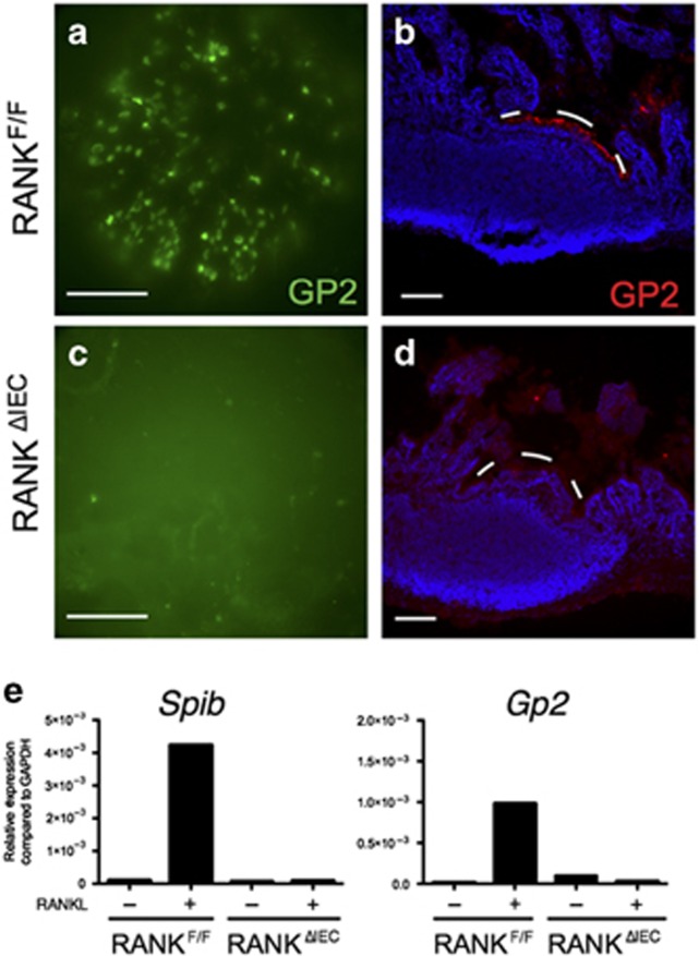
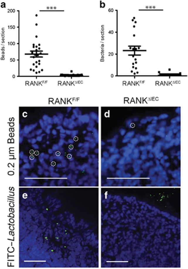
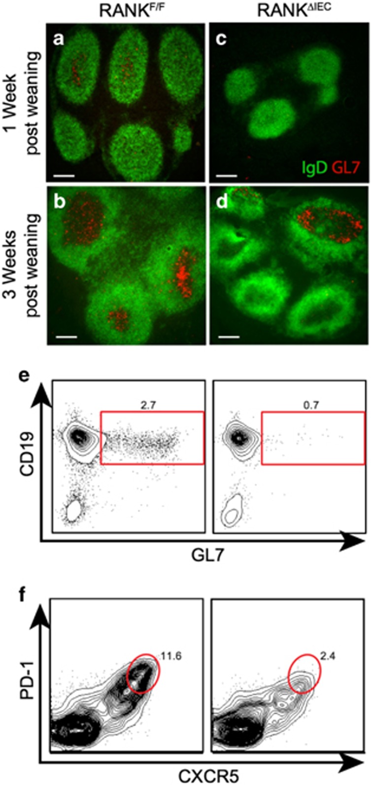
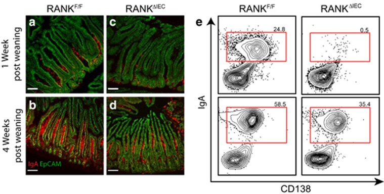
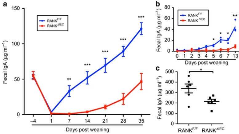
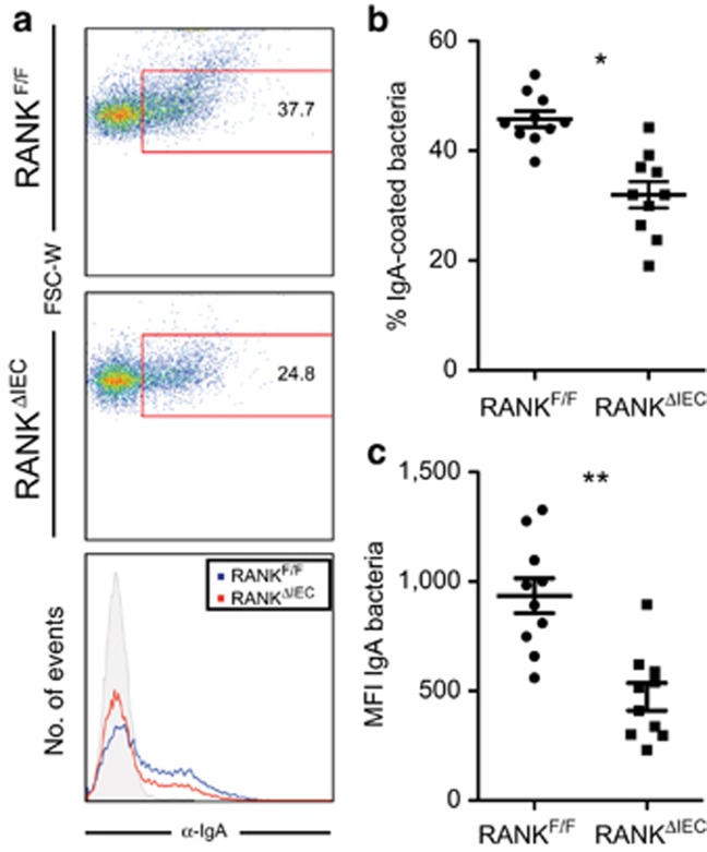
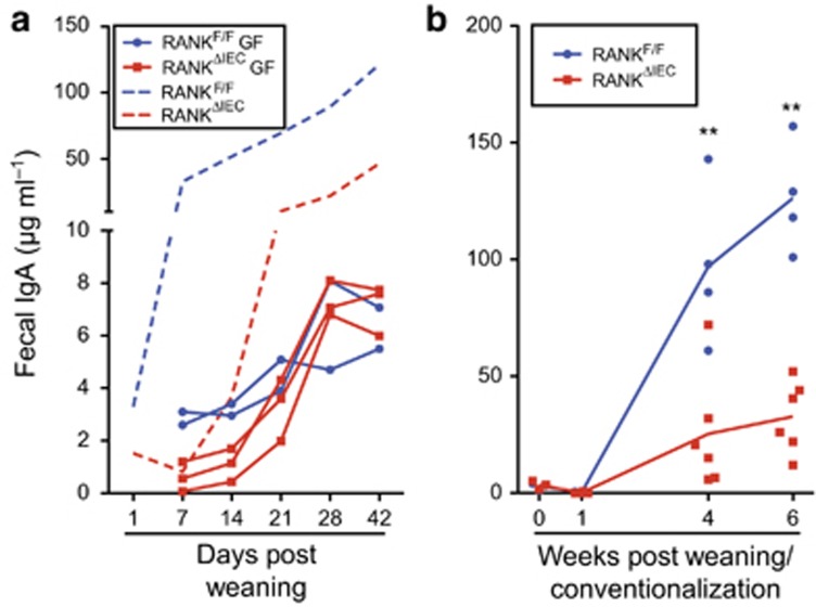
Secretory IgA (SIgA) directed against gut resident bacteria enables the mammalian mucosal immune system to establish homeostasis with the commensal gut microbiota after weaning. Germinal centers (GCs) in Peyer's patches (PPs) are the principal inductive sites where naive B cells specific for bacterial antigens encounter their cognate antigens and receive T-cell help driving their differentiation into IgA-producing plasma cells. We investigated the role of antigen sampling by intestinal M cells in initiating the SIgA response to gut bacteria by developing mice in which receptor activator of nuclear factor-κB ligand (RANKL)-dependent M-cell differentiation was abrogated by conditional deletion of Tnfrsf11a in the intestinal epithelium. Mice without intestinal M cells had profound delays in PP GC maturation and emergence of lamina propria IgA plasma cells, resulting in diminished levels of fecal SIgA that persisted into adulthood. We conclude that M-cell-mediated sampling of commensal bacteria is a required initial step for the efficient induction of intestinal SIgA.


Figures

Figure 1
Figure 1
RANKΔIEC mice lack Peyer's patch (PP) M cells. Representative images showing the distribution of GP2+ M cells in PPs from RANKF/F and RANKΔIEC mice as observed on whole mounts (a,c) and cryosections (b,d). The white dashes in b and d form an arc just above the apical surface of the follicle-associated epithelium (FAE). Bar = 100 μM. (e) Enteroid cultures of crypt cells from RANKF/F and RANKΔIEC mice were cultured for 3 days in the presence or absence of RANKL. The level of expression of M-cell-associated genes Spib and Gp2 was determined by quantitative PCR and the results normalized to Gapdh. Data are representative of three experiments done with independently derived enteroid cultures. See also Supplementary Figure 1 for additional information on the floxed RANK allele and Supplementary Figure 2 for a histologic comparison of goblet cell and Paneth cell differentiation in RANKF/F and RANKΔIEC mice.
Figure 2
Figure 2
Peyer's patches (PPs) lacking M cells have reduced capacity to phagocytose particulate antigens. RANKF/F and RANKΔIEC mice were gavage fed with either 1 × 10¹¹ 0.2-μm diameter fluorescein isothiocyanate (FITC)-labeled polystyrene beads (a) or 1 × 10⁹ colony-forming unit (CFU) FITC-labeled L. rhamnosus strain GG (LGG) (b), followed by excision of the PPs after 6 h (a) or 24 h (b). Individual points on the scatter plots represent the number of beads or bacteria manually counted in cryosections of a single PP follicle (a) or the entire PP (b). (c,d) Representative images showing 0.2 μm diameter beads within PPs from RANKF/F (c) and RANKΔIEC (d) mice. The dashed circles show individual phagocytic cells containing multiple beads. (e,f) Representative images showing FITC-LGG within PPs from RANKF/F (e) and RANKΔIEC (f) mice. Bar = 100 μM. Data from each group are summarized as mean±s.e.m. and are representative of two experiments with three mice per group. ***P<0.001 (t-test).
Figure 3
Figure 3
Germinal center (GC) formation is delayed in Peyer's patches (PPs) lacking M cells. Representative images of horizontal sections of PPs from RANKF/F and RANKΔIEC mice at 1 week (a,c) and 3 weeks (b,d) after weaning, showing the density of GL7+ GC B cells within the IgD region of follicles. Bars =100 μM. (e,f) Representative flow plots of PP lymphocytes from RANKF/F and RANKΔIEC mice at 2 weeks after weaning, stained for CD19 and GL7 to detect GC B cells (e) or PD-1 and CXCR5 to detect follicular helper T (Tfh) cells (f). Cells analyzed were gated on live CD45+ singlets that were also B220+ (e) or CD4+ (f). Data are representative of two experiments with three mice per group. See also Supplementary Figures 3 and 4.
Figure 4
Figure 4
Frequency of lamina propria IgA+ plasma cells is reduced in RANKΔIEC mice. Representative cryosections of small intestine from RANKF/F (a,b) or RANKΔIEC mice (c,d) at 1 week (a,c) or 4 weeks (b,d) after weaning were stained with anti-EpCAM (green) and anti-IgA (red). Bars = 100 μM. (e) Representative flow plots from cells isolated from small intestinal lamina propria at 1 and 4 weeks after weaning. Cells analyzed were gated on live CD45+ singlets that were also B220 and IgD. Data are representative of two experiments with three mice per group. See also Supplementary Figure 5.
Figure 5
Figure 5
Production of fecal IgA is delayed and reduced in RANKΔIEC mice. (a) Fecal IgA concentrations of RANKF/F and RANKΔIEC mice were determined at multiple time points starting at 4 days before weaning. Data are summarized as mean±s.e.m. and are representative of four experiments with four mice of each genotype. (b) Fecal IgA concentrations of RANKF/F and RANKΔIEC mice at daily time points beginning at weaning. Data are representative of two experiments with three mice of each genotype. (c) Fecal IgA concentrations in adult mice 8–10 weeks after weaning. Data are summarized as mean±s.e.m. and are from a single experiment with six mice of each genotype. *P<0.05, **P<0.01 (t-test). See also Supplementary Figure 6.
Figure 6
Figure 6
In vivo IgA coating of commensal microbiota is decreased in RANKΔIEC mice. (a) Representative flow plots of fecal bacteria isolated from RANKF/F and RANKΔIEC mice 4 weeks after weaning and stained with anti-IgA. (b,c) The percentage of IgA+ fecal bacteria (b) and the mean fluorescence intensity of IgA binding (c) were determined using fecal bacteria from RANKF/F and RANKΔIEC mice. Data are summarized as mean±s.e.m. and are from 1 experiment with 10 mice per group. *P<0.05, **P<0.01 (t-test).
Figure 7
Figure 7
M cells are required to initiate IgA responses to the intestinal microbiota. (a) The solid lines show fecal IgA concentrations in individual RANKF/F and RANKΔIEC littermates housed in a germ-free isolator from birth through 9 weeks of age. Data are from one of two litters that yielded similar results. The dashed lines show for comparison purposes the mean fecal IgA concentrations for RANKF/F and RANKΔIEC littermates housed under standard specific pathogen-free (SPF) conditions (data from Figure 5). (b) A single germ-free litter consisting of four RANKF/F mice and six RANKΔIEC mice was conventionalized by exposure to SPF fecal microbiota at weaning. The concentration of fecal IgA was determined at weaning and several time points after weaning. **P<0.01 (unpaired t-test). See also Supplementary Figure 9.

References

    1. Brandtzaeg, P. Secretory IgA: designed for anti-microbial defense. Front. Immunol. 4, 222 (2013). - PMC - PubMed
    1. Gutzeit, C., Magri, G. & Cerutti, A. Intestinal IgA production and its role in host-microbe interaction. Immunol. Rev. 260, 76–85 (2014). - PMC - PubMed
    1. Cebra, J.J., Periwal, S.B., Lee, G., Lee, F. & Shroff, K.E. Development and maintenance of the gut-associated lymphoid tissue (GALT): the roles of enteric bacteria and viruses. Dev. Immunol. 6, 13–18 (1998). - PMC - PubMed
    1. Macpherson, A.J., McCoy, K.D., Johansen, F.E. & Brandtzaeg, P. The immune geography of IgA induction and function. Mucosal Immunol. 1, 11–22 (2008). - PubMed
    1. Cerutti, A. The regulation of IgA class switching. Nat. Rev. Immunol. 8, 421–434 (2008). - PMC - PubMed

Publication types

MeSH terms

Substances