. 2016 Jan 15;291(3):1137-47.
doi: 10.1074/jbc.M115.698589. Epub 2015 Nov 24.

The Tomato Nucleotide-binding Leucine-rich Repeat Immune Receptor I-2 Couples DNA-binding to Nucleotide-binding Domain Nucleotide Exchange

Affiliations

The Tomato Nucleotide-binding Leucine-rich Repeat Immune Receptor I-2 Couples DNA-binding to Nucleotide-binding Domain Nucleotide Exchange

Stepan Fenyk et al. J Biol Chem. .

Abstract

Plant nucleotide-binding leucine-rich repeat (NLR) proteins enable plants to recognize and respond to pathogen attack. Previously, we demonstrated that the Rx1 NLR of potato is able to bind and bend DNA in vitro. DNA binding in situ requires its genuine activation following pathogen perception. However, it is unknown whether other NLR proteins are also able to bind DNA. Nor is it known how DNA binding relates to the ATPase activity intrinsic to NLR switch function required to immune activation. Here we investigate these issues using a recombinant protein corresponding to the N-terminal coiled-coil and nucleotide-binding domain regions of the I-2 NLR of tomato. Wild type I-2 protein bound nucleic acids with a preference of ssDNA ≈ dsDNA > ssRNA, which is distinct from Rx1. I-2 induced bending and melting of DNA. Notably, ATP enhanced DNA binding relative to ADP in the wild type protein, the null P-loop mutant K207R, and the autoactive mutant S233F. DNA binding was found to activate the intrinsic ATPase activity of I-2. Because DNA binding by I-2 was decreased in the presence of ADP when compared with ATP, a cyclic mechanism emerges; activated ATP-associated I-2 binds to DNA, which enhances ATP hydrolysis, releasing ADP-bound I-2 from the DNA. Thus DNA binding is a general property of at least a subset of NLR proteins, and NLR activation is directly linked to its activity at DNA.

Keywords: ATPases associated with diverse cellular activities (AAA); DNA binding protein; Nod-like receptor (NLR); cellular immune response; nucleotide; plant biochemistry.


Figures

FIGURE 1.
FIGURE 1.
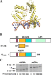
The I-2 CC-NB-ARC domains bind nucleic acids in vitro. A, structural homology model for amino acids 175–519 encompassing the NB-ARC domain of I-2, with associated ADP, bound to DNA. B, proteins used in this study. The top bar represents a generic CC-NB-LRR type R protein. Conserved domains are highlighted. The NB subdomain of the NB-ARC domain containing the P-loop is shown in orange, and the tandem ARC subdomains are shown in cyan. The bottom bars depict the domain composition of the proteins used, with amino acid positions delineating the relevant regions cloned. C, EMSA for I-21–519WT using 100 ng of ΦX174 DNA (ssDNA) or ΦX174 RF I DNA (dsDNA). For dsDNA, two bands are visible. The upper band represents relaxed circular DNA, whereas the lower band represents supercoiled circular DNA. Note that both bands shift in the presence of I-21–519WT. Molecular weight markers are indicated with arrows.
FIGURE 2.
FIGURE 2.
The I-2 CC-NB-ARC domain and the R1 NB domain bind nucleic acids in vitro. A, quantitative EMSA analysis giving affinities of I-21–519WT for synthetic oligonucleotides corresponding to different nucleic acids (n = 3–6, ±S.E.). B, overlay of a structural homology model for amino acids 197–334 encompassing the NB domain of R1-NB (blue) onto the I-2 NB-ARC domain model of Fig. 1A (yellow). C, EMSA for R1-NB using 100 ng of ΦX174 DNA (ssDNA) or ΦX174 RF I DNA (dsDNA). For dsDNA, the upper band represents relaxed circular DNA, whereas the lower band represents supercoiled circular DNA. Note that both bands shift upon incubation with the R1 NB domain. Molecular weight markers are indicated with arrows. D, quantitative EMSA analysis giving affinities of R1 NB for various synthetic oligonucleotides corresponding to different nucleic acids (n = 3–10, ±S.E.).
FIGURE 3.
FIGURE 3.
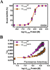
The I-2 K207R mutant shows an altered mode of interaction with dsDNA in comparison with wild type I-2. A, quantitative EMSA analysis giving comparative affinities of I-21–519WT and I-21–519K207R for synthetic dsDNA oligonucleotides (n = 3, ±S.E.). B, fluorescence anisotropy analysis giving distinct affinities of I-21–519WT and I-21–519K207R for dsDNA (n = 3, ±S.E.) suggestive for a different topology of the protein-DNA complex.
FIGURE 4.
FIGURE 4.
I-21–519 distorts and melts dsDNA. A, energy transfer efficiency in steady-state FRET in response to I-21–519 binding (n = 3, ±S.E.). The control is BSA. B, energy transfer efficiency in time resolved FRET in response to I-21–519 binding. C, P1 nuclease sensitivity of ssDNA and dsDNA following incubation with BSA (negative control), I-21–519, or ORC (positive control). *, p < 0.01 compared with control by one-way analysis of variance with post hoc Dunnett test.
FIGURE 5.
FIGURE 5.
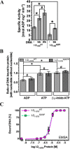
DNA binding is coupled to the nucleotide binding state and ATPase activity of I-21–519. A, the influence of 2 μm DNA on the intrinsic ATPase activity of I-21–519WT and I-21–519K207R (*, p < 0.05; #, p > 0.05; n = 4, ±S.E.). The source DNA is identical to that used for EMSA in Fig. 2A. B, double-stranded DNA binding by I-21–519WT and I-21–519K207R assessed by EMSA plotted as a ratio of binding in the presence of 5 μm nucleotide (ADP, ATP, or β,γ-imido ATP) compared with no nucleotide (*, p < 0.05; #, p > 0.05; n = 4–6, ±S.E.). C, quantitative EMSA analysis giving comparative affinities of I-21–519WT and I-21–519S233F for synthetic dsDNA oligonucleotides (n = 3, ±S.E.).

References

    1. Dangl J. L., and Jones J. D. (2001) Plant pathogens and integrated defence responses to infection. Nature 411, 826–833 - PubMed
    1. Jones J. D., and Dangl J. L. (2006) The plant immune system. Nature 444, 323–329 - PubMed
    1. Maekawa T., Kufer T. A., and Schulze-Lefert P. (2011) NLR functions in plant and animal immune systems: so far and yet so close. Nat. Immunol. 12, 817–826 - PubMed
    1. Medzhitov R. (2007) Recognition of microorganisms and activation of the immune response. Nature 449, 819–826 - PubMed
    1. Eitas T. K., and Dangl J. L. (2010) NB-LRR proteins: pairs, pieces, perception, partners, and pathways. Curr. Opin. Plant Biol. 13, 472–477 - PMC - PubMed

Publication types

MeSH terms

LinkOut - more resources