. 2016 Feb;137(2):591-600.e1.
doi: 10.1016/j.jaci.2015.07.050. Epub 2015 Oct 23.

Intrinsic functional defects of type 2 innate lymphoid cells impair innate allergic inflammation in promyelocytic leukemia zinc finger (PLZF)-deficient mice

Affiliations

Intrinsic functional defects of type 2 innate lymphoid cells impair innate allergic inflammation in promyelocytic leukemia zinc finger (PLZF)-deficient mice

Philip A Verhoef et al. J Allergy Clin Immunol. 2016 Feb.

Abstract

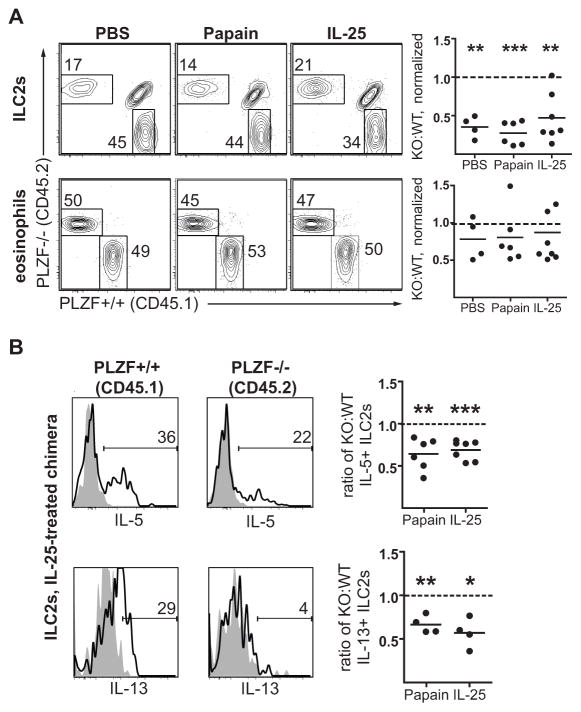
Background: The transcription factor promyelocytic leukemia zinc finger (PLZF) is transiently expressed during development of type 2 innate lymphoid cells (ILC2s) but is not present at the mature stage. We hypothesized that PLZF-deficient ILC2s have functional defects in the innate allergic response and represent a tool for studying innate immunity in a mouse with a functional adaptive immune response.

Objective: We determined the consequences of PLZF deficiency on ILC2 function in response to innate and adaptive immune stimuli by using PLZF(-/-) mice and mixed wild-type:PLZF(-/-) bone marrow chimeras.

Methods: PLZF(-/-) mice, wild-type littermates, or mixed bone marrow chimeras were treated with the protease allergen papain or the cytokines IL-25 and IL-33 or infected with the helminth Nippostrongylus brasiliensis to induce innate type 2 allergic responses. Mice were sensitized with intraperitoneal ovalbumin-alum, followed by intranasal challenge with ovalbumin alone, to induce adaptive TH2 responses. Lungs were analyzed for immune cell subsets, and alveolar lavage fluid was analyzed for ILC2-derived cytokines. In addition, ILC2s were stimulated ex vivo for their capacity to release type 2 cytokines.

Results: PLZF-deficient lung ILC2s exhibit a cell-intrinsic defect in the secretion of IL-5 and IL-13 in response to innate stimuli, resulting in defective recruitment of eosinophils and goblet cell hyperplasia. In contrast, the adaptive allergic inflammatory response to ovalbumin and alum was unimpaired.

Conclusions: PLZF expression at the innate lymphoid cell precursor stage has a long-range effect on the functional properties of mature ILC2s and highlights the importance of these cells for innate allergic responses in otherwise immunocompetent mice.

Keywords: Allergic mechanisms; innate lymphoid cells; mouse models.


Conflict of interest statement

Conflicts of interest: none

Figures

Figure 1
Figure 1. PLZF−/− mice have reduced type 2 inflammation in response to papain exposure
A. Schematic for intranasal papain treatment of PLZF+/+ versus PLZF−/− littermate mice. B. Histology at 50X magnification of lungs treated as indicated, with counts of the percentage of PAS+ epithelial cells. C. Flow cytometry plots showing cell percentages within each plot, quantified cell counts, and quantified cell percentages in lung digests from mice treated as indicated for eosinophils (Siglec F+, CD11c), basophils (Siglec F, CD11c, FcER1+, DX5+), ILC2s (lineage, CD90+, ICOS+, Sca1+, CD25+, CD127+), and CD4+ T cells (CD19, TCRβ+, CD4+). *p≤0.05, **p≤0.01, *** p≤0.001. Experiments are pooled data from at least 2 separate experiments.
Figure 2
Figure 2. PLZF−/− mice have reduced type 2 inflammation and ILC2 activation in response to IL-25 or IL-33 inhalation
A. Schematic for intranasal IL-25 or IL-33 treatment of PLZF+/+ versus PLZF−/− mice. B and C. Histology at 50X magnification of lungs treated with IL-25 (B), or IL-33 (C) with counts of PAS+ epithelial cells, and flow cytometry plots and cell percentages of eosinophils (Siglec F+, CD11c) in lung digests from mice treated as indicated. D. Bronchoalveolar lavage fluid from mice treated with the indicated stimuli for 3 days was tested for either IL-5 or IL-13 by cytometric bead array. Samples were tested in duplicate, with each experiment using at least 3 mice, repeated 2–3 times. E. Sort-purified ILC2s were treated for 3 days in media containing IL-7 and either PBS, IL-33, or PMA/ionomycin (P/I). Media was then collected and tested for levels of IL-5 and IL-13 by ELISA. Results represent pooled results from 4 different samples, repeated twice.
Figure 3
Figure 3. PLZF−/− mice have an impaired response to infection with Nippostrongylus brasiliensis
Flow cytometry plots, cell counts, and cell percentages from the lung digests of PLZF−/− mice and PLZF+/+ littermates 5 days after infection with 500 L3 Nippostrongylus brasiliensis for A. eosinophils (Siglec F+, CD11c), basophils (Siglec F, CD11c, FcER1+, DX5+), B. ILC2s (lineage, Thy1+, ICOS+, Sca1+, CD25+, CD127+), or C. CD4+ T cells (CD19, TCRβ+, CD4+). Bars indicate the mean, data are pooled from 2 different experiments (2–4 mice/condition/experiment), which were repeated twice.
Figure 4
Figure 4. Mixed bone marrow chimeras reveal cell-intrinsic reconstitution and activation defects in ILC2s from PLZF−/− mice
Mixed bone marrow chimeras were generated using CD45.1 PLZF+/+ (WT) and CD45.2 PLZF−/− (KO) bone marrow and, 6 weeks after reconstitution, treated for 3 days with papain, IL-25, or PBS intranasally, followed by analysis of lung digests. A. Flow cytometry plots and KO:WT ratios (normalized to splenic B cells) for ILC2s (CD25+, CD127+, Thy1+, ICOS+, lineage) and eosinophils (Siglec F+, CD11c). The dashed line indicates the predicted reconstitution ratio if no reconstitution defect were present. B. Flow cytometry histograms and quantitation of ILC2s from a representative IL-25-treated mixed chimera mouse comparing the percentage of CD45.2+, PLZF−/− ILC2s which are IL-5+ or IL-13+ (solid line) to CD45.1+, PLZF+/+ ILC2s. The negative control (filled grey) was derived by pre-incubating the ILC2s with excess unlabeled anti-cytokine antibody prior to staining with the indicated anti-cytokine antibody. Quantitation plots represent the ratio of CD45.2+, PLZF−/−, cytokine-positive ILC2s to CD45.1+, PLZF+/+, cytokine-positive ILC2s for either papain or IL-25 treatment. The dashed line indicates the predicted ratio if no activation defect were present. Experiments were repeated 2–3 times with 4–6 mice per condition. Statistical analysis with student’s t test for variation from a predicted ratio of 1.
Figure 5
Figure 5. PLZF−/− mice generate normal adaptive Th2 responses to ovalbumin
PLZF−/− or PLZF+/+ littermate mice were sensitized and challenged with ovalbumin. A. Flow cytometry plots and percentages of lung digests for eosinophils and CD4+ T cells, as indicated. Bars indicate the mean. Summary data are pooled from 2 separate experiments. B. Mixed bone marrow chimeras were generated using CD45.1 PLZF+/+ and CD45.2 PLZF−/− bone marrow and sensitized to ovalbumin as described above. Eosinophils, ILC2s, and CD4+ T cells were normalized to splenic B cell numbers and expressed as a ratio of CD45.2 PLZF−/− to CD45.1 PLZF+/+ to determine if there were cell intrinsic defects in cells participating in lung inflammation in response to ovalbumin. The dashed lines indicate the predicted PLZF−/−:PLZF+/+ ratio if no cell intrinsic defect were present. C. Flow cytometry plots of CD4+ T cells (CD4+, TCRβ+, CD8, CD19) from ova-sensitized mixed chimera mice challenged with ovalbumin, gated as either CD45.2 PLZF−/− vs CD45.1 PLZF+/+ or as IL-5+ vs IL-13+. The cytokine positive cells are then gated for CD45.2 PLZF−/− vs CD45.1 PLZF+/+. The quantitation plot shows the ratio of KO:WT IL-5+/IL-13+ CD4+ T cells to total CD4+ T cells, and the dashed line indicates the predicted ratio of KO:WT IL-5+/IL-13+ CD4+ T cells if no activation defect were present. Experiments were repeated 2–3 times with 4–6 mice per condition. Statistical analysis with student’s t test for variation from a ratio of 1.

References

    1. Brennan PJ, Brigl M, Brenner MB. Invariant natural killer T cells: an innate activation scheme linked to diverse effector functions. Nature reviews Immunology. 2013;13(2):101–17. - PubMed
    1. Bendelac A, Savage PB, Teyton L. The biology of NKT cells. Annu Rev Immunol. 2007;25:297–336. - PubMed
    1. Fuchs A, Colonna M. Innate lymphoid cells in homeostasis, infection, chronic inflammation and tumors of the gastrointestinal tract. Curr Opin Gastroenterol. 2013;29(6):581–7. - PubMed
    1. Vantourout P, Hayday A. Six-of-the-best: unique contributions of gammadelta T cells to immunology. Nature reviews Immunology. 2013;13(2):88–100. - PMC - PubMed
    1. Zeissig S, Blumberg RS. Commensal microbiota and NKT cells in the control of inflammatory diseases at mucosal surfaces. Current opinion in immunology. 2013;25(6):690–6. - PMC - PubMed

Publication types

MeSH terms