Review
. 2015 Dec;15(12):731-44.
doi: 10.1038/nri3920.

Regulation of macrophage development and function in peripheral tissues

Affiliations
Review

Regulation of macrophage development and function in peripheral tissues

Yonit Lavin et al. Nat Rev Immunol. 2015 Dec.

Abstract

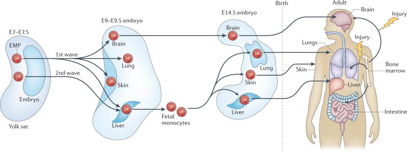
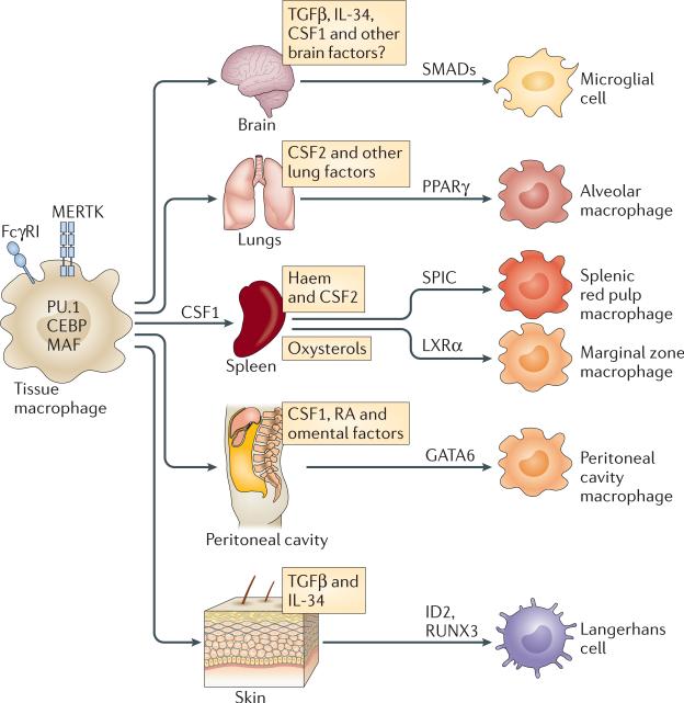
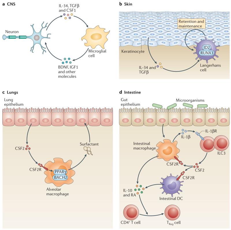
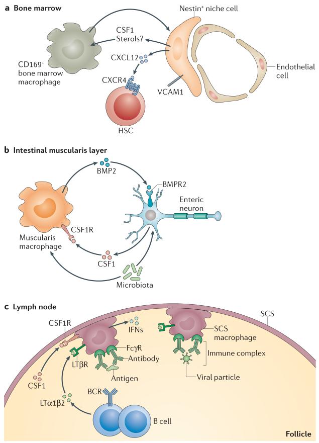
Macrophages are immune cells of haematopoietic origin that provide crucial innate immune defence and have tissue-specific functions in the regulation and maintenance of organ homeostasis. Recent studies of macrophage ontogeny, as well as transcriptional and epigenetic identity, have started to reveal the decisive role of the tissue stroma in the regulation of macrophage function. These findings suggest that most macrophages seed the tissues during embryonic development and functionally specialize in response to cytokines and metabolites that are released by the stroma and drive the expression of unique transcription factors. In this Review, we discuss how recent insights into macrophage ontogeny and macrophage-stroma interactions contribute to our understanding of the crosstalk that shapes macrophage function and the maintenance of organ integrity.


Figures

Figure 1
Figure 1. Origin of tissue-resident macrophages
Macrophages are maintained in most healthy tissues in mice by embryonic precursors (embryonic macrophages) independently of monocytes, with the exception of intestinal macrophages, dermal macrophages and a subset of cardiac macrophages. Two hypotheses dominate our thinking about the origin of embryonic macrophages. The first hypothesis suggests that all embryonic macrophages derive from yolk sac-derived erythromyeloid precursors (EMPs) that develop around embryonic day 7.5 (E7.5). The second hypothesis suggests that yolk sac-derived EMPs arise in two waves that differentially contribute to adult microglia and other macrophages: an early wave of yolk sac-derived EMPs that appear around E7.5 in the yolk sac colonize the brain and other fetal tissues around E9 to give rise to all tissue macrophages, and a later wave of yolk sac-derived EMPs that colonize and expand in the fetal liver to differentiate into fetal liver monocytes, which subsequently replace fetal macrophages, with the exception of microglia, and maintain them in adulthood.
Figure 2
Figure 2. The tissue microenvironment determines macrophage differentiation cues
During embryonic development, macrophages enter the tissues where they self-renew and proliferate. Macrophages in all tissues are characterized by expression of the cell surface marker FcγRI (also known as CD64), tyrosine-protein kinase MER (MERTK) and the transcription factors PU.1, CCAAT/enhancer-binding protein (CEBP) family members, MAF and MAFB. In the tissues, macrophage identity and functions are shaped by cytokines and metabolites that are produced in the local environment and drive specific transcription factor expression. In the brain, incoming yolk sac-derived cells are exposed to locally expressed transforming growth factor-β (TGFβ), which drives SMAD phosphorylation and the expression of genes that are unique to microglia. In the lungs, fetal monocytes that are exposed to colony-stimulating factor 2 (CSF2) express peroxisome proliferator-activated receptor-γ (PPARγ), which drives their differentiation into alveolar macrophages. In the spleen, haem drives SPIC expression, which controls the differentiation and maintenance of red pulp macrophages and the expression of key splenic red pulp macrophage-specific molecules, including vascular cell adhesion molecule 1 (VCAM1). In the marginal zone of the spleen, macrophage maintenance depends on liver X receptor-α (LXRα)-mediated signals. Retinoic acid (RA) and omental factors induce the expression of GATA-binding protein 6 (GATA6), which promotes the differentiation of peritoneal cavity macrophages. ID2, inhibitor of DNA binding 2; IL-34, interleukin-34; RUNX3, runt-related transcription factor 3.
Figure 3
Figure 3. IL-34 and CSF2 regulate specific tissue macrophage maintenance
a | In the brain, interleukin-34 (IL-34) is produced by neurons in the cortex, hippocampus and striatum and is necessary for microglial cell maintenance. Transforming growth factor-β (TGFβ) may be important for microglia maintenance, but it is still unclear which cells produce it in the brain. Conversely, microglia produce brain-derived neurotrophic factor (BDNF), which is important for learning-dependent synapse formation, and insulin-like growth factor 1 (IGF1), which is crucial for survival of layer V cortical neurons. b | In the skin, keratinocytes produce IL-34 and TGFβ, which are required for Langerhans cell homeostasis in the epithelium. TGFβ drives the expression of the transcription factors inhibitor of DNA binding 2 (ID2) and runt-related transcription factor 3 (RUNX3) in Langerhans cells and promotes their retention in the epidermis. The exact mechanism by which IL-34 drives Langerhans cell maintenance in the epidermis is unclear. c | In the lungs, epithelial cell-derived colony-stimulating factor 2 (CSF2) promotes peroxisome proliferator-activated receptor-γ (PPARγ) expression, and final maturation of alveolar macrophages is required for surfactant catabolism, which clears excess surfactant and maintains lung homeostasis. d | In the intestine, microbial products drive macrophage production of IL-1β that stimulates group 3 innate lymphoid cells (ILC3s) to produce CSF2. ILC3-mediated CSF2 production promotes the survival of intestinal macrophages and dendritic cells (DCs) and their production of IL-10 and retinoic acid (RA), which are required for the maintenance of regulatory T(TReg) cell homeostasis in the intestine. CSF2R, CSF2 receptor.
Figure 4
Figure 4. Macrophage–tissue crosstalk
a | Bone marrow macrophages are important for stimulating nestin+ niche cells to produce haematopoietic stem cell (HSC) retention factors, such as CXC-chemokine ligand 12 (CXCL12) and vascular cell adhesion molecule 1 (VCAM1). In turn, niche cells may produce colony-stimulating factor 1 (CSF1) to help to maintain macrophages in the niche. b | Intestinal muscularis macrophages produce bone morphogenetic protein 2 (BMP2), which signals through BMP receptor type 2 (BMPR2) expressed on enteric neurons and promotes neuronal control of peristaltic activity of the gut muscularis layer. In turn, enteric neurons, in response to microbial signals, produce CSF1, which is required to maintain muscularis macrophage homeostasis in vivo. c | In the lymph nodes, macrophages are situated directly beneath the subcapsular sinus (SCS) surrounding the B cell zone. They present large particulate antigen to B cells in the form of immune complexes and are important for trapping viral particles. In turn, B cells help to retain the SCS macrophage layer through the secretion of lymphotoxin α1β2 (LTα1β2). SCS macrophages also depend on local production of CSF1, although its source is unknown. BCR, B cell receptor; CSF1R, CSF1 receptor; FcγR, Fc receptor for IgG; IFNs, interferons; LTβR, lymphotoxin-β receptor.

References

    1. Gautier EL, et al. Gene-expression profiles and transcriptional regulatory pathways that underlie the identity and diversity of mouse tissue macrophages. Nat. Immunol. 2012;13:1118–1128. [This ImmGen consortia study shows that tissue macrophages have a distinct transcriptional profile that depends on the tissue in which they reside, highlighting the potential contribution of tissue imprinting to macrophage identity.] - PMC - PubMed
    1. Zagorska A, Traves PG, Lew ED, Dransfield I, Lemke G. Diversification of TAM receptor tyrosine kinase function. Nat. Immunol. 2014;15:920–928. - PMC - PubMed
    1. Canton J, Neculai D, Grinstein S. Scavenger receptors in homeostasis and immunity. Nat. Rev. Immunol. 2013;13:621–634. - PubMed
    1. Kawai T, Akira S. The role of pattern-recognition receptors in innate immunity: update on Toll-like receptors. Nat. Immunol. 2010;11:373–384. - PubMed
    1. Hussell T, Bell TJ. Alveolar macrophages: plasticity in a tissue-specific context. Nat. Rev. Immunol. 2014;14:81–93. - PubMed

Publication types

MeSH terms