. 2016 Feb 1;371(1):48-61.
doi: 10.1016/j.canlet.2015.10.020. Epub 2015 Nov 18.

Bovine milk-derived exosomes for drug delivery

Affiliations

Bovine milk-derived exosomes for drug delivery

Radha Munagala et al. Cancer Lett. .

Abstract

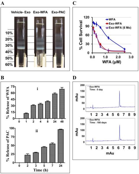
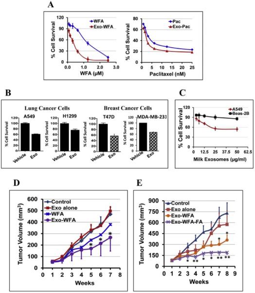
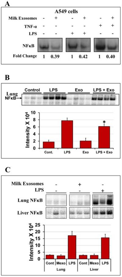
Exosomes are biological nanovesicles that are involved in cell-cell communication via the functionally-active cargo (such as miRNA, mRNA, DNA and proteins). Because of their nanosize, exosomes are explored as nanodevices for the development of new therapeutic applications. However, bulk, safe and cost-effective production of exosomes is not available. Here, we show that bovine milk can serve as a scalable source of exosomes that can act as a carrier for chemotherapeutic/chemopreventive agents. Drug-loaded exosomes showed significantly higher efficacy compared to free drug in cell culture studies and against lung tumor xenografts in vivo. Moreover, tumor targeting ligands such as folate increased cancer-cell targeting of the exosomes resulting in enhanced tumor reduction. Milk exosomes exhibited cross-species tolerance with no adverse immune and inflammatory response. Thus, we show the versatility of milk exosomes with respect to the cargo it can carry and ability to achieve tumor targetability. This is the first report to identify a biocompatible and cost-effective means of exosomes to enhance oral bioavailability, improve efficacy and safety of drugs.

Keywords: Chemopreventive agents; Chemotherapeutic drugs; Drug delivery; Milk-derived exosomes; Tumor-targeting.


Figures

Fig. 1
Fig. 1. Isolation and characterization of milk-exosomes
(A) Schematic representation of the major steps involved in the isolation of exosomes from bovine raw milk. (B) The exosome suspension (6 mg/ml protein concentration) was diluted 20-50 fold in phosphate-buffer-saline (PBS), pH 7.4 and a total of 200 μl was analyzed by NanoSight. (C) Bovine milk-derived exosomes were analyzed using 1 ml of the diluted suspension (1 mg/ml) in disposable cuvettes and milk exosomes size distribution was measured by Zetasizer. (D) Diluted exosomal suspension was loaded on a cleaned silicon wafers and air-dried for 30 min. Asylum MF-3D (Asylum Research, Oxford Instruments) AFM in 3d and tapping mode, and silicon probes coated with aluminum (Force Constant = 40 Nm-1; Resonant Frequency = 300 kHz, Budget Sensors.com) were used for imaging. Topographic and amplitude images were captured concurrently with a fixed force (<1 nN) with a scanning rate of 1Hz. (E) Exosomal suspension was filtered through 0.22 μm and loaded over clean silicon wafers and air-dried for 30 min. Silicon wafers were grounded using copper adhesive tape for conductivity. Exosomes were imaged in Zeiss Supra 35 SEM under beam energies (5kV). 142,000 × magnification. (F) Milk exosomes were layered onto Opti-prep gradient (10-60%) and centrifuged at 150,000×g for 16 h using 41 Ti swing rotor. The densities and refractive index values for the corresponding fractions are indicated in the table.
Fig. 2
Fig. 2. Exosomal markers and cargo
(A) Exosomes isolated in four different batches (numbered 1 - 4) were analyzed for the indicated exosomal and plasma membrane proteins. (B) Total RNA was isolated from 1 ml exosomal suspension (6 mg exosomal protein) by Trizol method (n=3), and reverse transcription and PCR were performed using bovine specific Taqman probes and primers (Applied Biosystems) for indicated immune-related miRNAs (Top) and exosome-related mRNAs (Bottom). Ct (threshold cycle) is plotted on y-axis and comparison between milk and colostrum exosomes were made. Student's t-test was performed to determine statistical significance; p<0.05=*, p<0.01= ** & p<0.001= ***.
Fig. 3
Fig. 3. Uptake of milk exosomes
(A) Dose-dependent uptake of milk exosomes by human lung cancer H1299 cells in vitro. Left: PKH-67-labeled milk exosomes were added to H1299 cells in 8-well chamber slide at 0-500 μg/ml and the uptake was monitored after 6 h. Cells were fixed and visualized under confocal microscope. Nuclei were visualized by staining with DAPI. Representative confocal images are shown (left panel). Scale bar, 100 μm. Fluorescent intensity of the PKH-67-labelled exosomes was quantified using ImageJ software (right panel). (B) PKH-67-labeled milk exosomes were added to H1299 in 8-well chamber slide at 50 μg/ml and the uptake was monitored after 1, 2, 4, 8 and 16 h. Cells were fixed and visualized. Confocal images are show in left panel and quantification of fluorescent images in right panel. (C) 500 μg/ml of the PKH67-labelled milk exosomes were added per 40,000 cells and incubated at 37 °C for 4 h. The uptake of the labeled exosomes was detected by confocal microscopy. Alexa fluor-phalloidin 594 (red) was used to detect actin filaments and DAPI for nucleus (blue) and PKH-67 to label the exosomes (green). Scale bar, 20 μm.
Fig. 4
Fig. 4. Biodistribution, stability and immunological response of milk exosomes
(A) Blood from nude mice treated with a single dose of 60 mg DiR-labeled exosomes by gavage (p.o.) was drawn and imaged at indicated times. Untreated control mice had baseline intensity value of < 1.25 × 10⁸ units. (B) Female nude mice were administered a single dose of 60 mg DiR-labeled exosomes/kg body weight by gavage (p.o.) or intravenously (i.v.). Top: Ex vivo-imaging of the tissues after 4 days of exosome administration were performed using Biospace lab Photon Imager Optima. Representative images are shown. Bottom: Fluorescent intensity of exosomes distributed in various tissues quantified using photon optima software. Figure shows average ± standard deviation of four animals. (C) Female Sprague-Dawley rats were treated with a single dose (short-term toxicity) of exosomes (25 mg exosomal protein/kg, b.wt.) by oral gavage or once daily for 15 days (long-term toxicity) and the serum was analyzed for cytokine levels using bio-plex cytokine Th1/Th2 assay. Acute toxicity observations were made at 1h, 3h and 6h post treatment. Vehicle-treated control (vehicle) animals were used as reference for baseline cytokine levels.
Fig. 5
Fig. 5. in vitro release profile and stability of drug-loaded exosomes
(A) Separation of vehicle- and drug-loaded milk exosomes by Opti-prep density gradient. Indicated agents were mixed with exosome in the presence of 10% ethanol. Unbound drug was removed by centrifugation at 10,000×g for 10 min. The drug loaded exosomes were layered onto Opti-prep gradient (10 - 60%) and separated by centrifugation at 150,000×g for 16 h using SW 41 Ti swing rotor. (B) The release study was done of Exo-WFA (top) and Exo- PAC (bottom) formulations was using dialysis tubes against buffer containing the surfactant, Tween-80 (0.02%) at 37°C. (C) Anti-proliferative activity of exosomal-WFA formulation of fresh preparation (red) and after 6 months of storage (purple) against A549 cells. Free WFA was included for reference (blue) (mean ± SD). (D) UPLC profile of WFA extracted from exosomal formulation at day 0 and after 180 days.
Fig. 6
Fig. 6. Increased drug bioavailability and enhanced anti-cancer effects
(A) Antiproliferative activity of drug-loaded milk exosomes versus free drugs [withaferin A (WFA) and paclitaxel (PAC)] in human lung cancer A549 cells. For Exo-drug treatments, exosomal protein concentration was maintained constant (50 μg/ml). (B) Anti-proliferative activity of milk exosomes per se against human lung (A549 and H1299), and breast (T47D and MDA-MB-231) cancer cells. Cells were treated with 50 μg/ml exosomal protein for 72 h. The percent cell survival was analyzed by MTT assay. Data represent average ± SD (n=3). (C) Anti-proliferative activity of milk exosomes per se at concentrations 0- 50 μg/ml for 72 h against human normal lung Beas-2b and lung cancer (A549) cells. Data represent average ± SD (n=3). (D) Following inoculation with human lung cancer A549 cells (2.5 × 10⁶ cells), when tumor xenografts grew to over 80 mm3, animals were treated i.p. three times a week with Exo-WFA (4 mg/kg WFA and 25 mg/kg b. wt.). Two other groups were treated i.p. with Exo alone (25 mg/kg b. wt.) or WFA (4 mg/kg). Data represent average ± SE (n = 6–8); SE is not shown in WFA alone for clarity. Statistical analysis was done using student's t-test; *, p < 0.05; **, p < 0.005. (E) Animals bearing A549 xenografts were treated with oral gavage three times a week with FA-Exo-WFA (8 mg/kg WFA and 25 mg/kg b. wt. exo protein) to achieve tumor targeting. Two other groups were treated with Exo alone (25 mg/kg b. wt.) or vehicle. Data represent average ± SE (n = 8–10). Statistical analysis was done using student's t-test; *, p < 0.05; **, p < 0.005.
Fig. 7
Fig. 7. Anti-inflammatory effects of milk exosomes
(A) Human lung A549 cancer cells were pre-treated with Exo (90 μg/ml Exo protein), for 6 h followed by treatment with or without tumor necrosis factor (TNF)-α (10 ng/ml) or LPS (1 μg/ml) to induce NF-κB activation. NF-κB levels were determined by electrophoretic mobility shift assay (EMSA). (B) Female Sprague-Dawley rats were treated with milk exosomes daily (25 mg Exo protein/kg b.wt.) by gavage for 15 days. Top: Lung and liver tissues were analyzed for NF-κB by EMSA. Animals treated with LPS served as a positive control. Data shown are from 3 individual animals. Bottom: Bar graph represents intensity quantification of NF-κB levels. Cont- vehicle control; Mexo- milk exosomes and LPS- Lipopolysaccharide. (C) Anti-inflammatory activity of milk exosomes in the lung tissue of female Sprague-Dawley rats treated with LPS as measured NF-κB activation by EMSA. Rats were treated with LPS (10 mg/kg b.wt.) and milk exosomes (25 mg Exo protein/kg b. wt.), both intraperitoneally, alone or in combination for 6 h. Data from 3-4 animals are shown for each group. Free probe was in excess in all samples and not shown for clarity. Bar graph represents intensity quantification, p<0.01.

References

    1. Jabir NR, Tabrez S, Ashraf GM, Shakil S, Damanhouri GA, Kamal MA. Nanotechnology-based approaches in anticancer research. International journal of nanomedicine. 2012;7:4391–4408. - PMC - PubMed
    1. Lim EK, Jang E, Lee K, Haam S, Huh YM. Delivery of cancer therapeutics using nanotechnology. Pharmaceutics. 2013;5:294–317. - PMC - PubMed
    1. Barenholz Y. Doxil(R)--the first FDA-approved nano-drug: lessons learned. Journal of controlled release : official journal of the Controlled Release Society. 2012;160:117–134. - PubMed
    1. Ciruelos E, Jackisch C. Evaluating the role of nab-paclitaxel (Abraxane) in women with aggressive metastatic breast cancer. Expert review of anticancer therapy. 2014;14:511–521. - PubMed
    1. Saif MW. U.S. Food and Drug Administration approves paclitaxel protein-bound particles (Abraxane(R)) in combination with gemcitabine as first-line treatment of patients with metastatic pancreatic cancer. JOP : Journal of the pancreas. 2013;14:686–688. - PubMed

Publication types

MeSH terms