

Rescue of neurodegeneration in the Fig4 null mouse by a catalytically inactive FIG4 transgene
- PMID: 26604144
- PMCID: PMC4706117
- DOI: 10.1093/hmg/ddv480
Rescue of neurodegeneration in the Fig4 null mouse by a catalytically inactive FIG4 transgene
Abstract
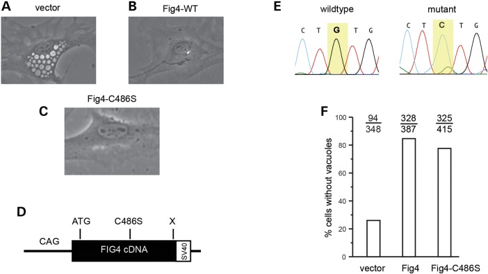
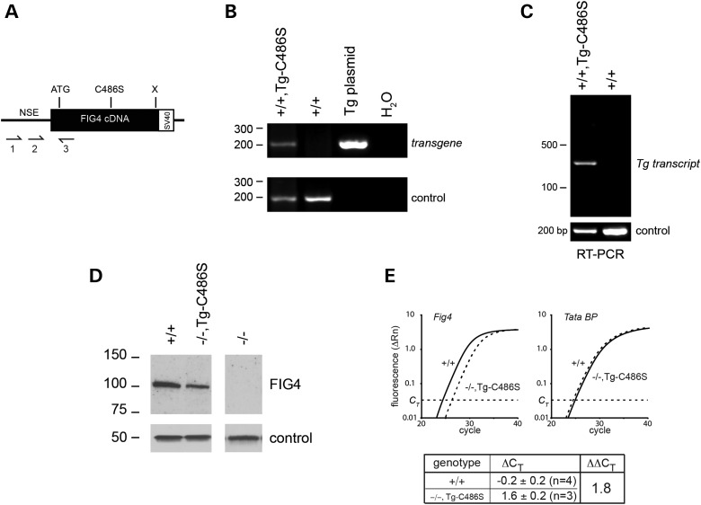
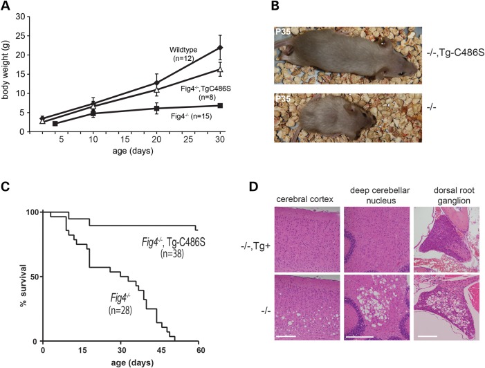
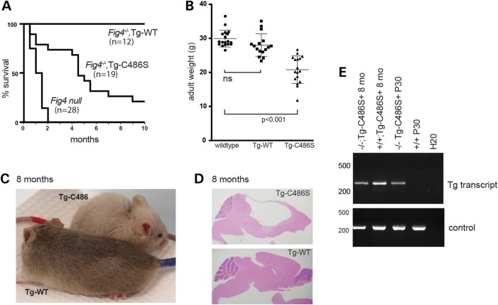
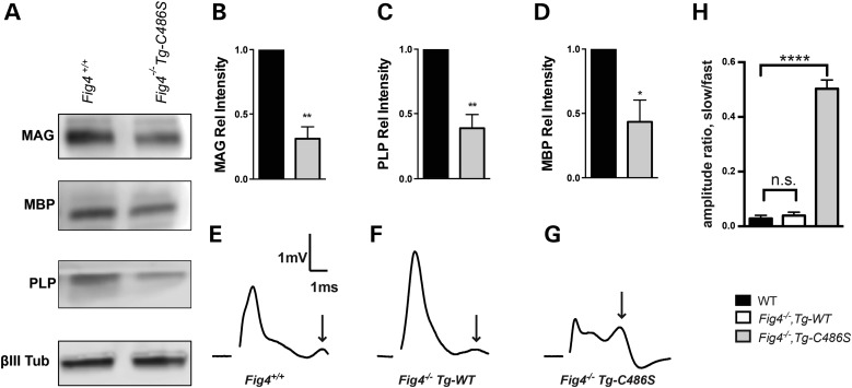
The lipid phosphatase FIG4 is a subunit of the protein complex that regulates biosynthesis of the signaling lipid PI(3,5)P2. Mutations of FIG4 result in juvenile lethality and spongiform neurodegeneration in the mouse, and are responsible for the human disorders Charcot-Marie-Tooth disease, Yunis-Varon syndrome and polymicrogyria with seizures. We previously demonstrated that conditional expression of a wild-type FIG4 transgene in neurons is sufficient to rescue most of the abnormalities of Fig4 null mice, including juvenile lethality and extensive neurodegeneration. To evaluate the contribution of the phosphatase activity to the in vivo function of Fig4, we introduced the mutation p.Cys486Ser into the Sac phosphatase active-site motif CX5RT. Transfection of the Fig4(Cys486Ser) cDNA into cultured Fig4(-/-) fibroblasts was effective in preventing vacuolization. The neuronal expression of an NSE-Fig4(Cys486Ser) transgene in vivo prevented the neonatal neurodegeneration and juvenile lethality seen in Fig4 null mice. These observations demonstrate that the catalytically inactive FIG4 protein provides significant function, possibly by stabilization of the PI(3,5)P2 biosynthetic complex and/or localization of the complex to endolysosomal vesicles. Despite this partial rescue, later in life the NSE-Fig4(Cys486Ser) transgenic mice display significant abnormalities that include hydrocephalus, defective myelination and reduced lifespan. The late onset phenotype of the NSE-Fig4(Cys486Ser) transgenic mice demonstrates that the phosphatase activity of FIG4 has an essential role in vivo.
© The Author 2015. Published by Oxford University Press. All rights reserved. For Permissions, please email: journals.permissions@oup.com.
Figures
Similar articles
-
Protective role of the lipid phosphatase Fig4 in the adult nervous system.Hum Mol Genet. 2018 Jul 15;27(14):2443-2453. doi: 10.1093/hmg/ddy145. Hum Mol Genet. 2018. PMID: 29688489 Free PMC article.
-
Yunis-Varón syndrome is caused by mutations in FIG4, encoding a phosphoinositide phosphatase.Am J Hum Genet. 2013 May 2;92(5):781-91. doi: 10.1016/j.ajhg.2013.03.020. Epub 2013 Apr 25. Am J Hum Genet. 2013. PMID: 23623387 Free PMC article.
-
FIG4 mutations leading to parkinsonism and a phenotypical continuum between CMT4J and Yunis Varón syndrome.Parkinsonism Relat Disord. 2020 May;74:6-11. doi: 10.1016/j.parkreldis.2020.03.021. Epub 2020 Mar 28. Parkinsonism Relat Disord. 2020. PMID: 32268254
-
FIG4-Related Parkinsonism and the Particularities of the I41T Mutation: A Review of the Literature.Genes (Basel). 2024 Oct 21;15(10):1344. doi: 10.3390/genes15101344. Genes (Basel). 2024. PMID: 39457468 Free PMC article. Review.
-
Fig4 deficiency: a newly emerged lysosomal storage disorder?Prog Neurobiol. 2013 Feb-Mar;101-102:35-45. doi: 10.1016/j.pneurobio.2012.11.001. Epub 2012 Nov 16. Prog Neurobiol. 2013. PMID: 23165282 Free PMC article. Review.
Cited by
-
FIG4 regulates lysosome membrane homeostasis independent of phosphatase function.Hum Mol Genet. 2016 Feb 15;25(4):681-92. doi: 10.1093/hmg/ddv505. Epub 2015 Dec 11. Hum Mol Genet. 2016. PMID: 26662798 Free PMC article.
-
A New Mutation in FIG4 Causes a Severe Form of CMT4J Involving TRPV4 in the Pathogenic Cascade.J Neuropathol Exp Neurol. 2017 Sep 1;76(9):789-799. doi: 10.1093/jnen/nlx062. J Neuropathol Exp Neurol. 2017. PMID: 28859335 Free PMC article.
-
The endolysosomal pathway and ALS/FTD.Trends Neurosci. 2023 Dec;46(12):1025-1041. doi: 10.1016/j.tins.2023.09.004. Epub 2023 Oct 10. Trends Neurosci. 2023. PMID: 37827960 Free PMC article. Review.
-
Protective role of the lipid phosphatase Fig4 in the adult nervous system.Hum Mol Genet. 2018 Jul 15;27(14):2443-2453. doi: 10.1093/hmg/ddy145. Hum Mol Genet. 2018. PMID: 29688489 Free PMC article.
-
The role of autophagy in the pathogenesis and treatment of amyotrophic lateral sclerosis (ALS) and frontotemporal dementia (FTD).Autophagy Rep. 2025 Mar 20;4(1):2474796. doi: 10.1080/27694127.2025.2474796. eCollection 2025. Autophagy Rep. 2025. PMID: 40395983 Free PMC article. Review.
References
-
- Ho C.Y., Alghamdi T.A., Botelho R.J. (2012) Phosphatidylinositol-3,5-bisphosphate: no longer the poor PIP2. Traffic, 13, 1–8. - PubMed
-
- Takasuga S., Sasaki T. (2013) Phosphatidylinositol-3,5-bisphosphate: metabolism and physiological functions. J. Biochem., 154, 211–218. - PubMed
-
- Michell R.H. (2013) Inositol lipids: from an archaeal origin to phosphatidylinositol 3,5-bisphosphate faults in human disease. FEBS J., 280, 6281–6294. - PubMed
Publication types
MeSH terms
Substances
Supplementary concepts
Grants and funding
LinkOut - more resources
Full Text Sources
Other Literature Sources
Medical
Molecular Biology Databases
Research Materials
Miscellaneous
