. 2016 Mar;29(1):40-53.
doi: 10.1177/0394632015615592. Epub 2015 Nov 24.

Age and diabetes related changes of the retinal capillaries: An ultrastructural and immunohistochemical study

Affiliations

Age and diabetes related changes of the retinal capillaries: An ultrastructural and immunohistochemical study

Enrica Bianchi et al. Int J Immunopathol Pharmacol. 2016 Mar.

Abstract

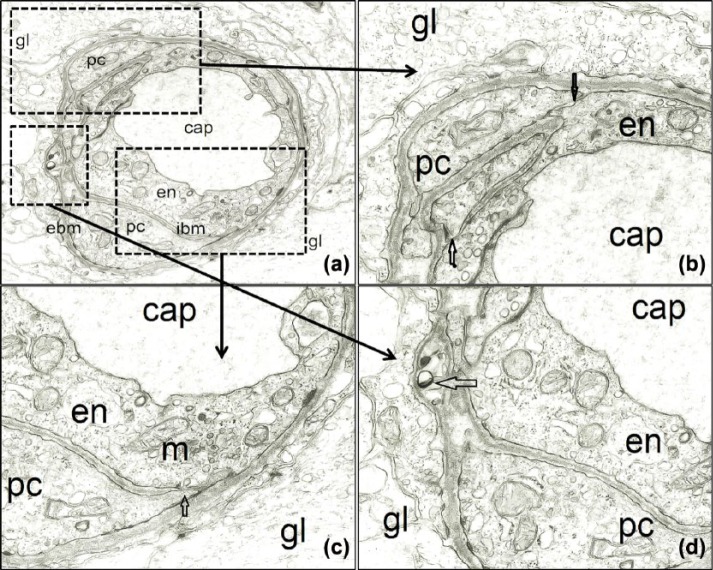
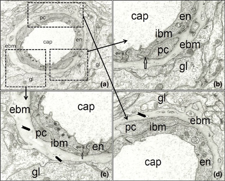
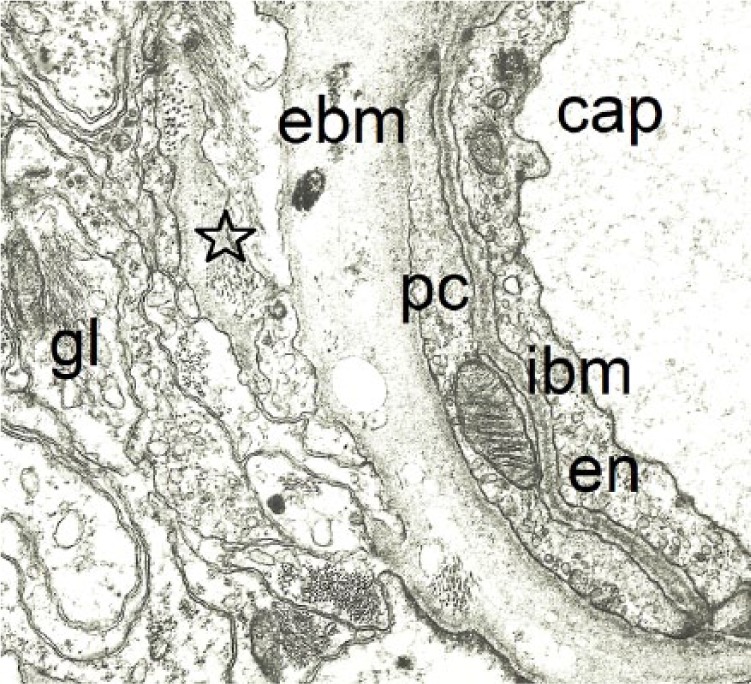
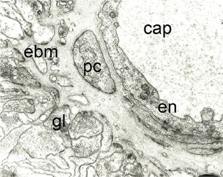
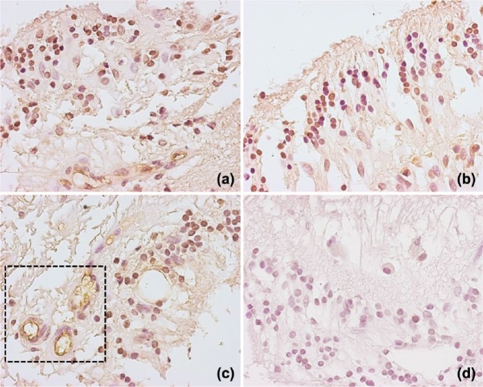
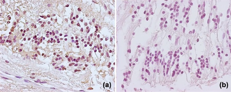
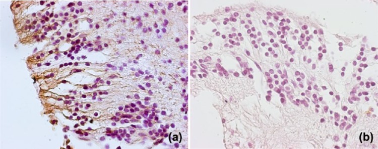
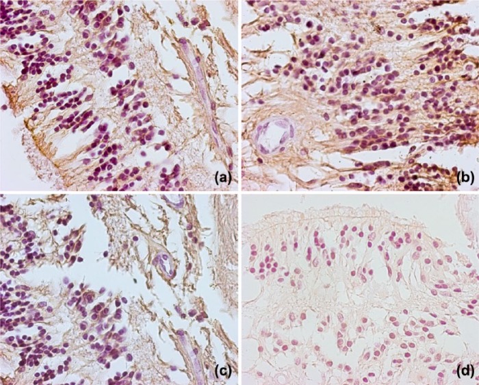
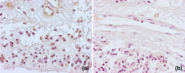
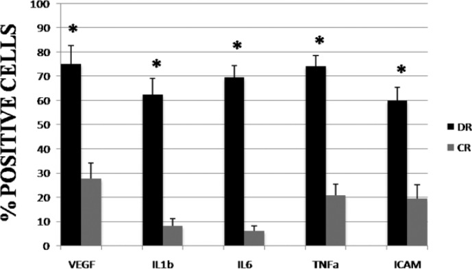
Normal human aging and diabetes are associated with a gradual decrease of cerebral flow in the brain with changes in vascular architecture. Thickening of the capillary basement membrane and microvascular fibrosis are evident in the central nervous system of elderly and diabetic patients. Current findings assign a primary role to endothelial dysfunction as a cause of basement membrane (BM) thickening, while retinal alterations are considered to be a secondary cause of either ischemia or exudation. The aim of this study was to reveal any initial retinal alterations and variations in the BM of retinal capillaries during diabetes and aging as compared to healthy controls. Moreover, we investigated the potential role of vascular endothelial growth factor (VEGF) and pro-inflammatory cytokines in diabetic retina.Transmission electron microscopy (TEM) was performed on 46 enucleated human eyes with particular attention to alterations of the retinal capillary wall and Müller glial cells. Inflammatory cytokines expression in the retina was investigated by immunohistochemistry.Our electron microscopy findings demonstrated that thickening of the BM begins primarily at the level of the glial side of the retina during aging and diabetes. The Müller cells showed numerous cytoplasmic endosomes and highly electron-dense lysosomes which surrounded the retinal capillaries. Our study is the first to present morphological evidence that Müller cells start to deposit excessive BM material in retinal capillaries during aging and diabetes. Our results confirm the induction of pro-inflammatory cytokines TNF-α and IL-1β within the retina as a result of diabetes.These observations strongly suggest that inflammatory cytokines and changes in the metabolism of Müller glial cells rather than changes in of endothelial cells may play a primary role in the alteration of retinal capillaries BM during aging and diabetes.

Keywords: Müller cells; age; basement membrane (BM); growth factors; inflammatory cytokines; pericytes; retinal capillaries.


Conflict of interest statement

Declaration of conflicting interests: The author(s) declared no potential conflicts of interest with respect to the research, authorship, and/or publication of this article.

Figures

Figure 1.
Figure 1.
The retinal capillary wall of a 6-year-old boy. (a) Retinal capillaries (cap) are formed by two distinct layers: the inner layer (ibm) placed between the endothelial cells (en) and the pericytes (pc), and the external layer (ebm) interposed between the pericytes and the Müller glial cells (gl). Magnification 8000×. (b) Endothelial cells-pericyte contact sites (arrows). Magnification 24,000×. (c) Another contact site between pericyte (pc) and endothelial cell (en) with a pinocytotic vesicle indicating direct cytoplasmic communication between these cells. Both endothelial cells and pericytes have numerous mitochondria (m) and other cytoplasmic organelles. Magnification 24,000×. (d) In some places, we can see vacuoles in the external basement membrane (ebm). Magnification 24,000×.
Figure 2.
Figure 2.
The retinal capillary wall of an 81-year-old man. (a) The structure of endothelial cells (en) is well preserved. Pericytes (PC) form a discontinuous layer around the circumference of retinal capillary (cap) and a thin internal basement membrane can be seen between endothelial cells and pericytes. Significant thickening of the external basement membrane (ebm). Magnification 8000×. (b) Endothelial cell-pericyte contact site (arrow) can be identified in a photo at higher magnification. External basement membrane (ebm) contains some rudimentary processes of pericytes. Magnification 24,000×. (c) Endothelial cells (en) show numerous endosomes and cytoplasmatic organelles, while the cytoplasm of pericytes (pc) presents few organelles. The external basement membrane (ebm) contains some vacuoles (black arrowheads). The contact site between endothelial cell and pericyte can be observed (arrow). Magnification 24,000×. (d) The border between external basement membrane (ebm) and Müller cells (gl) is clearly visible. The external basement membrane (ebm) contains some vacuoles (black arrowheads). Magnification 24,000×.
Figure 3.
Figure 3.
Retinal capillary wall of a 62-year-old man affected by diabetes. Normal appearance of endothelial cells (en) and pericytes (pc). Internal basement membrane (ibm) is very thin and the external basement membrane (ebm) is significantly thickened with some electro-dense granules and vacuoles. Note that the border between basement membrane and Müller cells and adjacent Müller cells (star) is irregular. Magnification 20,000×.
Figure 4.
Figure 4.
Retinal capillary wall of an 83-year-old woman affected by diabetes. External basement membrane is significantly thickened and root-like extensions formed deeply in between Müller cells. Magnification 20,000×.
Figure 5.
Figure 5.
Immunohistochemical staining of human retina for vascular endothelial growth factor (VEGF). In diabetic retina (a, b) VEGF immunoreactivity was observed in retinal pigment epithelial cells, retinal microvascular endothelial cells, retinal pericytes, and Müller cells. As it is visible in (c) VEGF immunostaining has been found in vascularized areas (boxed area). VEGF immunoreactivity was weakly noted in the inner nuclear layer in the normal control retina (d) (magnification 40×).
Figure 6.
Figure 6.
Immunohistochemical staining of human retina for interleukin-1beta (IL-1β). In diabetic retina (a) immunostaining for IL-1β was observed in the inner nuclear layer and in the retinal pigment epithelium. No immunoreactivity for IL-1β was detected in the negative control sample (b) (magnification 40×).
Figure 7.
Figure 7.
Immunohistochemical staining of human retina for interleukin-6 (IL-6). Immunoreactivity for IL-6 was stronger in RPE cells from diabetic eyes (a). IL-6 immunoreactivity was not observed in the normal retina (b) (magnification 40×).
Figure 8.
Figure 8.
Immunohistochemical analysis of human retina for TNF-α. In diabetic retinas immunoreactivity for TNF-α was observed in the glial cells (a), in the nerve fiber and retinal ganglion cell layers (b, c). In control eyes immunostaining for TNF-α was observed in few glial cells (d). Magnification 40×.
Figure 9.
Figure 9.
Immunohistochemical analysis of human retina for intracellular adhesion molecule-1 (ICAM-1). In diabetic retinas (a) immunoreactivity for ICAM-1 was localized in the inner layer and in blood vessels of the retina. Only faint immunoreactivity was seen in the retinas from control eyes (b). Magnification 40×.
Figure 10.
Figure 10.
Pro-inflammatory cytokines and VEGF positive cell index in the diabetic and control retina. The bar graph indicates the mean % positive cells to growth factors and proinflammatory cytokines ± SD. Statistical analysis is performed using Student’s t-test. *P <0.05.

Similar articles

Cited by

References

    1. Xi Y, Nette EG, King DW, et al. (1982) Age-related changes in normal human basement membrane. Mechanisms of Ageing and Development 19: 315–324. - PubMed
    1. Cavallotti C, Artico M, Cavallotti D. (1999) Occurrence of adrenergic nerve fibers and of noradrenaline in thymus gland of juvenile and aged rats. Immunology Letters 70: 53–62. - PubMed
    1. Cogan DG, Kuwabara T, Friedman E. (1968) Retinal vasculature. Microvascular Research 1: 115–132.
    1. Aiello LP, Gardner TW, King GI, et al. (1998) Diabetic retinopathy. Diabetes Care 21: 143–156. - PubMed
    1. Liu Y, Leo LF, McGregor C, et al. (2012) PEDF peptide eye drops reduce inflammation, cell death, and vascular leakage in diabetic retinopathy in the Ins2akita mice. Molecular Medicine 18: 1387–1401. - PMC - PubMed

Publication types

MeSH terms

Substances

LinkOut - more resources