. 2016 Jan 1;129(1):51-64.
doi: 10.1242/jcs.176701. Epub 2015 Nov 24.

LPS impairs oxygen utilization in epithelia by triggering degradation of the mitochondrial enzyme Alcat1

Affiliations

LPS impairs oxygen utilization in epithelia by triggering degradation of the mitochondrial enzyme Alcat1

Chunbin Zou et al. J Cell Sci. .

Abstract

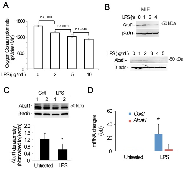
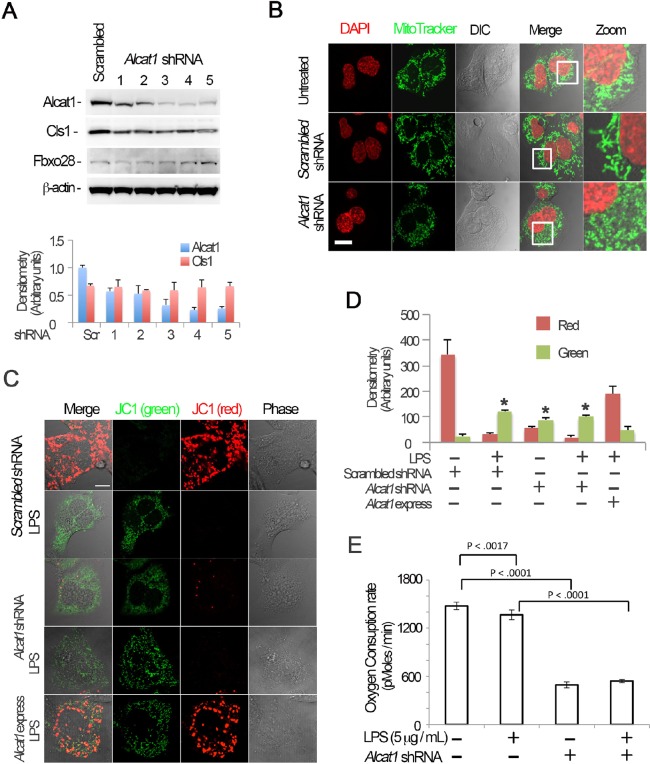
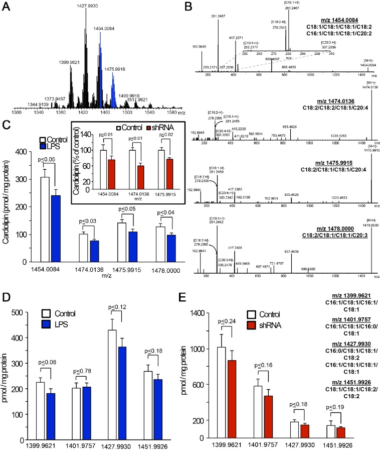
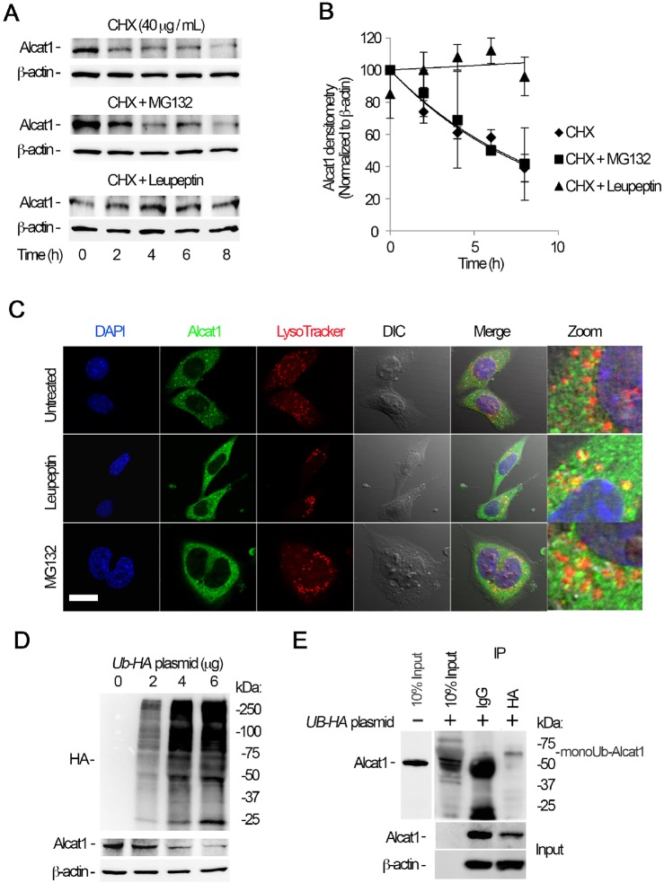
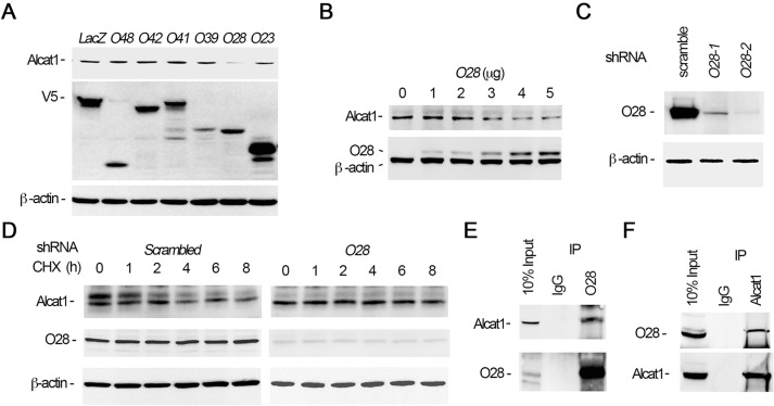
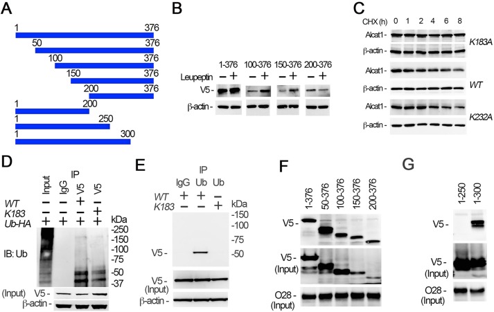
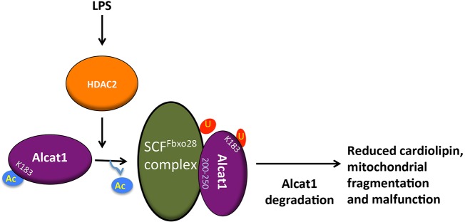
Cardiolipin (also known as PDL6) is an indispensable lipid required for mitochondrial respiration that is generated through de novo synthesis and remodeling. Here, the cardiolipin remodeling enzyme, acyl-CoA:lysocardiolipin-acyltransferase-1 (Alcat1; SwissProt ID, Q6UWP7) is destabilized in epithelia by lipopolysaccharide (LPS) impairing mitochondrial function. Exposure to LPS selectively decreased levels of carbon 20 (C20)-containing cardiolipin molecular species, whereas the content of C18 or C16 species was not significantly altered, consistent with decreased levels of Alcat1. Alcat1 is a labile protein that is lysosomally degraded by the ubiquitin E3 ligase Skp-Cullin-F-box containing the Fbxo28 subunit (SCF-Fbxo28) that targets Alcat1 for monoubiquitylation at residue K183. Interestingly, K183 is also an acetylation-acceptor site, and acetylation conferred stability to the enzyme. Histone deacetylase 2 (HDAC2) interacted with Alcat1, and expression of a plasmid encoding HDAC2 or treatment of cells with LPS deacetylated and destabilized Alcat1, whereas treatment of cells with a pan-HDAC inhibitor increased Alcat1 levels. Alcat1 degradation was partially abrogated in LPS-treated cells that had been silenced for HDAC2 or treated with MLN4924, an inhibitor of Cullin-RING E3 ubiquitin ligases. Thus, LPS increases HDAC2-mediated Alcat1 deacetylation and facilitates SCF-Fbxo28-mediated disposal of Alcat1, thus impairing mitochondrial integrity.

Keywords: Degradation; Mitochondria; Ubiquitin.


Conflict of interest statement

Competing interests

The authors declare no competing or financial interests.

Figures

Fig. 1.
Fig. 1.
LPS triggers Alcat1 degradation. (A–C) MLE cells were treated with LPS at varying concentrations (B, lower panels) or times (B, upper panel; fixed LPS concentration of 4 µg/ml). The oxygen consumption rate of cells was analyzed with a seahorse XF analyzer (A), or cell lysates were immunoblotted for Alcat1 and β-actin (B). (C) C57BL6 mice were treated with LPS (5 mg/kg) intratracheally overnight, and whole-lung tissue lysates were analyzed by immunoblotting for Alcat1 and β-actin. (D) Total cellular RNA was isolated from untreated or LPS-treated MLE cells in B (4 µg/ml for 16 h), and quantitative PCR analysis was conducted to determine steady-state mRNA levels using Alcat1-specific primers. Cox2 was used as a positive control. *P<0.05, Cox2 mRNA changes in untreated versus LPS-treated cells (Student's t-test). Data in each panel represents n=3 separate experiments. Means±s.e.m. are shown.
Fig. 2.
Fig. 2.
Alcat1 is required for maintenance of mitochondrial function and morphology. (A) Alcat1 was depleted using one of several (numbered 1–5) candidate lentiviral shRNA constructs for 48 h in MLE cells. Cell lysates were analyzed by immunoblotting for Alcat1 or for one of several additional control proteins (Cls1, cardiolipin synthase1; Fbxo28, F-box only protein 28; β-actin). The lower panel shows plots of the densitometry analysis of blots in A. The densitometry analysis values were normalized to those of β-actin. Scr, scrambled. (B) shRNA against Alcat1 (Alcat1 shRNA, shRNA construct 3 shown in Fig. 2A) or scrambled RNA constructs were introduced into cells for 48 h, and the cells were subjected to MitoTracker staining, or DAPI in order to visualize the nucleus. Scale bar: 10 μm. (C) JC-1 staining of Alcat1-shRNA- and LPS-treated MLE cells. Cells were electroporated with scrambled RNA or Alcat1 shRNA plasmid for 48 h with or without LPS. A plasmid encoding Alcat1 was also overexpressed in cells (Alcat1 express). Cells were treated with LPS for 5 min before staining with JC-1 (2 µM) for 20 min. Cells were washed with warm PBS five times before analysis by using confocal microscopy. Scale bar: 10 μm. (D) Densitometry analysis of images detailed in C. *P<0.05, green versus red staining (Student's t-test). (E) Cells were infected with lentiviral Alcat1 shRNA or scrambled RNA with or without LPS treatment, and the oxygen consumption rate was measured using a seahorse XF analyzer. Data in each panel represents n=3 separate experiments. Means±s.e.m. are shown.
Fig. 3.
Fig. 3.
Silencing of Alcat1 or treatment with LPS decreases C20-containing cardiolipin molecular species in MLE cells. (A) Typical mass spectrum of cardiolipins obtained from MLE cells. Signals from C20-containing molecular species of cardiolipins are highlighted in blue. (B) Fragmentation patterns from tandem mass spectrometry analyses of C20-containing molecular species of cardiolipin [m/z 1454.0084, 1474.0136, 1475.9915 and 1478.0000 (top to bottom)] in MLE cells. Upper panel inset, a zoomed segment of a tandem spectrometry spectrum of cardiolipin molecular species with m/z 1454.0084 in the m/z range 250 to 310 showing the signals from [C18:2-H] and [C18:1-H] [C16:1-H] and [C20:2-H] ions. (C) Quantitative assessments of C20-containing molecular species of cardiolipin in MLE cells that had been treated with LPS (4 µg/ml, 16 h, 37°C) or shRNA (inset). Quantitative LC-MS analysis of the cardiolipin species with the indicated m/z (x-axis) in MLE cells that had been treated with LPS (D) or Alcat1 shRNA (E). Data are mean±s.d., n=3.
Fig. 4.
Fig. 4.
Alcat1 is monoubiquitylated and degraded through a lysosomal pathway. (A) MLE cells were treated with cycloheximide (CHX), CHX with MG132, or CHX with leupeptin, and cells were harvested at a variety of time points; lysates were analyzed by immunoblotting for Alcat1 and β-actin. (B) The bands shown in A were quantified, and the densitometry results are shown graphically. (C) Alcat1 colocalizes with lysosomes. Cells were treated with leupeptin or MG132 for 6 h and then immunostained for Alcat1 or with LysoTracker. The nucleus was visualized with DAPI staining. Untreated cells were used as a control. Scale bar: 10 μm. (D) Ectopically expressed HA–ubiquitin stimulates Alcat1 degradation. Various amounts of HA–ubiquitin (UB-HA) plasmids were introduced into MLE cells for 48 h. Cell lysates were processed for immunostaining of HA, Alcat1 and β-actin. (E) Cell expressing HA–ubiquitin were subjected to immunoprecipitation of HA, followed by immunoblotting for Alcat1 and β-actin. Data in each panel represent n=3 separate experiments. IP, immunoprecipitation; monoUb-Alact1, monoubiquitylated Alcat1. Means±s.e.m. are shown.
Fig. 5.
Fig. 5.
Alcat1 is selectively targeted by the ubiquitin E3 ligase SCF-Fbxo28. (A) Overexpression of Fbxo28 degrades Alcat1. Various V5-tagged F-box-encoding plasmids were expressed in MLE cells for 48 h (O48, Fbxo48; O42, Fbxo42; O41, Fbxo41; O39, Fbxo39; O28, Fbxo28; O23, Fbxo23). Cell lysates were analyzed by immunoblotting for Alcat1, V5 or β-actin. LacZ was used as a negative control. (B) Fbxo28 mediates Alcat1 degradation. Different amounts of V5–Fbxo28 expression plasmid were introduced into cells for 24 h. Cell lysates were analyzed by immunoblotting for Alcat1, V5 or β-actin. (C) Fbxo28 was knocked down by using shRNA (Fbxo28 shRNA). Lentiviral shRNA constructs against Fbxo28 (O28-1 and O28-2) were introduced into cells for 48 h. Cell lysates were analyzed by immunoblotting for Fbxo28 and β-actin. (D) Depletion of Fbxo28 by using shRNA stabilizes Alcat1 levels. An Fbxo28 shRNA construct (O28-2 in C) was introduced into cells for 48 h. Cells were treated with CHX for various time points. Cell lysates were analyzed by immunoblotting for Alcat1, Fbxo28 or β-actin. A scrambled lentiviral shRNA construct was used as a control. (E,F) Fbxo28 associates with Alcat1. MLE cell lysates were immunoprecipitated (IP) with antibodies against Fbxo28 (E) or Alcat1 (F), and the precipitates were subjected to immunoblotting as indicated. Data in each panel represents n=3 separate experiments.
Fig. 6.
Fig. 6.
SCF-Fbxo28 selectively ubiquitylates Alcat1 at residue K183 and docks within the protein. (A) Schematic presentation of Alcat1 truncation mutants, which were tagged with V5. Amino acid positions are numbered. (B) The wild type (WT) (residues 1–376) and Alcat1 truncated mutants were expressed in MLE cells for 48 h. The cells were treated with leupeptin for 6 h. Cell lysates were analyzed by immunoblotting for V5 or β-actin. (C) WT V5–Alcat1, V5–K183A or V5-K232A Alcat1 mutants were expressed in cells for 48 h, and cells were then treated with CHX for various times. Cell lysates were subjected to immunoblotting for V5 and β-actin. (D) WT Alcat1 or the K183A mutant were co-expressed with HA–ubiquitin (Ub-HA) in cells for 48 h. Equal amounts of cell lysate (1 mg total protein) were subjected to immunoprecipitation with an antibody against V5; the precipitates were analyzed by immunoblotting for ubiquitin (Ub). The input is 5% of the total cell lysate. (E) WT V5–Alcat1 or a V5–K183A mutant was expressed in cells for 48 h. Equal amounts of cell lysate (1 mg total protein) were subjected to immunoprecipitated with an antibody against ubiquitin; the precipitates were analyzed by immunoblotting for V5. (F,G) Pull-down assays were conducted with truncated mutants to map the region within Alcat1 with which Fbxo28 interacts. Precipitates were then immunoblotted for V5 or Fbxo28 (O28).
Fig. 7.
Fig. 7.
HDAC2 promotes Alcat1 degradation. (A) V5-tagged WT Alcat1, or the K183A or K232A mutants were separately expressed in MLE cells for 48 h. Cell lysates were subjected to immunoprecipitation of V5 followed by immunoblotting for acetyl-lysine, V5 or Alcat1. (B) Cells were treated with an acetylation inhibitor, anacardic acid (AA), or a deacetylation inhibitor, trichostatin A (TSA), overnight. Cell lysates were immunoblotted for Alcat1 and β-actin. (C,D) Alcat1 interacts with HDAC2. Cell lysates were subjected to immunoprecipitation of HDAC2 (C) or Alcat1 (D), and precipitates were immunoblotted for the indicated proteins. (E) MLE cells or Alcat1-silenced cells (Alcat1 sh) were treated with LPS (4 μg/ml) in the presence of the Cullin-1 inhibitor MLN. Cell lysates were subjected to immunoprecipitation with an antibody against Alcat1; the precipitates were analyzed by immunoblotting for acetyl lysine or Alcat1. (F) Cells were treated with different concentrations of LPS with or without the deacetylase inhibitor TSA. Cell lysates were analyzed by immunoblotting for Alcat1 and β-actin. (G) A plasmid encoding HDAC2 was expressed (HDAC2 express), or HDAC2 was silenced by using retroviral shRNA constructs (HDAC2 shRNA) (48 h). Alcat1 degradation was assayed by using CHX, and lysates were immunoblotted for Alcat1, HDAC2 and β-actin. Vector-only-transfected cells were used as control. Data in each panel represent n=2 separate experiments.
Fig. 8.
Fig. 8.
LPS impacts mitochondrial morphology and function by destabilizing Alcat1. Treatment with LPS activates the HDAC2 deacetylase, which targets Alcat1 at K183. SCF-Fbxo28 docks with deacetylated Alcat1 (in the region of Alcat1 comprising residues 200–250) and catalyzes monoubiquitylation of K183 within Alcat1. Monoubiquitylated Alcat1 is degraded through a lysosomal pathway. Reduction in the cellular concentration of Alcat1 leads to altered cardiolipin content, and impaired mitochondrial structure and bioenergetics. Ac, acetylation; U, ubiquitin.

References

    1. Aapola U., Liiv I. and Peterson P. (2002). Imprinting regulator DNMT3L is a transcriptional repressor associated with histone deacetylase activity. Nucleic Acids Res. 30, 3602-3608. 10.1093/nar/gkf474 - DOI - PMC - PubMed
    1. Acehan D., Khuchua Z., Houtkooper R. H., Malhotra A., Kaufman J., Vaz F. M., Ren M., Rockman H. A., Stokes D. L. and Schlame M. (2009). Distinct effects of tafazzin deletion in differentiated and undifferentiated mitochondria. Mitochondrion 9, 86-95. 10.1016/j.mito.2008.12.001 - DOI - PMC - PubMed
    1. Acehan D., Vaz F., Houtkooper R. H., James J., Moore V., Tokunaga C., Kulik W., Wansapura J., Toth M. J., Strauss A. et al. (2011). Cardiac and skeletal muscle defects in a mouse model of human Barth syndrome. J. Biol. Chem. 286, 899-908. 10.1074/jbc.M110.171439 - DOI - PMC - PubMed
    1. Au P. Y., Argiropoulos B., Parboosingh J. S. and Micheil Innes A. (2014). Refinement of the critical region of 1q41q42 microdeletion syndrome identifies FBXO28 as a candidate causative gene for intellectual disability and seizures. Am. J. Med. Genet. A 164A, 441-448. 10.1002/ajmg.a.36320 - DOI - PubMed
    1. Baile M. G., Sathappa M., Lu Y.-W., Pryce E., Whited K., McCaffery J. M., Han X., Alder N. N. and Claypool S. M. (2014). Unremodeled and remodeled cardiolipin are functionally indistinguishable in yeast. J. Biol. Chem. 289, 1768-1778. 10.1074/jbc.M113.525733 - DOI - PMC - PubMed

Publication types

MeSH terms

LinkOut - more resources