

Stem Cell Ophthalmology Treatment Study (SCOTS) for retinal and optic nerve diseases: a case report of improvement in relapsing auto-immune optic neuropathy
- PMID: 26604914
- PMCID: PMC4625519
- DOI: 10.4103/1673-5374.165525
Stem Cell Ophthalmology Treatment Study (SCOTS) for retinal and optic nerve diseases: a case report of improvement in relapsing auto-immune optic neuropathy
Abstract
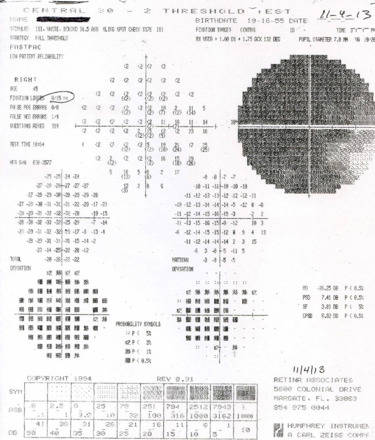
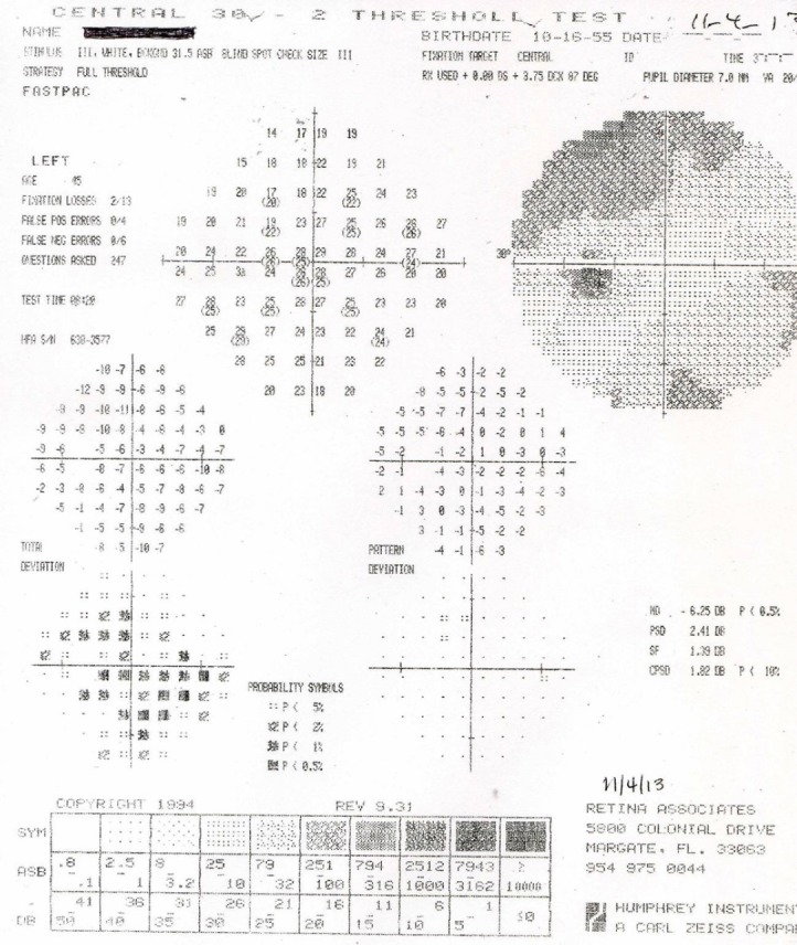
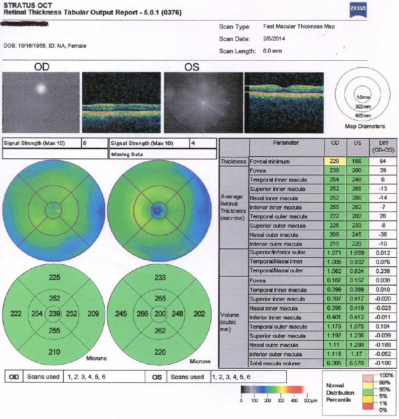
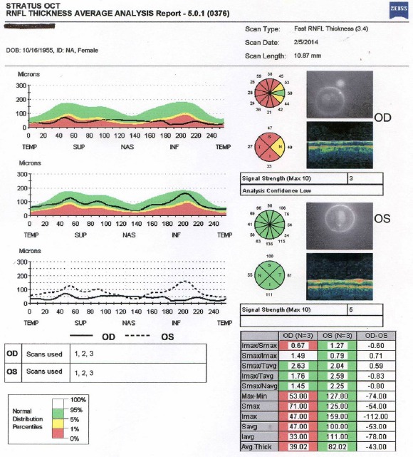
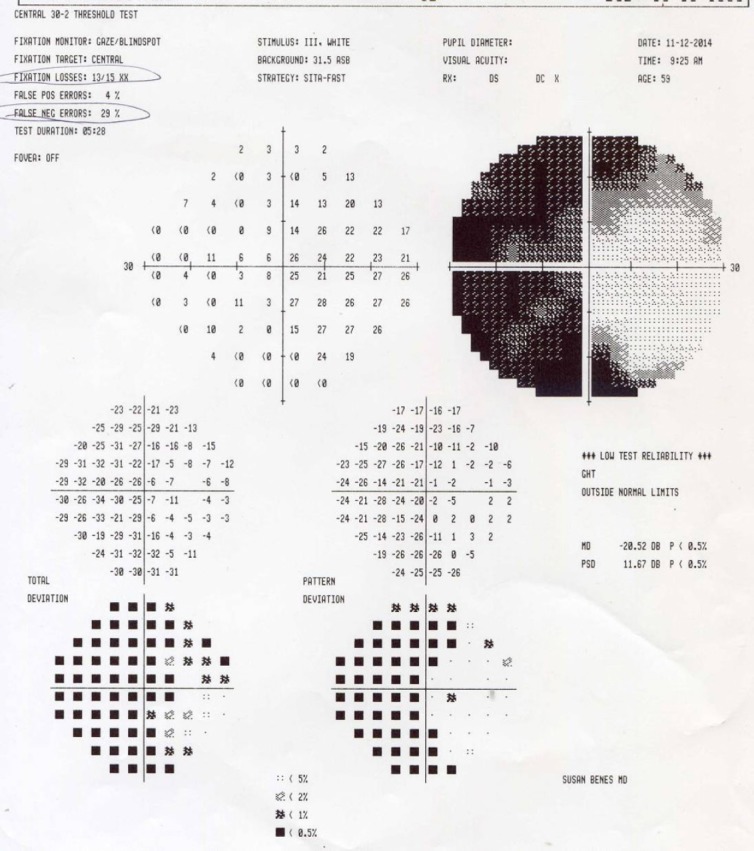
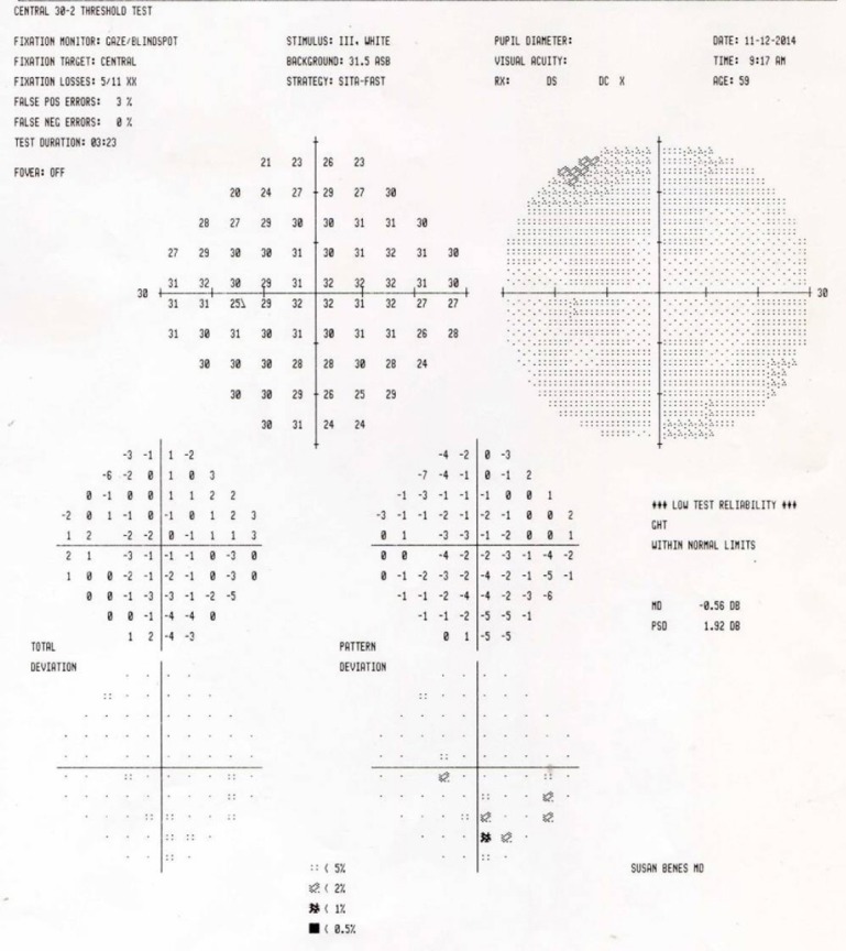
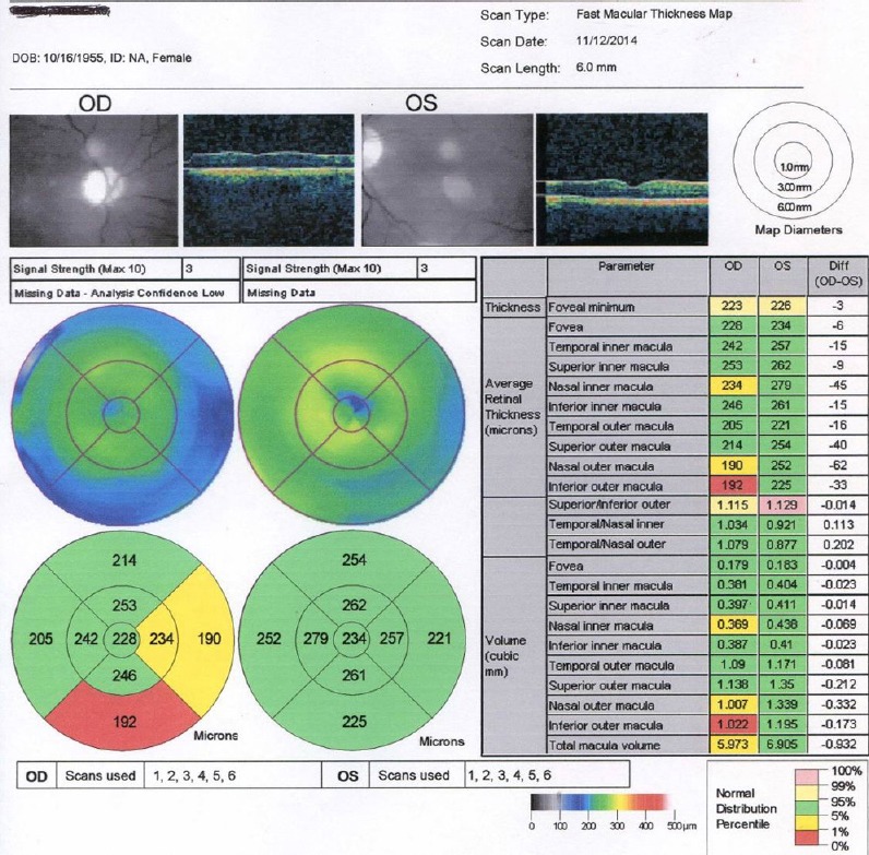
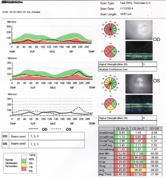
We present the results from a patient with relapsing optic neuropathy treated within the Stem Cell Ophthalmology Treatment Study (SCOTS). SCOTS is an Institutional Review Board approved clinical trial and has become the largest ophthalmology stem cell study registered at the National Institutes of Health to date (www.clinicaltrials.gov Identifier NCT 01920867). SCOTS utilizes autologous bone marrow-derived stem cells (BMSCs) for treatment of retinal and optic nerve diseases. Pre-treatment and post-treatment comprehensive eye exams of a 54 year old female patient were performed both at the Florida Study Center, USA and at The Eye Center of Columbus, USA. As a consequence of a relapsing optic neuritis, the patient's previously normal visual acuity decreased to between 20/350 and 20/400 in the right eye and to 20/70 in the left eye. Significant visual field loss developed bilaterally. The patient underwent a right eye vitrectomy with injection of BMSCs into the optic nerve of the right eyeand retrobulbar, subtenon and intravitreal injection of BMSCs in the left eye. At 15 months after SCOTS treatment, the patient's visual acuity had improved to 20/150 in the right eye and 20/20 in the left eye. Bilateral visual fields improved markedly. Both macular thickness and fast retinal nerve fiber layer thickness were maximally improved at 3 and 6 months after SCOTS treatment. The patient also reduced her mycophenylate dose from 1,500 mg per day to 500 mg per day and required no steroid pulse therapy during the 15-month follow up.
Keywords: Stem Cell Ophthalmology Treatment Study; autoimmune; blindness; bone marrow-derived stem cells; nerve regeneration; neural regeneration; ophthalmology; optic nerve; optic neuropathy; stem cells; visual loss.
Conflict of interest statement
Figures
References
-
- Collongues N, Marignier R, Zéphir H, Papeix C, Blanc F, Ritleng C, Tchikviladzé M, Outteryck O, Vukusic S, Fleury M, Fontaine B, Brassat D, Clanet M, Milh M, Pelletier J, Audoin B, Ruet A, Lebrun-Frenay C, Thouvenot E, Camu W, et al. Neuromyelitis optica in France: a multicenter study of 125 patients. Neurology. 2010;74:736–742. - PubMed
-
- Kok PH, van den Berg TJ, van Dijk HW, Stehouwer M, van der Meulen IJ, Mourits MP, Verbraak FD. The relationship between the optical density of cataract and its influence on retinal nerve fibre layer thickness measured with spectral domain optical coherence tomography. Acta Ophthalmol. 2013;91:418–424. - PubMed
-
- Lennon VA, Wingerchuk DM, Kryzer TJ, Pittock SJ, Lucchinetti CF, Fujihara K, Nakashima I, Weinshenker BG. A serum autoantibody marker of neuromyelitis optica: distinction from multiple sclerosis. Lancet. 2004;364:2106–2112. - PubMed
-
- Mwanza JC, Bhorade AM, Sekhon N, McSoley JJ, Yoo SH, Feuer WJ, Budenz DL. Effect of cataract and its removal on signal strength and peripapillary retinal nerve fiber layer optical coherence tomography measurements. J Glaucoma. 2011;20:37–43. - PubMed
LinkOut - more resources
Full Text Sources
Other Literature Sources
Medical
Research Materials