Giant sialoliths of Wharton duct: Report of two rare cases and review of literature
- PMID: 26604966
- PMCID: PMC4630716
- DOI: 10.4103/1735-3327.166238
Giant sialoliths of Wharton duct: Report of two rare cases and review of literature
Abstract
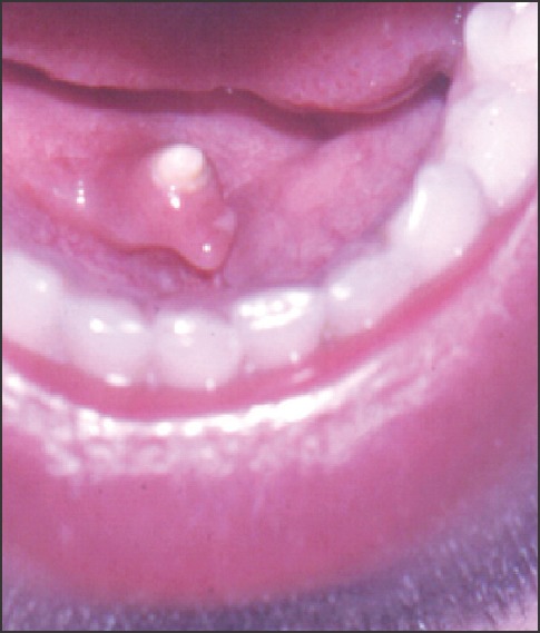
Sialolithiasis is a common disease of the major salivary glands, characterized by the obstruction of a salivary gland or its excretory duct due to the formation of calcareous concretions. Sialoliths usually measure from 1 mm to <10 mm. They rarely measure more than 15 mm, and infrequently giant salivary gland calculi >15 mm have been reported in the literature. The submandibular gland and its duct appear to be the most susceptible sites for this disease. In this article, we report two unique cases, including a giant bilateral case, measuring 50 mm in length and 5 mm in width on the right side and one, 30 mm in length, and 5 mm in width on the left side; and another case, measuring 83 mm in length. The diagnostic and therapeutic approaches consisted of transocclusal radiography with the conservative transoral surgical technique in both cases. The follow-up showed the normal function of the relevant salivary glands. To the best of our knowledge and belief, similar cases have not been reported in the literature.
Keywords: Salivary duct; Sialoliths; Submandibular Glands; Wharton duct.
Figures


Similar articles
-
Giant Salivary Gland Calculi (GSGC): Report Of Two Cases.Open Dent J. 2011;5:90-5. doi: 10.2174/1874210601105010090. Epub 2011 Jul 7. Open Dent J. 2011. PMID: 21760861 Free PMC article.
-
An Unusually Large Submandibular Sialolith: A Case Report.Cureus. 2024 Sep 27;16(9):e70356. doi: 10.7759/cureus.70356. eCollection 2024 Sep. Cureus. 2024. PMID: 39469404 Free PMC article.
-
Submandibular giant sialoliths-2 case reports and review of the literature.Indian J Otolaryngol Head Neck Surg. 2009 Jan;61(Suppl 1):55-8. doi: 10.1007/s12070-009-0019-3. Epub 2009 Mar 21. Indian J Otolaryngol Head Neck Surg. 2009. PMID: 23120671 Free PMC article.
-
Submandibular giant sialoliths: report of two cases and review of the literature.Ear Nose Throat J. 2010 Jun;89(6):E1-4. Ear Nose Throat J. 2010. PMID: 20556723 Review.
-
[Narrative review of imaging studies of calcifications of the submandibular gland].Rev Cient Odontol (Lima). 2023 Mar 26;11(1):e143. doi: 10.21142/2523-2754-1101-2023-143. eCollection 2023 Jan-Mar. Rev Cient Odontol (Lima). 2023. PMID: 38303738 Free PMC article. Review. Spanish.
Cited by
-
Removal of stones from the superficial lobe of the submandibular gland (SMG) via an intraoral endoscopy-assisted sialolithotomy.Clin Oral Investig. 2019 Nov;23(11):4145-4156. doi: 10.1007/s00784-019-02853-9. Epub 2019 Mar 5. Clin Oral Investig. 2019. PMID: 30834990
-
Untreated submandibular megalith for over 60 years.Saudi Med J. 2018 Jul;39(7):729-732. doi: 10.15537/smj.2018.7.22265. Saudi Med J. 2018. PMID: 29968898 Free PMC article.
-
Removal of Large Wharton's Duct Salivary Stones Using a CO2 Laser: A Report of Two Cases.J Lasers Med Sci. 2021 May 10;12:e19. doi: 10.34172/jlms.2021.19. eCollection 2021. J Lasers Med Sci. 2021. PMID: 34733742 Free PMC article.
-
Giant Sialolith of the Submandibular Gland.J Clin Diagn Res. 2017 Aug;11(8):ZJ03-ZJ04. doi: 10.7860/JCDR/2017/29383.10391. Epub 2017 Aug 1. J Clin Diagn Res. 2017. PMID: 28969298 Free PMC article. No abstract available.
-
A Misleading Stone: Case Report of a Giant Sialolithiasis and Review of Literature.Indian J Otolaryngol Head Neck Surg. 2025 Jul;77(7):2704-2710. doi: 10.1007/s12070-025-05507-3. Epub 2025 May 12. Indian J Otolaryngol Head Neck Surg. 2025. PMID: 40503114
References
-
- Ledesma-Montes C, Garcés-Ortíz M, Salcido-García JF, Hernández-Flores F, Hernández-Guerrero JC. Giant sialolith: Case report and review of the literature. J Oral Maxillofac Surg. 2007;65:128–30. - PubMed
-
- Brusati R, Fiamminghi L. Large calculus of the submandibular gland: Report of case. J Oral Surg. 1973;31:710–1. - PubMed
-
- Marchal F, Dulguerov P. Sialolithiasis management: The state of the art. Arch Otolaryngol Head Neck Surg. 2003;129:951–6. - PubMed
-
- Angiero F, Benedicenti S, Romanos GE, Crippa R. Sialolithiasis of the submandibular salivary gland treated with the 810- to 830-nm diode laser. Photomed Laser Surg. 2008;26:517–21. - PubMed
Publication types
LinkOut - more resources
Full Text Sources