Review
. 2015 Oct 5:3:21.
doi: 10.1186/s40364-015-0041-4. eCollection 2015.

Paediatric acute myeloid leukaemia with the t(7;12)(q36;p13) rearrangement: a review of the biological and clinical management aspects

Affiliations
Review

Paediatric acute myeloid leukaemia with the t(7;12)(q36;p13) rearrangement: a review of the biological and clinical management aspects

Sabrina Tosi et al. Biomark Res. .

Abstract

The presence of chromosomal abnormalities is one of the most important criteria for leukaemia diagnosis and management. Infant leukaemia is a rare disease that affects children in their first year of life. It has been estimated that approximately one third of infants with acute myeloid leukaemia harbour the t(7;12)(q36;p13) rearrangement in their leukaemic blasts. However, the WHO classification of acute myeloid leukaemia does not yet include the t(7;12) as a separate entity among the different genetic subtypes, although the presence of this chromosomal abnormality has been associated with an extremely poor clinical outcome. Currently, there is no consensus treatment for t(7;12) leukaemia patients. However, with the inferior outcome with the standard induction therapy, stem cell transplantation may offer a better chance for disease control. A better insight into the chromosome biology of this entity might shed some light into the pathogenic mechanisms arising from this chromosomal translocation, that at present are not fully understood. Further work is needed to improve our understanding of the molecular and genetic basis of this disorder. This will hopefully open some grounds for possible tailored treatment for this subset of very young patients with inferior disease outcome. This review aims at highlighting the cytogenetic features that characterise the t(7;12) leukaemias for a better detection of the abnormality in the diagnostic setting. We also review treatment and clinical outcome in the cases reported to date.

Keywords: Acute myeloid leukaemia; Chromosomal abnormalities; Clinical outcome; HLXB9 gene; Paediatric leukaemia; t(7;12) translocation.


Figures
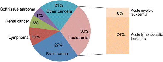
Fig. 1
Fig. 1
Main types of childhood cancers. The ideogram shows the different proportions of cancers affecting paediatric patients. Acute leukaemia is the most commonly reported cancer in children, with ALL affecting approximately 80 % of patients and AML diagnosed in 15–20 % of patients. Forms of chronic leukaemia and myelodysplastic syndromes are very rare in children and their incidence has been omitted from this graph (based on cancers statistics collected from the National Registry of Childhood Tumours, years 2009–2011, accessed through the Cancer Research UK website)
Fig. 2
Fig. 2
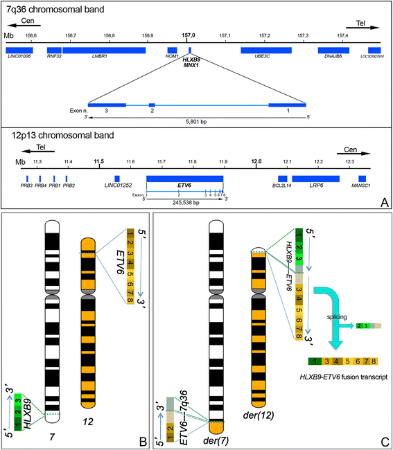
Schematic representation of the t(7;12)(q36;p13) and fusion transcript formation. a Representation of the 7q36 and 12p13 regions spanning 1 Mb around the genes of interests. The breakpoints are proximal to the HLXB9 gene on chromosome 7 and at the 5′ end of the ETV6 gene on chromosome 12. HLXB9 is a small gene composed of three exons. ETV6 is a larger gene composed of eight exons. In both cases the direction of transcription is from the telomeric to the centromeric end. b The gene location and direction of transcription of both HLXB9 and ETV6 are shown on the ideograms of chromosomes 7 and 12. For each gene all the exons are indicated. c Derivative chromosomes 7 and 12: the der(12) harbours the whole HLXB9 gene and the 3′ portion of ETV6 including exons 3–8. If a fusion transcript arose from this translocation, splicing of HLXB9 exons 2 and 3 as well as any genomic material from chromosome 7 downstream to HLXB9 that was translocated on the der(12) should take place
Fig. 3
Fig. 3
Simplified model of radial chromosome and gene organisation in the cell nuclei. Upper left: In the cell nucleus, chromosomes composed of gene dense and gene poor bands are arranged in a zig-zag manner, with the gene poor regions (blue segments) close to the nuclear envelope, in a compact, generally not-transcribed, chromatin organisation. The gene dense regions (red segments) are located in a more internal position, with an open chromatin structure, that favour gene transcription. The interaction of cis-acting or trans-acting sequences, as well as the presence of specific regulatory factors consents gene activation. Upper right: a chromosomal translocation that involves regions normally positioned in different areas of the nucleus could determine an ectopic activation of translocated genes, on the basis of the new nuclear environment where they are repositioned. Bottom panel: Schematic representation of the distribution of FISH signals relative to the regions involved in the t(7;12) rearrangement. Left: Both copies of HLXB9 occupy a peripheral positioning, whereas both copies of ETV6 are localised towards the interior in the nucleus of normal cells. Right: The HLXB9 gene translocated on the der(12) is repositioned towards the nuclear interior, whereas the remaining portion of the ETV6 gene translocated on the der(7) is repositioned towards the nuclear periphery
Fig. 4
Fig. 4
Fluorescence in situ hybridisation (FISH) performed on metaphase chromosomes harbouring the t(7;12)(q36;p13). a FISH using a three colour approach enables the detection of both normal chromosomes 7 (harbouring only blue hybridisation signals) and 12 (harbouring green and orange fluorescent signals) and their derivatives (green signals on the der(7) and blue and orange signals on the der(12)). The DAPI counterstaining of the chromosomes has been converted into grey scale to simulate a G-banding pattern (figure taken from Naiel et al., 2013 [17]). b Schematic representation showing localisation and colour-code of the FISH probes relative to the three colour probe set used. c FISH using a two colour approach enables the detection of both normal chromosomes 7 (harbouring only orange hybridisation signals) and 12 (harbouring only green fluorescent signals) and their derivatives carrying orange and green fusion signals. d Schematic representation showing localisation and colour-code of the FISH probes relative to the two colour probe set used. It should be noted that the two colour set does not allow to discriminate between the two derivatives based on colour pattern only. The size and morphology of chromosomes 7 and 12 compared to their respective derivatives are very similar, making the identification of the rearrangement difficult without the aid of FISH. Both probe sets have been provided by MetaSystems Gmbh, Altlussheim, Germany

References

    1. Cancer statistics for the UK: Cancer research UK. [http://www.cancerresearchuk.org/cancer-info/cancerstats/].
    1. Howlader N, Noone AM, Krapcho M, Garshell J, Neyman N, Altekruse SF, et al. SEER Cancer Statistics Review, 1975–2010, Bethesda, MD; National Cancer Institute. http://seer.cancer.gov/csr/1975_2010/
    1. De Rooij J, Zwaan C, van den Heuvel-Eibrink M. Pediatric AML: from biology to clinical management. J Clin Med. 2015;4:127–49. doi: 10.3390/jcm4010127. - DOI - PMC - PubMed
    1. Ward E, DeSantis C, Robbins A, Kohler B, Jemal A. Childhood and adolescent cancer statistics, 2014. CA Cancer J Clin. 2014;64:83–103. doi: 10.3322/caac.21219. - DOI - PubMed
    1. Vardiman JW, Thiele J, Arber DA, Brunning RD, Borowitz MJ, Porwit A, et al. The 2008 revision of the World Health Organization (WHO) classification of myeloid neoplasms and acute leukemia: rationale and important changes. Blood. 2009;114:937–51. doi: 10.1182/blood-2009-03-209262. - DOI - PubMed

LinkOut - more resources