. 2015 Oct;3(2):122-6.
doi: 10.14791/btrt.2015.3.2.122. Epub 2015 Oct 30.

Multimodal Treatment of Skull Base Inflammatory Pseudotumor: Case Report

Affiliations

Multimodal Treatment of Skull Base Inflammatory Pseudotumor: Case Report

Sang Gun Lee et al. Brain Tumor Res Treat. 2015 Oct.

Abstract

lnflammatory pseudotumor (IPT) is a rare, non-neoplastic inflammatory process. It is most commonly occurs in the orbit, but extension into brain parenchyma is uncommon. In a confirmed case of IPT, most cases show good improvement with steroid theraphy. A 50-year-old man with progressive left-eye visual disturbance and mass lesion was admitted in a hospital. A left orbital mass biopsy revealed what was highly suspected as an inflammatory pseudotumor. Steroid pulse therapy with dexamethasone, radiation therapy, and chemotherapy with amphotericin B were performed, but they were not effective in improving the condition of the patient. Revision open surgery was then performed. A follow-up brain enhancement computerized tomography showed an enlarged mass volume and hydrocephalus with periventricular enhancement. As an additional procedure, ventriculoperitoneal shunt and tuberculosis medication were administered. About 2 weeks later, clinical symptoms and radiologic findings improved. We present a case of intra-cranial IPT and discuss further treatment methods.

Keywords: Cavernous sinus; Central nervous system; Granuloma; Sphenoid sinus.


Conflict of interest statement

The authors have no financial conflicts of interest.

Figures
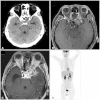
Fig. 1
Fig. 1. Neuroimaging findings on admission. A: Pre-operative brain computed tomography (CT). Spindle shaped, slightly high density is noted in the left anterior cavernous sinus and orbital apex. Pressure erosion of the adjacent bones. B and C: Pre-operative brain magnetic resonance image (MRI) T2-weighted image (B). Pre-operative brain MRI T1-weighted enhanced image (C). Mixed intensity lesion at the left pituitary fossa, orbital apex, and middle cranial fossa, with strong enhancement. D: Whole body positron emission tomography-CT. Hypermetabolic, infiltrative lesion at the left anterior skull base, but there is no other definite metastatic lesion.
Fig. 2
Fig. 2. Pathological findings. A: Bone marrow fibrosis. B: Bone marrow cell infiltration. Photomicrograph shows infiltrating small lymphocytes and uninucleated histiocytes in the fibrous stroma. Lymphoid follicle with surrounding lymphocytes (hematoxylin and eosin stain; A and B: ×100). C: Keratin negative (no carcinoma). In the keratin finding stain, there are no definitive carcinoma. D: Left coronary artery (LCA) positive (infiltration). LCA positive findings prove there are multiple infiltrations (C and D: ×40).
Fig. 3
Fig. 3. Neuroimaging findings at post operative periods. A: First post-operative brain computed tomography (CT) (craniotomy with tumor removed). Decreased high density lesion at the left anterior cavernous sinus and orbital apex with hyodrocephalus. B: Second post-operative brain CT (ventriculoperitoneal shunt). Ventriculoperitoneal shunt state. C: Follow-up brain magnetic resonance image T1-weighted enhanced image at post-operative period. Hyper intensity with strong enhancement lesion was markedly decreased. D: Follow-up brain CT at post-operative period 6 months later. Decreased high density lesion with ventricle size.

References

    1. Gandhi RH, Li L, Qian J, Kuo YH. Intraventricular inflammatory pseudotumor: report of two cases and review of the literature. Neuropathology. 2011;31:446–454. - PubMed
    1. Saifudheen K, Jose J, Gafoor VA. Inflammatory pseudotumor of the head presenting with hemiparesis and aphasia. Case Rep Neurol Med. 2011;2011:176546. - PMC - PubMed
    1. Ohue S, Kohno S, Matsui S, Kumon Y, Ohnishi T. Inflammatory pseudotumor in the lateral ventricle. Case report. Neurol Med Chir (Tokyo) 2012;52:599–602. - PubMed
    1. Lin YJ, Yang TM, Lin JW, Song MZ, Lee TC. Posterior fossa intracranial inflammatory pseudotumor: a case report and literature review. Surg Neurol. 2009;72:712–716. discussion 716. - PubMed
    1. Lee EJ, Jung SL, Kim BS, et al. MR imaging of orbital inflammatory pseudotumors with extraorbital extension. Korean J Radiol. 2005;6:82–88. - PMC - PubMed